
Abstract
Prevalence and type of neoplastic disease were determined in 551 camelid submissions (368 alpacas [Lama pacos], 180 llamas [Lama glama], and 3 cases in which species was not identified) over a 5-year period. Forty neoplasms were identified in 38 animals (6.9%). Prevalence of neoplasia in llamas was higher (11%) than in alpacas (4.9%). Mean age of camelids with neoplasia was 9.42 ± 4.9 years. Mean age of alpacas with neoplasia (5.48 ± 3.7 years) was significantly less than of llamas with neoplasia (12.53 ± 3.2 years; P < 0.001). Cutaneous and mucocutaneous fibroma/fibropapilloma was most common (10 animals), followed by cutaneous and mucocutaneous squamous cell carcinoma (6 animals), disseminated lymphoma (5 animals), and fibrosarcoma (4 animals). Four of 5 animals with lymphoma were alpacas, aged 0.21 to 4 years. Lymphoma occurred in 1 aged llama (15 years). Disseminated carcinoma and adenocarcinoma occurred in 4 llamas and 2 alpacas, and included biliary (2), gastrointestinal (2), mammary gland (1), and unknown (1) origin. Mean age of camelids with any type of carcinoma or adenocarcinoma (12.36 ± 2.8 years) was significantly greater than that of camelids with lymphoma (4.24 ± 6.2 years; P = 0.02). Results indicate that neoplasia is relatively common in camelids and that there are differences between llamas and alpacas as regards prevalence of neoplasia, tumor types, and age at diagnosis.
There are numerous reports of neoplasia in South American camelids, including lymphoma, 3,10,12,16,21,24,25 urogenital neoplasia, 3,4,7,8,15,24,25 cutaneous and mucocutaneous neoplasia, 17,20,23,25 oral neoplasia, 1,2,14,24,26 intraocular neoplasia, 3,5,11 malignant gastrointestinal neoplasia, 22,25 mammary carcinoma, 3,13,25 pulmonary neoplasia, 18,19 neuroendocrine neoplasia, 21 congenital hepatic neoplasia, 27 osseous neoplasia, 9 and neoplasia of the brain. 6 Disseminated lymphoma is the most commonly reported tumor in llamas and alpacas. 3,10,12,16,21,24,25 Only 2 prior studies have looked at overall prevalence and types of neoplasia in camelids. 3,24 Excluding abortions, the prevalence of neoplastic disease in 72 llamas and alpacas submitted for necropsy examination at 1 laboratory over a 7-year period was 8.3%. 24 Biopsy samples were not included in that study. A 1% prevalence of neoplasia was reported in a teaching hospital population of camelids, but that study was limited to malignant neoplasia. 3
The purpose of this report is to describe the prevalence and types of neoplasia diagnosed by routine histopathologic evaluation in camelid biopsy and necropsy samples examined at the Veterinary Diagnostic Laboratory at Oregon State University, which provides service to an area in which llamas and alpacas are common livestock, over a 5-year period. Microscopic sections from all cases were reviewed by one author (BAV). Features of neoplasia in llamas were compared to those in alpacas. Tumor prevalence and mean age ± SD were calculated, and comparisons were performed using the Student's t-test assuming unequal variance. One case in this study has been previously reported. 7
Excluding abortions, from July 2001 to July 2006 there were 551 camelid submissions for biopsy or necropsy examination. Submissions were from 368 alpacas (Lama pacos) and 180 llamas (Lama glama); species was not identified in 3 cases. Forty neoplasms were identified in 38 animals (overall prevalence of 6.9%). Results are summarized in Table 1. Thirty-four animals were from the Pacific Northwest, 3 were from the Midwest, and 1 was from Colorado. Neoplasia was identified in 20 llamas (11% prevalence) and 18 alpacas (4.9% prevalence). Neoplasia occurred in 21 females and 14 males (intact and castrated); gender of 3 animals was not specified. Excluding tumors of mammary gland and genital tract, no gender bias was apparent. Mean age of all camelids with neoplasia was 9.42 ± 4.9 years (range 0.21-19 years). Mean age of alpacas with neoplasia (5.48 ± 3.7 years; range 0.21-12 years) was significantly less than that of llamas with neoplasia (12.53 ± 3.2 years; range 5–19 years; P < 0.001).
Cutaneous and mucocutaneous neoplasms were most common (22 animals), particularly fibroma/fibropapilloma (12 tumors from 10 animals), squamous cell carcinoma (6 animals), and fibrosarcoma (4 animals). Four animals with fibroma/fibropapilloma had multiple lesions. Only 1 of these tumors, from the face of a 4-year-old alpaca with multiple lesions, exhibited convincing viral change (koilo-cytes). Mean age of animals with fibroma/fibropapilloma was 6.9 ± 3.5 years, range 4–12 years. Mean age of alpacas with fibroma/fibropapilloma (5.6 ± 2.6 years, range 4–11 years) was significantly less than that of llamas with fibroma/fibropapilloma (11.5 ± 0.7 years, range 11–12 years; P < 0.001). Cutaneous and mucocutaneous squamous cell carcinoma was more common in llamas (5 animals) than in alpacas (1 animal). Squamous cell carcinoma involved haired skin as well as perineal and ocular tissue. Fibrosarcoma was distinguished from fibroma/fibropapilloma based on high tumor cellularity, tumor cell pleomorphism, and presence of mitotic activity; it has not been previously reported in camelids. All tumors diagnosed as fibrosarcoma involved cutaneous or mucocutaneous tissues of the head.
Summary of 40 tumors in 20 llamas and 18 alpacas in a 5-year period.
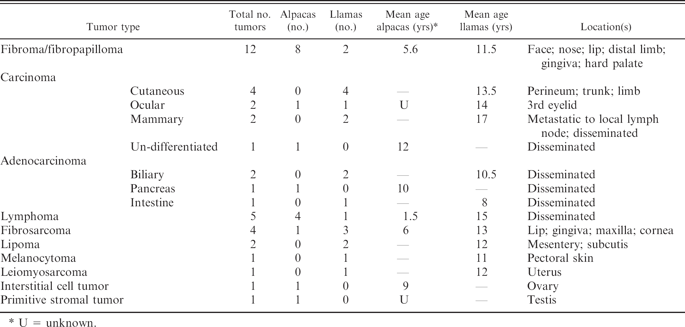
U = unknown.
Disseminated lymphoma was diagnosed in 5 animals (4 alpacas and 1 llama), mean age 4.24 ± 6.2 years (range 0.21-15 years). All alpacas with lymphoma were 4 years of age or less. Disseminated carcinoma or adenocarcinoma occurred in 4 llamas and 2 alpacas, and included tumors of biliary (2), gastrointestinal (2), mammary gland (1), and unknown (1) origin. Mean age of camelids with any type of carcinoma or adenocarcinoma (12.36 ± 2.8 years, range 8–17 years) was significantly greater than that of camelids with lymphoma (P = 0.02).
Cutaneous and oral fibroma/fibropapilloma was the most common neoplasm identified in this study, and was diagnosed in animals from 4 to 12 years of age. Multiple lesions, either submitted or reported in the history, were common (4 of 10 animals). Schulman et al. 23 described mucocutaneous fibropapillomas with histopathologic features similar to equine sarcoid in five 6-year-old camelids with single to multiple lesions. Papillomavirus DNA was detected by polymerase chain reaction (PCR), and the causative virus was determined to be a unique papilloma-virus. 23 Several cases in this current study also had features similar to equine sarcoid, and most occurred on the head as previously reported. 23 Others exhibited interlacing bundles of dense collagen more consistent with fibroma. This was particularly true of 2 of 3 tumors occurring on the distal limb and of the lesion on the hard palate. Overlying epithelium was not present for evaluation in all cases diagnosed as fibroma, and PCR testing for papillomavirus was not performed. Therefore, in this study all such lesions were grouped together with tumors diagnosed as fibropapilloma. It is suspected, however, that nonviral-associated fibroma may also occur in camelids.
Cutaneous and mucocutaneous squamous cell carcinoma was the most frequent malignant neoplasm identified in this population. Gastric squamous cell carcinoma has been reported in 5 South American camelids, 22 but none were found in this study or in a prior survey. 24 The prevalence of camelid squamous cell carcinoma in the Colorado study was also high, just slightly less than that of lymphoma, but tumor site was not specified. 3
Lymphoma, the most common malignant neoplasm in camelids in 2 previous studies, 3,24 was the second most common malignant neoplasm in this current study, and frequently occurred in young animals. The mean age of the 5 camelids with lymphoma (4.24 years) was very similar to that in a previous report of 10 cases (4.6 years). 3 The diagnosis of lymphoma in this current study was based on gross pathologic and routine histopathologic findings of disseminated malignant round cell neoplasia. Sartin et al. 21 found that 1 of 4 alpacas diagnosed as lymphoma based on gross pathologic and routine histopathologic examination did not express lymphocyte markers, and expression of neuron specific enolase and synaptophysin suggested neuroendocrine neoplasia. It is possible that some cases diagnosed as lymphoma in this study are neuroendocrine in nature, and additional immunohistochemical studies are needed.
Reports of disease processes typically combine data from llamas and alpacas. This study indicates that, as regards neoplasia, llamas and alpacas differ in prevalence, tumor types, and age affected. Overall prevalence of neoplasia was higher in llamas than in alpacas. Mean age of alpacas with neoplasia was, however, significantly lower than that of llamas with neoplasia. Regional differences may also exist regarding prevalence and type of neoplasia in llamas and alpacas.
Acknowledgements. The authors thank Rocky Baker for assistance with data retrieval and Dr. Chris Cebra for helpful input.