
Abstract
Gangrenous dermatitis (GD) is a disease of chickens and turkeys that causes severe economic losses in the poultry industry worldwide. Clostridium septicum, Clostridium perfringens type A, and occasionally Clostridium sordellii are considered the main causes of GD, although Staphylococcus aureus and other aerobic bacteria may also be involved in some cases of the disease. GD has become one of the most significant diseases of commercial turkeys in the United States. Several infectious and/or environmental immunosuppressive factors can predispose to GD. Skin lesions are considered to be the main portal of entry of the microorganism(s) involved. GD is characterized by acute onset of mortality associated with gross skin and subcutaneous tissue lesions consisting of variable amounts of serosanguineous exudate together with emphysema and hemorrhages. The underlying skeletal muscle can also be involved. Ulceration of the epidermis may be also noticed in cases complicated with S. aureus. Microscopically, necrosis of the epidermis and dermis, and subcutaneous edema and emphysema are commonly observed. Gram-positive rods can be identified within the subcutis and skeletal muscles, usually associated with minimal inflammatory infiltrate. A presumptive diagnosis of GD can be made based on history, clinical signs, and gross anatomic and microscopic lesions. However, confirmation should be based on demonstration of the causative agents by culture, PCR, immunohistochemistry, and/or fluorescent antibody tests.
Introduction
Gangrenous dermatitis (GD) is a disease that affects primarily commercial broiler chickens and turkeys, and it is responsible for severe economic losses in the poultry industry worldwide. 42 The disease is also called blue wing disease in chickens and cellulitis in turkeys. The condition is characterized by congestion, hemorrhage, and necrosis of the skin and subcutaneous tissue, associated with edema and/or emphysema, which sometimes extends into the underlying musculature. The most significantly affected areas include breast, back, abdomen, thighs, tail and wings (USDA-APHIS, 2011, Clostridial dermatitis in U.S. commercial broilers and turkeys, https://goo.gl/Xnirdc).10,53,68,73
GD is primarily caused by Clostridium septicum and Clostridium perfringens type A, acting singly or in combination (Table 1).8,53,68,71,73 However, cases of GD may also be caused by a myriad of anaerobic and aerobic bacteria including Clostridium sordellii, Clostridium novyi, Staphylococcus aureus, Staphylococcus epidermidis, Staphylococcus xylosus, Escherichia coli, Enterococcus faecalis, Pasteurella multocida, Gallibacterium anatis biovar haemolytica, Proteus spp., Pseudomonas aeruginosa, Bacillus spp., and Erysipelothrix rhusiopathiae (Table 1; Andrada M, et al. Dermatitis gangrenosa en pollos de engorde: caso clínico [Gangrenous dermatitis in broilers: a clinical case]. Proc XVIII Reunión de la Sociedad Española de Anatomía Patológica Veterinaria; 2006 Jun 28–30; Rabat, Morocco. Spanish, https://goo.gl/DwKifa; https://goo.gl/Xnirdc).1,9,10,17,53,55,56,67,68,73
Anaerobic and aerobic bacterial species involved in gangrenous dermatitis of chickens and turkeys.
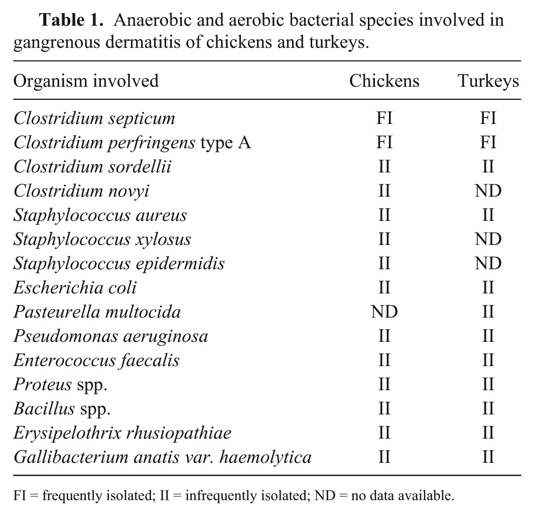
FI = frequently isolated; II = infrequently isolated; ND = no data available.
Although GD has been recognized for many years as a sporadic disease, 73 the prevalence and severity of this condition has increased over the past two decades in the United States and elsewhere. GD is currently considered one of the 3 most significant diseases of commercial turkeys in the US (USDA-APHIS, 2012, Role of intestinal pathology and clostridial species in clostridial dermatitis on U.S. turkey-grower farms, https://goo.gl/tiuETr), and it was listed among the most frequently diagnosed diseases in commercial broiler chickens in California in January 2010–December 2012 (Carnaccini S, et al. Summary of diseases diagnosed in broiler chickens submitted to the California Animal Health and Food Safety Laboratory System, 2010–2012. Proc 62nd West Poult Dis Conf; 2013 Mar 25–27; Sacramento, CA, https://goo.gl/vg4Xwf).
GD was first reported in the United States in the early 1930s 48 ; Clostridium welchii (now C. perfringens) was isolated from heart blood and liver of 2 chickens. The disease was reproduced experimentally in chickens by intramuscular inoculation of this isolate, causing severe necrosis of the skeletal muscles and subcutaneous tissue. 48 Later, GD was diagnosed in chickens suffering heavy mortality. C. welchii, C. septicum, and C. novyi were isolated from the tissues of the dead birds. 13
GD has received different names over the years, including necrotic dermatitis, gangrenous cellulitis, gangrenous dermatomyositis, spontaneous clostridial myonecrosis, poultry gangrene, avian malignant edema, gas edema disease, subcutaneous emphysema, tailitis, blue wing disease, and wing rot.32,35,53,68,73 However, as of 2017, gangrenous dermatitis is the preferred and most widely used name (https://goo.gl/Xnirdc; https://goo.gl/tiuETr).10,35
Etiology
Clostridia
Genus Clostridium, which belongs to phylum Firmicutes, class Clostridia, order Clostridiales, family Clostridiaceae, 59 is composed of anaerobic, mostly gram-positive, spore-forming rods.12,53,59
C. perfringens type A is the most frequently reported toxinotype of this bacterial species involved in GD outbreaks.53,68 All type A isolates produce alpha toxin (CPA); some strains may also produce one or more additional toxins including necrotic enteritis B–like toxin (NetB) and enterotoxin (CPE). 58 The phylogenetic relation of 139 C. perfringens strains isolated from chickens and turkeys with necrotic enteritis or GD was studied by multilocus sequence typing (MST). 32 The study demonstrated that GD-associated C. perfringens isolates are significantly different from isolates obtained from cases of necrotic enteritis. 32 The role of specific toxins in the pathogenesis of GD is still unknown, although CPA has been suggested to play the most critical role. 68
The main virulence factor of C. septicum is alpha toxin (ATX), a necrotizing pore-forming toxin (PFT), which is unrelated to the alpha toxin of C. perfringens. ATX induces increased membrane permeability, which leads to cell necrosis. C. septicum also produces septicolysin, another PFT, which is thought to have a synergistic effect with ATX in the pathogenesis of gas gangrene lesions. 57 C. septicum isolates (n = 109) obtained from turkeys and chickens with GD were analyzed by MST. Most of the C. septicum isolates belonged to a predominant clonal population composed of 2 clusters with little genetic diversity. 47 Based on this finding, it has been hypothesized that only certain strains of C. septicum are implicated in cases of GD in poultry. Several authors have suggested that ATX plays a critical role in the pathogenesis of GD.10,53,68 This is supported by the ATX effect on endothelial cells, causing fluid extravasation, and the possible synergistic effect that septicolysin has with ATX. 57
C. sordellii produces 2 main toxins, namely lethal toxin (TcsL) and hemorrhagic toxin (TcsH); both of which are glycosylating. In addition, most strains of this microorganism produce sordellilysin, phospholipase, neuraminidase, and collagenase. 57 No information is currently available on the role that any of the C. sordellii toxins have in the pathogenesis of GD.
Staphylococcus spp
Genus Staphylococcus is composed of gram-positive, coccoid-shaped, aerobic bacteria, which are commonly seen as clusters when grown in solid media and short chains when cultured in liquid media. 2 S. aureus is the most common non-clostridial bacterial species associated with GD. 2 This microorganism is able to cause GD alone or in combination with one or more clostridial species. Other species of this genus that have been found in outbreaks of GD in broiler chickens include S. xylosus and S. epidermidis (Andrada M, et al. Dermatitis gangrenosa en pollos de engorde: caso clínico [Gangrenous dermatitis in broilers]. Proc XVIII Reunión de la Sociedad Española de Anatomía Patológica Veterinaria; 2006 Jun 28–30; Rabat, Morocco. Spanish). 73 However, these microorganisms can also be found in skin and nares of healthy chickens, and isolation from chickens with GD does not necessarily confirm a causative role in this disease. 2 S. aureus can produce several toxins, including hyaluronidase, deoxyribonuclease, fibrinolysin, lipase, protease, leucocidin, hemolysins, epidermolytic toxin, and dermonecrotic toxin. 31 Following intradermal inoculation in poultry and rabbits, 9 dermonecrotic toxin induced severe dermal inflammation and skin necrosis at the injection site, which supports a possible role of this toxin in the pathogenesis of GD.
Other aerobic bacteria
E. coli isolates obtained from cases of cellulitis, swollen head syndrome, and colisepticemia in chickens produce a vacuolating cytotoxin. This toxin is specific for avian cells and appears to be similar to the one produced by Helicobacter pylori. 64 The role of this toxin in cases of GD is unknown. Serogroup D P. multocida have been associated with dermal lesions in poultry with GD. Strains containing a heat-labile protein have been isolated from turkeys. Dermal inoculation of sonicated suspensions of these strains produced necrotic lesions in turkey skin.61,62 Facial cellulitis associated with P. multocida in turkeys has also been described. 37 P. aeruginosa is characterized by a wide virulence repertoire, including extracellular and cellular components. Most strains produce exotoxin A, responsible for tissue necrosis with a mechanism similar to that of diphtheria toxin. 11 , 19 The most important virulence factor of E. rhusiopathiae is a neuraminidase that promotes adhesion and spreading of the pathogen. 74 Whether this toxin plays a role in the pathogenesis of GD is not known.
Pathogenesis
The proposed pathogenesis of GD in chickens and turkeys is summarized in Figures 1 and 2, respectively. Several authors suggested that immunosuppression may be the key predisposing factor for GD in chickens and turkeys.10,33,53,68,73 A retrospective study of GD in broiler chickens, including cases that occurred in 1995–2006 in Alabama, described severe lymphocytic depletion of the thymus and bursa of Fabricius, associated with the onset of GD (Hoerr F. Case reports from Alabama. Proc 56th West Poult Dis Conf; 2007 Mar 26–29; Las Vegas, NV, https://goo.gl/vg4Xwf). Experimentally, intramuscular administration of dexamethasone successfully predisposed to the development of GD after subcutaneous challenge with C. septicum or C. perfringens in turkey poults. 72
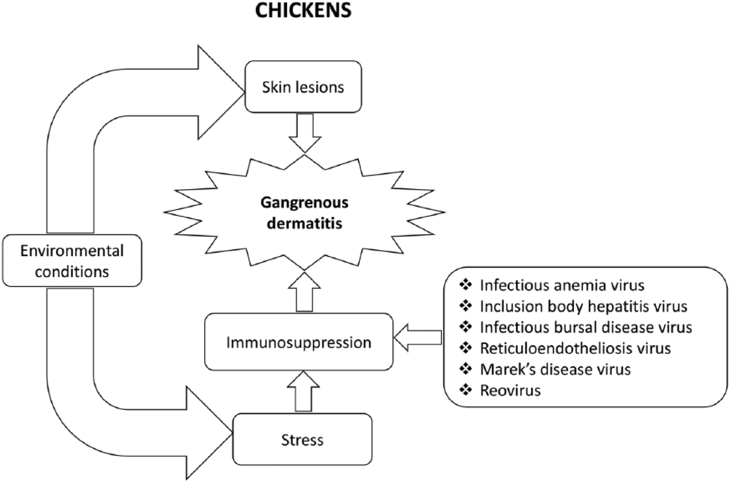
Proposed pathogenesis of gangrenous dermatitis in chickens.
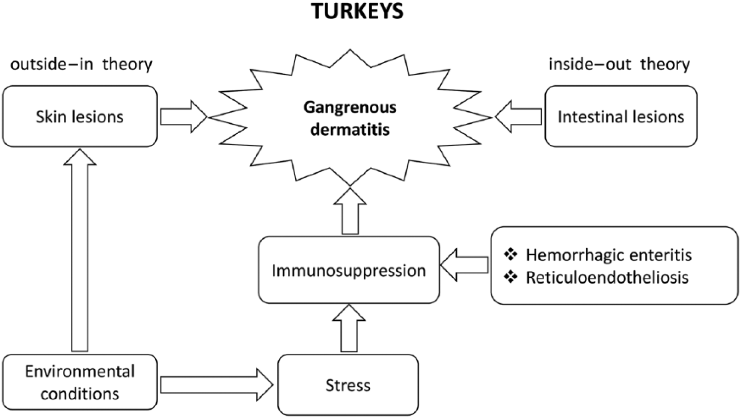
Proposed pathogenesis of gangrenous dermatitis in turkeys.
Under natural conditions, immunosuppression can be triggered by a wide range of infectious and environmental factors in both chickens and turkeys.10,33,53,68,73 Immunosuppressive viral agents that may predispose to GD in chickens and turkeys include Marek’s disease virus, infectious bursal disease virus, chicken anemia virus, several reoviruses, and reticuloendotheliosis virus.10,20,33,34,53,67,68,73 Other viral infectious agents such as inclusion body hepatitis virus53,73 in chickens and hemorrhagic enteritis virus in turkeys have also been suggested as possible immunosuppressive agents that may trigger GD.10,33
Environmental factors that can predispose chickens and turkeys to GD10,33,53 are: 1) traumatic lesions of the skin associated with cannibalism and/or fighting (the latter being more common in turkeys); overcrowding; feed outages; deficient diets; and 2) wet and poor litter conditions; contaminated feed, water, equipment, and vaccines; high ammonia levels; and mycotoxins (e.g., aflatoxins, trichothecenes, fumonisins, and ochratoxins) in feed.10,16,33,44,53,68
In broiler chickens, GD is mainly predisposed to by traumatic damage of the skin, usually associated with cannibalism and overcrowding. Such skin lesions provide a port of entry for bacteria.49,53,65,75 However, GD was also reported, albeit infrequently, in heavy broiler chickens with grossly intact skin. In those cases, the predisposing factor was considered to be anaerobiosis generated by subcutaneous tissue necrosis associated with trauma of the pectoral region, as a result of prolonged ventral recumbency. Chaotic multiplication of the intestinal flora followed by absorption into the bloodstream promoted bacteremia, which was thought to be the origin of some of the GD lesions.34,66 There is anecdotal evidence of a similar pathogenesis of GD in broiler chickens as a result of the use of ionophores in the feed associated with toxic muscle damage (authors’ unpublished observations).
The pathogenesis of GD in turkeys is not fully understood.10,43,68 As of 2017, 2 models are being investigated in this species. The first model is the so-called “inside-out” model, which considers that the first step in pathogenesis is intestinal overgrowth and/or loss of integrity of the intestinal mucosa allowing the clostridia responsible for GD to reach the bloodstream. These organisms then reach the muscle and subcutaneous tissue hematogenously (https://goo.gl/tiuETr).10,43 Transmission of these microorganisms from one animal to another would occur via the orofecal route. The second model, the so-called “outside-in” model, resembles the pathogenesis of GD in chickens, and suggests the entry of microorganisms into the subcutaneous tissue through moist or damaged skin.10,68
Several experimental challenge models have been used to clarify the role of C. septicum, C. perfringens, and S. aureus in the pathogenesis of GD.70,71,76 Four-wk-old broiler chickens were inoculated simultaneously by subcutaneous and intramuscular routes with C. septicum and/or S. aureus; mortality rates were much higher in chickens challenged with both microorganisms than in those inoculated with either isolate separately. 76 GD lesions and death were reproduced in 10-wk-old breeder turkeys by intravenous administration of C. septicum or C. perfringens (untyped). Three- and 7-wk-old turkeys were challenged subcutaneously with C. septicum and C. perfringens type A, separately or in combination. 71 Although both C. perfringens and C. septicum caused cellulitis and mortality when inoculated combined or separately, C. septicum was found to be more effective than C. perfringens in causing cellulitis and mortality. 71 Oral challenge with either C. septicum or C. perfringens showed only limited success reproducing GD, supporting the idea that skin lacerations are the main port of entry for most or all of the microorganisms involved in GD. 43
Epidemiology and clinical signs
GD has been described in chickens in several countries, including Argentina, 7 Australia, 5 , 60 Bulgaria, 16 Egypt, 4 Hungary, 36 India, 39 Japan, 66 New Zealand, 44 Nigeria, 51 Spain (Andrada M, et al. Gangrenous dermatitis in broilers; a clinical case), United Kingdom, 22 and the United States (https://goo.gl/Xnirdc; https://goo.gl/vg4Xwf).23,30,33,34,42,65,75,77 In turkeys, GD has been reported only in the United States (https://goo.gl/Xnirdc)8,43
GD is commonly observed in close-to-market age (>35 d) broiler chickens and turkeys (>13–16 wk). 68 GD has been associated with increased condemnation rates and downgrading of chicken and turkey carcasses at slaughter. 68 Affected flocks typically show daily mortality ranging from a few birds to 3%. 68 However, mortality of up to 60% was reported in some flocks.53,68
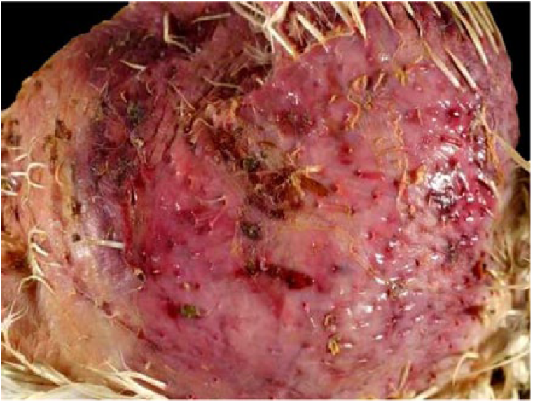
The disease can occur without clinical signs being observed. However, high fever, depression, anorexia, ataxia, leg weakness, and lateral recumbency are usually seen in both chickens and turkeys. 68 The lower abdomen and inner thighs are frequently affected by the accumulation of subcutaneous edema. 68 The skin over affected areas is usually featherless and can show dark-red, purple, green, or green-blue discoloration (Fig. 3). The most frequently affected areas of the body are breast, abdomen, back, thighs, legs, and wings (Fig. 4).10,16,42,53,68
Affected featherless and wet skin area showing diffuse dark-red to purple discoloration in a chicken with gangrenous dermatitis.
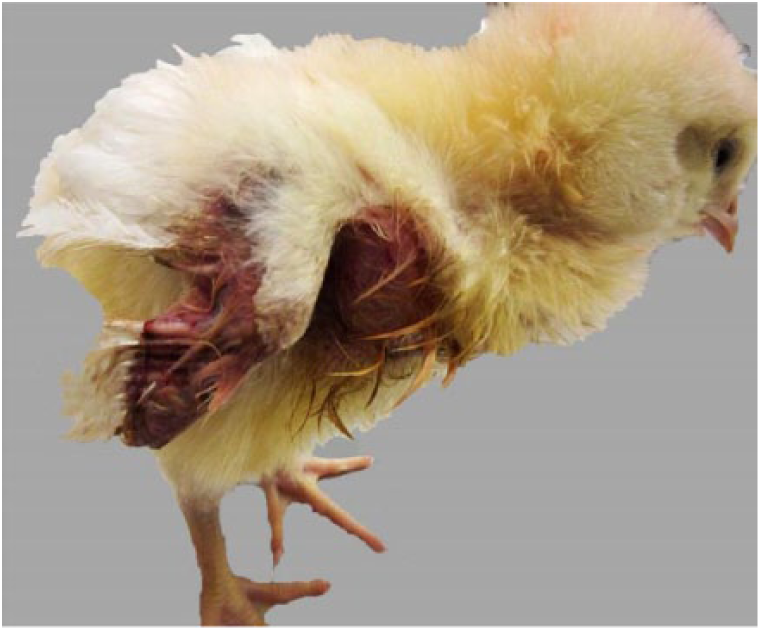
Unilateral blue wing disease showing wet, edematous, hyperemic skin and caking feathers in a chick with gangrenous dermatitis.
Gross anatomic lesions
Rapid autolysis is a common and predominant feature of carcasses affected by GD, particularly in cases of sudden death. Feathers can be easily removed from the affected skin areas. 67 Extensive amounts of edema mixed with gas, and multifocal-to-coalescent hemorrhages can be present in the subcutaneous tissue (Fig. 5).10,16,42,53,68 Abrasions are usually present in the overlying skin of affected birds, although cases without obvious pre-existing skin lesions have also been reported. 53 The underlying skeletal muscle can show gray or tan discoloration, hemorrhages, edema, and gas between muscle bundles (Fig. 6).42,53 Vesicle-like lesions and edema, together with soft, blood-filled or broken feather shafts, have been described in the tail region of turkeys. 53
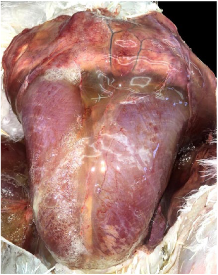
Severe subcutaneous edema, emphysema, and hemorrhage in a turkey with gangrenous dermatitis.
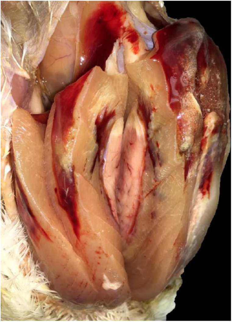
Pale areas of discoloration and multifocal-to-coalescing hemorrhagic areas in breast muscles in a turkey with gangrenous dermatitis.
Microscopic lesions
In uncomplicated cases of GD associated with C. perfringens, necrotic changes are usually seen in the epidermis and dermis. 67 Cases coinfected with S. aureus are usually characterized by ulceration of the epidermis and necrosis in dermis and subcutis (Fig. 7). 68 Subcutaneous tissue has accumulations of serofibrinous exudate and emphysema (Figs. 8, 9).5,67,68 Examination of the underlying skeletal muscle reveals variable degrees of degeneration and necrosis together with congestion, hemorrhages, and mild inflammatory cell infiltrates (Fig. 10).53,68 Uncomplicated cases are characterized by the presence of numerous gram-positive, usually non-sporulated, bacilli, singly or in clusters, which are commonly observed within the areas of hemorrhage and subcutaneous edema (Fig. 11). The lack of a prominent inflammatory cell response is characteristic of such cases. 68 In cases of GD complicated with S. aureus coinfection, gram-positive cocci mixed with variable numbers of heterophils can be observed. 68 The liver and spleen of affected birds can show randomly scattered, small foci of coagulative necrosis associated with intralesional bacterial colonies secondary to hematogenous spread of bacteria from the skin, subcutis, and muscle.10,42,43,53,55,67,68
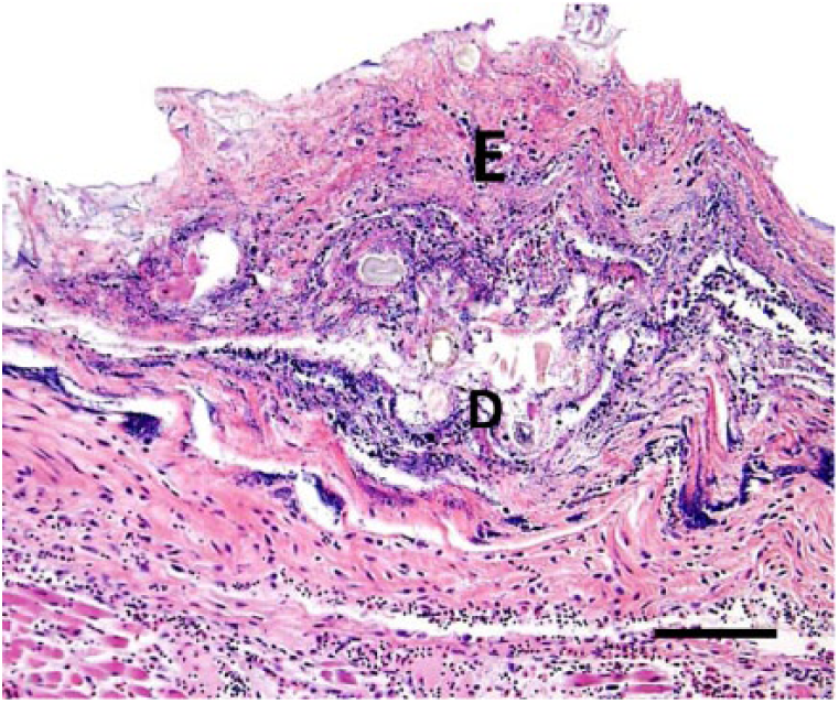
Focal necrosis of epidermis (E) and dermis (D) with moderate infiltration of mixed inflammatory cells in a chicken with gangrenous dermatitis. H&E. Bar = 50 μm.
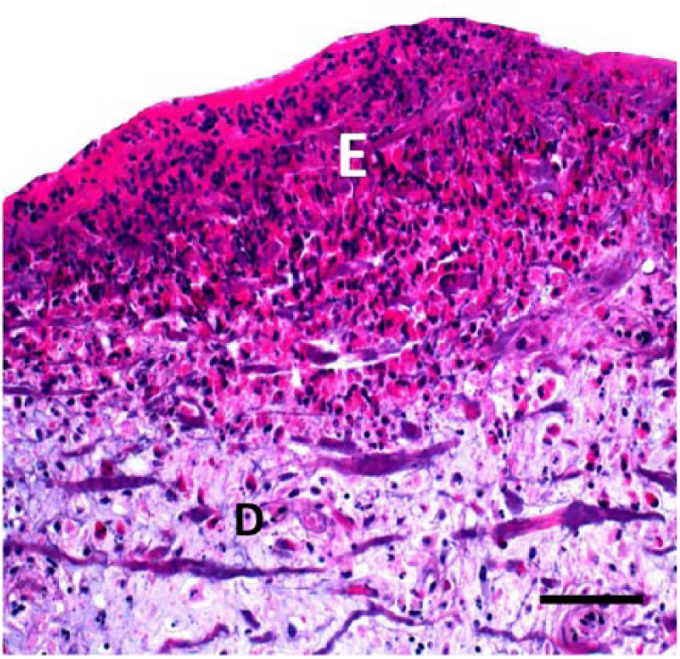
Focal necrosis of epidermis (E) and dermis (D) with inflammatory exudate and edema extending to subcutis in a chicken with gangrenous dermatitis. H&E. Bar = 50 μm.
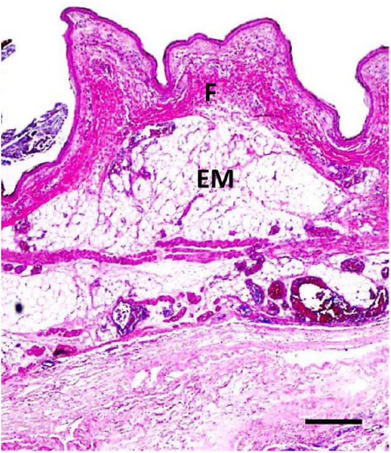
Marked fibrinous exudate (F) and emphysema (EM) in the subcutis of a chicken with gangrenous dermatitis. H&E. Bar = 70 μm.
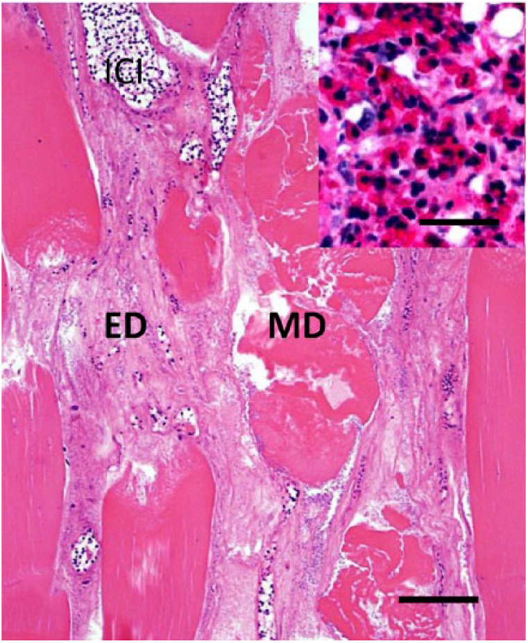
Skeletal myodegeneration (MD) and inflammatory cell infiltrate (ICI) and edema (ED) in a chicken with gangrenous dermatitis. H&E. Bar = 50 μm. Inset: higher magnification of inflammatory cells. Bar = 20 μm.
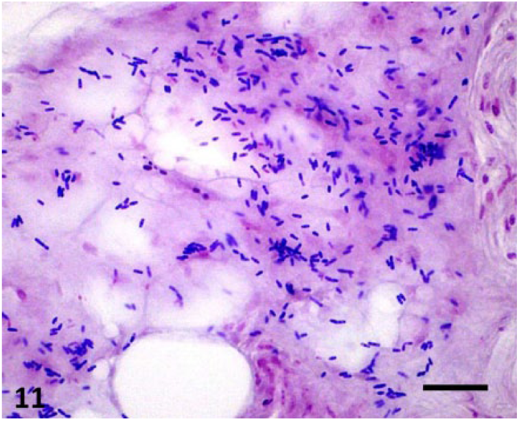
Numerous gram-positive, non-sporulated, bacilli within an area of subcutaneous edema in a chicken with gangrenous dermatitis. Gram. Bar = 10 μm.
Diagnosis
Epidemiology, clinical signs, and gross anatomic and microscopic changes are highly suggestive of GD and allow the establishment of a presumptive diagnosis. The observation of gram-positive rods on smears of the serosanguineous exudate, collected from affected skin and/or subcutaneous tissue, adds certainty to the presumptive diagnosis.53,68 Confirmation of a diagnosis of GD must be based, however, on demonstration of the microorganism(s) involved. Detection can be done by isolation or PCR demonstration of the bacterial species involved.10,53,68,73 Immunohistochemistry and fluorescent antibody tests are also helpful ancillary techniques to confirm the involvement of specific microorganisms. 68 GD must be differentiated from a wide range of infectious and non-infectious skin conditions of chickens and turkeys in which necrosis of the skin, subcutaneous tissues, and underlying skeletal muscles occurs. The most significant skin conditions resembling GD include contact dermatitis 26 ; mycotic dermatitis caused by Candida albicans 41 and Rhodotorula spp. infections 3 , 6 , 54 ; bacterial cellulitis caused by E. coli,14,18,38,40,45,50,52 Streptococcus dysgalactiae,45,52 E. rhusiopathiae, 15 Aeromonas hydrophila,1,52 and mixed aerobic bacteria 24 ; focal ulcerative dermatitis of turkeys 25 , 69 ; scabby hip dermatitis of broiler chickens 29 ; and skin neoplasms such as squamous cell carcinoma21,28,46,63 and avian keratoacanthoma.27,28
Prevention
GD can be prevented or controlled to a great extent by preventing cannibalism, reducing overcrowding, providing a balanced diet, decreasing the intensity of light, good ventilation, controlling humidity, controlling ectoparasites, providing perches and dust bathing on the floor, beak and toe trimming, and cleaning and disinfection of houses between each flock placement. Cannibalism is a natural behavioral trait exhibited by dominant birds, which is influenced by genetics and can therefore be difficult to prevent. A few practices that may help to control cannibalism include removing dead birds from the house 2 to 3 times per day, keeping the litter dry, acidifying drinking water and litter, adding iodine to the drinking water, and minimizing bird stress.
Conclusions
GD is considered a major disease in most poultry production areas of the world. 10 Although previous studies have filled some of the gaps in the knowledge about the pathogenesis of GD, particularly in commercial turkeys (https://goo.gl/tiuETr), more information is required to fully understand the pathogenesis of this complex disease. Our review will be useful for the development of prevention and control strategies for GD.
Footnotes
Declaration of conflicting interests
The authors declared no potential conflicts of interest with respect to the research, authorship, and/or publication of this article.
Funding
Funding for the current review was provided by the California Animal Health and Food Safety Laboratory, School of Veterinary Medicine, University of California, Davis.