
Abstract
We documented changes in serum amyloid A (SAA) and haptoglobin (Hp) concentration in goats during pregnancy, as measured by competitive ELISAs. Fifteen does (pregnant group) and 20 castrated males (control group) were enrolled in the study. Blood samples were collected on the same day from all 35 goats, 7 times throughout the study period: at mating, then once every month, during the week preceding kidding, and 1 mo after kidding. Baseline SAA and Hp concentrations at mating were identical in the 2 groups. In the pregnant group, SAA concentration rose significantly in the second month and remained elevated until the end of pregnancy, with the peak concentration at kidding. In the control group, SAA concentration remained unchanged compared to the baseline concentration throughout the study. SAA concentration was significantly higher in the pregnant than control group only at the end of the fourth month of pregnancy and at kidding. Hp concentration did not change during pregnancy or between groups. SAA concentration at kidding was affected only by parity of does – it was highest in does in the third and fourth pregnancy and gradually lower in older does.
Introduction
Acute-phase proteins (APPs) are a family of blood proteins, mainly α-globulins, whose changes in concentration reflect nonspecific systemic inflammatory response of animals to a range of extrinsic and intrinsic challenging factors. 10 Haptoglobin (Hp) and serum amyloid A (SAA) are considered the major APPs in goats, 11 similarly to cattle and sheep. 5 Several studies have noted the development of acute-phase reaction during pregnancy in women14,25; however, the situation in animals is unclear, and patterns of APP fluctuations during pregnancy seem to differ between species. Hp was shown to rise constantly in pregnant sows, 29 whereas in cattle the increase was observed only in the last trimester. 3 On the other hand, Hp was stable throughout the pregnancy in sows in 2 other studies,30,36 as it was also in dams 35 in which SAA seemed not to change during gestation. 34 Generally, the acute-phase reaction seems to develop soon after parturition with rapid and short-term increase in SAA concentration in women, 6 mares, 7 and cattle.1,24 Goats in the second trimester of gestation had lower Hp concentration than non-pregnant goats. 15 Simultaneously, no difference in SAA concentration was revealed. However, that study 15 was designed to determine reference intervals of APPs in goats, so the comparative analysis was carried out in a cross-sectional manner and based only on a few animals in each group. Therefore, we carried out a prospective study to document changes in SAA and Hp concentration in goats during pregnancy.
Materials and methods
Animals
The study was approved by the III Local Ethical Committee in Warsaw (approval 31/2013). Twenty-one does (pregnant group) and 20 castrated males (control group), were enrolled in the study. The goats were dairy goats of Polish White Improved (PWI) and Polish Fawn Improved (PFI) breeds kept on a farm located in central Poland belonging to the Polish Academy of Science. The groups were housed in one barn, in separate pens.
Six of 21 pregnant goats were dropped from the analysis because of barrenness (3), spontaneous abortion (2), and death (1). The pregnant group thus included 15 does: 10 PWI and 5 PFI, aged 3–9 y (median: 5 y; interquartile range: 5–7 y), with no difference between breeds (p > 0.05). Does were significantly older than males in the control group (p < 0.05). The does had been mated first at the age of at least 6 mo and then yearly. They were seropositive for small ruminant lentivirus (SRLV) for 1–5 y (median: 2 y) before the onset of the study.
The control group consisted of 20 castrated males (14 PWI and 6 PFI), all born 2 y before, and participating in a parallel observational study regarding the course of SRLV infection. The goats spent at least the first week of their life with dams and were allowed to consume colostrum and milk at will to ensure SRLV infection, which was then confirmed either by seroconversion or a positive PCR result.
All goats were apparently healthy, without any clinical signs of caprine arthritis-encephalitis on initial evaluation or throughout the pregnancy. All 15 does gave birth to live kids in good condition (8 twins, 4 triplets, 2 singles, and 1 quadruplet), and 14 of them remained apparently healthy for the next 3 mo following kidding and entered normal lactation with satisfactory milk yields. One goat was sold because of poor milk yield despite good general condition and appetite, and its future was unknown.
Sampling protocol
The study was carried out between September 2015 and March 2016. All goats were blood-sampled on the same day, 7 times: 1 wk after does had been comingled with a buck (mating), then once every month (end of the first through fourth month of pregnancy), 1–7 d before parturition (kidding), and 1 mo after parturition (1 mo postpartum). Samples were kept at room temperature overnight to allow for clot formation, then centrifuged (3,000 × g, 10 min.) and stored at −20°C until testing.
Determination of SAA and Hp levels
Competitive inhibition immunoenzymatic tests were used for determining Hp and SAA concentration in serum samples (goat haptoglobin ELISA kit, Cusabio Biotech, Wuhan, China; goat serum amyloid A ELISA kit, Cusabio Biotech). Intra- and inter-assay precision expressed as coefficient of variation were <8% and <10%, respectively, for the Hp ELISA, and both were <15% for the SAA ELISA. All of the samples were thawed at the same time and then screened for SAA a day later. Because of some technical issues, screening for Hp had to be postponed for another 3 wk, so the sera were refrozen, stored at −20°C, and then thawed again just before screening. Undiluted samples were used in the SAA test, whereas a 1:18,000 dilution was necessary to fit the standard curve in the Hp test.
Statistical analysis
Age was given as median and interquartile range and compared between groups with the Mann–Whitney U test. APP concentrations were given as an arithmetic mean with 95% confidence intervals (95% CIs) in Figures. To let readers appreciate the high individual-level variability of APP concentrations, results for all animals were presented in Figures. A repeated-measures analysis of variance (ANOVA) was used to analyze change of APP concentrations. Normality of data distribution was checked using the Shapiro–Wilk test, and sphericity assumption was verified using the Mauchly test. Because the sphericity assumption was violated in both SAA and Hp analysis (p < 0.05), a Greenhouse–Geisser correction was applied. If ANOVA yielded a significant result, pairwise comparisons were performed using the Dunnett test and the Fisher least significant difference (LSD) test with Bonferroni correction. The Dunnett test was used to compare concentration of APPs between the basal level at mating and each consecutive time point (i.e., mating, first through fourth month of pregnancy, kidding, 1 mo postpartum [pp]); the Fisher LSD test was used to compare APP concentrations between the pregnant and control group at each time point. To investigate the influence of doe breed, parity, and the number of kids born on SAA and Hp concentrations at kidding, a linear regression model was applied. A level of significance (α) of 0.05 was assumed. When a Bonferroni correction was applied, a p value obtained from each LSD test was multiplied by 7 because 7 pairwise comparisons were made in total. All of the statistical tests were two-tailed. Analyses and plots were performed with Statistica 12 software (StatSoft, Tulsa, OK).
Results
Baseline SAA and Hp concentrations at mating did not differ between groups (p > 0.05). SAA concentration changed significantly during pregnancy and between the 2 groups (p < 0.05). In the control group, the SAA concentration remained unchanged compared to the baseline concentration throughout the study. In the pregnant group, the SAA concentration remained at the baseline level at the end of the first month (p > 0.05); however, it rose significantly at the end of the second month (p < 0.05). It remained elevated at the end of the third and fourth month with the peak concentration at kidding, and reverted to basal level 1 mo pp (p > 0.05; Fig. 1). However, the SAA concentration was significantly higher in the pregnant group than in the control group only at the end of the fourth month of pregnancy and at kidding (Fig. 1).
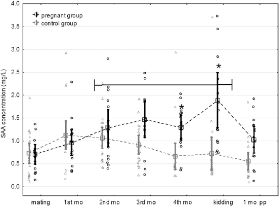
Changes of serum amyloid A (SAA) concentrations in pregnant (black) and control (gray) goats. Squares denote arithmetic means; vertical bars are 95% confidence intervals for the means; circles and triangles signify measurements of individual pregnant and control animals, respectively. Black horizontal line covers the time period when pregnant goats had SAA concentration significantly higher than at mating. Asterisks indicate time points when SAA concentration differed significantly between pregnant and control goats. p.p. = postpartum.
Hp concentration did not change during pregnancy or between groups (p > 0.05; Fig. 2). The SAA concentration at kidding was affected by doe parity (p < 0.05) but not by doe breed (p > 0.05) or number of kids born (p > 0.05). The SAA concentration was highest in does in the third and fourth pregnancy and gradually lower in goats with higher parities. None of these 3 factors was related to Hp concentration (Table 1).
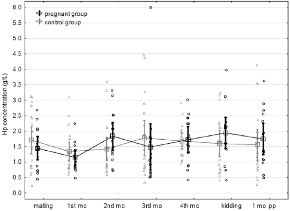
Haptoglobin (Hp) concentrations in pregnant (black) and control (gray) goats. Squares denote arithmetic means; vertical bars are 95% confidence intervals for the means; circles and triangles signify measurements of individual pregnant and control animals, respectively.
Linear regression for serum amyloid A and haptoglobin at kidding.
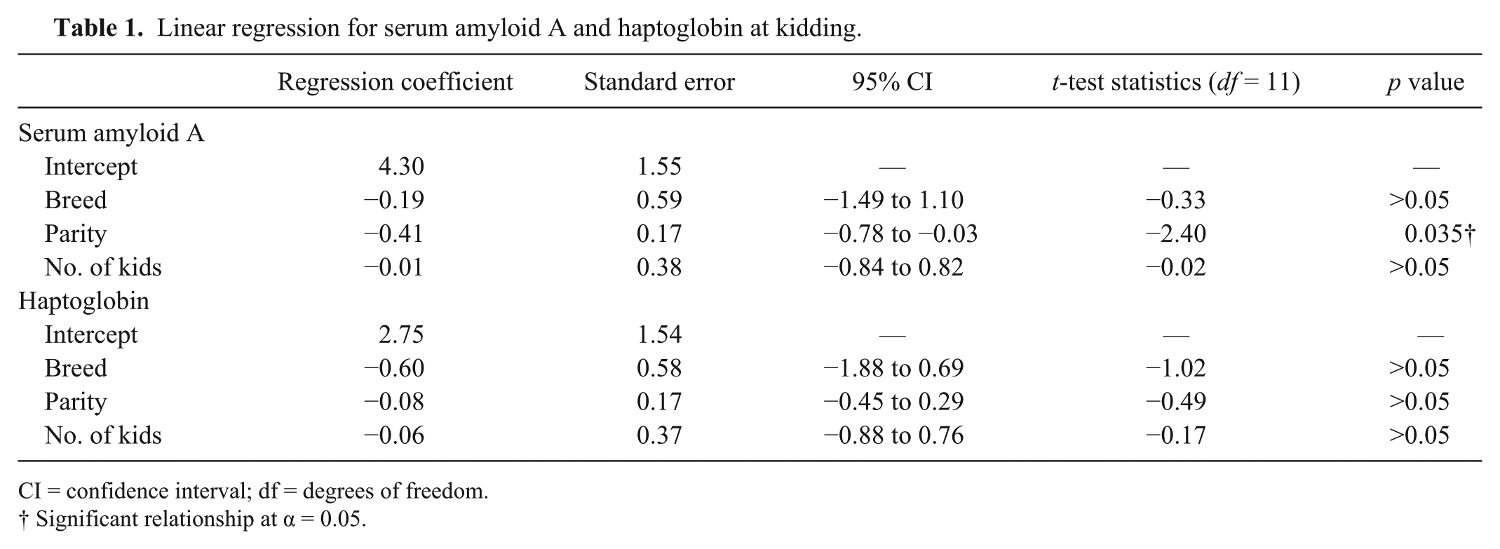
CI = confidence interval; df = degrees of freedom.
Significant relationship at α = 0.05.
Discussion
We observed that pregnancy in goats was associated with an increment of SAA concentration, with the peak concentration in the periparturient period, and stable Hp concentration. This contradicts the observation of others 16 that goats in the second trimester of pregnancy had a lower Hp concentration than non-pregnant goats. It is noteworthy that we found marked SAA elevation in pregnant goats during the week preceding parturition, whereas the acute-phase reaction in other animal species is usually observed soon after parturition.1,7,24 Higher SAA and Hp concentrations in the week preceding lambing versus the week following conception were observed by others 37 in sheep; however, their study had different objectives and the observation was incidental. Increase of SAA but not of Hp might be explained by distinct regulatory mechanisms of these 2 APPs. In humans, SAA is mainly stimulated by interleukin 1 (IL-1) and tumor necrosis factor–alpha (TNF-α), whereas Hp is stimulated by IL-6. 28 IL-1 and TNF concentrations were shown to rise in periparturient cattle, unlike IL-6, which remained unchanged. 9 A similar pattern of cytokine secretion may account for changes in major APP concentrations in pregnant goats.
Our observations may have practical application. An increase of Hp concentration during pregnancy or of SAA in the first trimester of pregnancy may warn a clinician of some insidiously developing disease, such as pregnancy toxemia, in which Hp has been shown to rise.12,33 On the other hand, 2- to 4-fold elevated or an increasing SAA concentration over the second half of pregnancy should not be the only basis for considering a goat as being at risk of pregnancy complications given that it seems to be a physiologic reaction.
The only factor associated with a SAA increase in the periparturient period was parity, with the SAA concentration lower in does that had experienced more pregnancies. This may indicate that the stress reaction associated with kidding weakens along with growing experience of a doe; however, it is also possible that does that control stress better are more likely to remain in a herd and kid more times. In any case, the influence of cortisol or prostaglandins on APP synthesis is likely to contribute to this phenomenon. 1 No effect of breed on SAA concentration was observed. The breed was included in the analysis because SAA concentration after stimulation proved to differ between 2 distinct beef cattle breeds. 4 Both goat breeds in our study were dairy goats and, although PWI goats are slightly larger, they have similar production characteristics 2 and no difference in the intensity of their stress reaction was expected. Number of kids born was not linked to SAA or Hp concentration, although number of fetuses is an important risk factor for pregnancy toxemia. 26 One study 33 that investigated Hp concentration in periparturient goats indicated the positive relationship between concentrations of Hp and β-hydroxybutyrate, a marker of subclinical pregnancy toxemia in small ruminants. This observation was later confirmed by a prospective study 12 in which β-hydroxybutyrate and Hp levels increased significantly in the course of pregnancy toxemia, whereas SAA remained unaffected. Lack of a relationship in our study might be explained by the fact that, in herds in which balanced and high-quality nutrition is practiced, the risk of pregnancy toxemia is minimal.
In both pregnant and control goats, SAA concentrations fell within the reference intervals established using the same ELISA as in our study 15 for goats (0.43–2.05 mg/L) with only a few exceptions, mainly in does at kidding. The concentrations were also consistent with basal levels determined in sheep,8,37 although, on average, 4-fold lower than basal concentrations reported by others in goats, 11 which implies that considerable variation may be expected in APP concentrations. The differences observed may, at least to some extent, result from different ELISAs used in these studies, as described for results of ELISA and spectrophotometric assay for feline Hp. 31 Hp concentrations in our study were roughly 2- to 4-fold higher than reported by others in goats 11 and in sheep 8 and considerably exceeded reference intervals established using the same ELISA 15 (0.40–1.24 g/L). This might be attributed to chronic SRLV infection. All goats in our study had been infected with SRLV for at least 2 y before the study began. SRLV infection is a commonplace goat health problem in Poland with most large herds extensively affected. 23 Published evidence on the occurrence of an acute-phase reaction in asymptomatic SRLV-infected goats is weak. 22 However, an increase in Hp was also observed to precede abscess formation in goats experimentally infected with Corynebacterium pseudotuberculosis, 21 whereas the SAA level remained unchanged. A significantly higher proportion of cattle with acute inflammation were found to have higher values of SAA and Hp than those with chronic inflammation,17,20 but in one study 32 both APPs were significantly elevated in calves with chronic respiratory disease. Obviously, a prospective study is warranted to determine changes of APPs in the course of chronic SRLV infection.
Our study has several limitations. First, enrolling a control group of non-pregnant does was not possible. All control goats were castrated males of the same age, which was significantly younger than pregnant does. We decided that maintaining the same environmental conditions in both groups was more important than matching the sex and age of animals in the study as long as they were all adults. No differences in Hp and SAA concentrations between bucks and does have so far been revealed, 15 whereas Hp concentration has been found to differ between sows coming from different herds. 36
Second, the pregnant group lacked does in their first and second pregnancy. We decided not to enroll uniparous does assuming that the stress reaction associated with the first pregnancy might be more intensive than is expected in multiparous does, as described by others 36 for Hp concentration in sows. Lack of second-parity goats was a consequence of an unfortunate coincidence – all 3 does in their second pregnancy were dropped from our study because of spontaneous abortion (2) or death (1).
Third, sera awaited screening longer and at higher temperature than is recommended by the manufacturer of the ELISA kit (between 8 mo [first samples collected at mating] and 2 mo [last sampling 1 mo pp] at −20°C), whereas it should have not been longer than 1 mo at −20°C or 2 mo at −80°C according to the manufacturer’s manual. Despite this fact, no linear increase in either SAA or Hp concentration could be observed between the oldest (from mating) and newest samples (from 1 mo pp), which indicates that caprine SAA or Hp are fairly stable at −20°C, and serum can be safely stored at such temperature for at least 6 mo. Human C-reactive protein and Hp maintained a reliable assay outcome for 1 y at −20°C18,19; porcine Hp was stable at −20°C for 7 mo. 13 Moreover, storage at room temperature did not affect stability of equine SAA after 2.5 wk 16 nor canine CRP after 1 mo. 27
Fourth, samples tested by Hp ELISA went through one additional freeze–thaw cycle. Although multiple freeze–thaw cycles were shown to increase APP concentration in saliva samples, 13 it is unlikely that one cycle was enough to have the same effect and bias our results. Nevertheless, we investigated the influence of gestation on APP concentration rather than determining basal (normal) SAA and Hp levels. Given that samples from both groups were handled in identical conditions, even if Hp concentration had falsely risen because of technical problems, this should not have interfered with the final conclusions of our study.
Statistical analysis was carried out using repeated-measures ANOVA instead of its nonparametric alternative (i.e., Friedman test), although at several times APP concentrations lacked normal distribution mainly because of extremely high values. The parametric approach was chosen for the broader range of comparisons and clearer visualization of results offered, as has been done by several other authors.21,36,37
Footnotes
Declaration of conflicting interests
The authors declared no potential conflicts of interest with respect to the research, authorship, and/or publication of this article.
Funding
This work was financially supported by the Polish National Science Center (grant 2013/09/B/NZ6/03514).