
Abstract
Salmonella enterica subsp. enterica serovar Dublin (Salmonella Dublin) is a host-adapted bacterium that causes high morbidity and mortality in dairy cattle worldwide. A retrospective search of archives at the New York Animal Health Diagnostic Center revealed 57 culture-confirmed Salmonella Dublin cases from New York and Pennsylvania in which detailed histology of multiple tissues was available. Tissues routinely submitted by referring veterinarians for histologic evaluation included sections of heart, lungs, liver, spleen, and lymph nodes. Of the 57 Salmonella Dublin–positive cases, all were Holstein breed, 53 were female (93%), and 49 (86%) were <6 mo of age. Specifically, in calves <6 mo of age, >90% (45 of 49) of lungs, 90% (28 of 31) of livers, 50% (11 of 22) of spleens, and 62% (18 of 29) of lymph nodes examined had moderate-to-severe inflammation with or without necrosis. Inconstant lesions were seen in 48% (10 of 21) of hearts examined, and consisted of variable inflammatory infiltrates and rare areas of necrosis. We propose a histopathology case definition of Salmonella Dublin in <6-mo-old Holstein cattle that includes a combination of pulmonary alveolar capillary neutrophilia with or without hepatocellular necrosis and paratyphoid granulomas, splenitis, and lymphadenitis. These findings will assist in the development of improved protocols for the diagnosis of infectious diseases of dairy cattle.
Salmonella enterica subsp. enterica serovar Dublin (Salmonella Dublin) is a rod-shaped, flagellated, aerobic, gram-negative, cattle-adapted bacterial pathogen of global clinical significance that causes high morbidity and mortality in both beef and dairy cattle. 5 In addition to the economic losses associated with the disease, consumption of raw cow milk has been linked with zoonotic transmission of Salmonella causing gastrointestinal illness in humans. 8 Prior to 1967, Salmonella Dublin in the United States was found primarily west of the Rocky Mountains and was generally uncommon outside California until the late 1980s when it emerged in the northeastern United States. 9
In adult cattle, the infection is primarily asymptomatic and can result in development of carriers that can shed up to 108 colony forming units/g of feces, urine, milk, and vaginal discharge 10 with sporadic abortion of pregnant cows. 4 By contrast, in naive herds experiencing their first outbreak or endemic herds with cyclical management breakdowns, the disease is primarily seen in young calves with acute systemic infection and clinical signs consistent with endotoxemia, including depression, swollen joints, respiratory distress and labored breathing, occasional diarrhea, and sudden death.5,9 Although several previous experimental infection studies mostly in young cattle have elucidated the pathogenesis and outlined histologic changes seen with Salmonella Dublin infection,2,3,6,11,12 there is a lack of detailed descriptions of lesions in naturally acquired disease. We characterized the histopathologic features of commonly submitted tissues obtained from Holstein cattle <6 mo of age with naturally occurring Salmonella Dublin infection in the northeastern United States to delineate a histopathology case definition of the disease.
Given that Salmonella Dublin infection often occurs with clinical signs of respiratory disease in young cattle, samples of alimentary tract may not be obtained during field autopsies performed by referring veterinarians. However, bacteria can be cultured from nearly every tissue taken from young calves with systemic salmonellosis caused by Salmonella Dublin. 3 Therefore, the sample population for the histologic case definition of Salmonella Dublin consisted of cattle <6 mo of age submitted to the Animal Health Diagnostic Center (AHDC) at Cornell University (Ithaca, New York) between 2008 and 2016 that were positive for Salmonella Dublin by routine bacterial culture, as described previously. 9 With the exception of 10 cases that were submitted for postmortem examination, selected tissues were collected by referring veterinarians during field autopsies and submitted by overnight courier for diagnostic investigation. Salmonella and aerobic culture of a standard set of tissues, fecal analysis, and fluorescent antibody tests (FATs) for viral antigens, including bovine respiratory syncytial virus (bovine orthopneumovirus), infectious bovine rhinotracheitis virus (bovine alphaherpesvirus 1), bovine viral diarrhea virus, bovine parainfluenza virus 3 (bovine respirovirus 3), rotavirus, and coronavirus were performed on fresh tissue when available.
For histopathology, sections of heart, lungs, liver, spleen, and lymph node were fixed in 10% neutral-buffered formalin, embedded in paraffin, sectioned at 5-µm thickness, and stained with hematoxylin and eosin. In addition, to confirm the presence of paratyphoid granulomas, which have been described in the livers of Salmonella Dublin experimentally infected cattle, 12 selected sections of liver were deparaffinized and processed for antigen retrieval and immunohistochemical (IHC) staining with rabbit polyclonal antibodies to ionized calcium-binding adaptor molecule 1 (Iba-1; Wako Chemicals, Richmond, VA), a marker for macrophages and dendritic cells that has been used for characterization of the macrophage response in cattle with paratuberculosis. 1 Briefly, a stainer (Leica Biosystems, Buffalo Grove, IL) was used with retrieval solution (Leica Biosystems) for 10 min, followed by hydrogen peroxide to quench endogenous peroxidase activity for 5 min, and incubation with a 1:3,000 dilution of the Iba-1 rabbit antibody for 15 min. This was followed by incubation with a detection kit (Leica Biosystems) that contains a proprietary polymer–horseradish peroxidase IgG reagent against rabbit antibodies for 10 min, substrate chromogen 3,3’-diaminobenzidine (DAB), and hematoxylin counterstain, as per the manufacturer’s directions. A positive control reference tissue was included on the same slide as the case, and a separate section of the case was stained with normal rabbit primary antibody as a negative control in each assay.
Using a semiquantitative histologic scoring scheme (Table 1), lung lesions were scored as mild, moderate, or severe based on increasing degrees of pulmonary alveolar capillary neutrophilia, with or without edema and fibrin exudation within alveolar lumens and interlobular septa. Similarly, liver lesions were scored from mild to severe according to increasing degrees of sinusoidal neutrophilia, with or without multifocal hepatocellular necrosis and the presence of paratyphoid granulomas. Lesions in sections of spleen were assessed for the presence and degree of marginal zone neutrophilia and red pulp necrosis. Sections of lymph nodes were assessed for the degree of sinus neutrophilia with or without multifocal necrosis. Sections of ventricular myocardium were assessed for the presence of circulating neutrophilia. The histologic changes in the lungs of calves <6 mo of age with naturally occurring Salmonella Dublin infection were compared with those of similar aged calves with various other fatal diseases. This included calves with naturally occurring Cryptosporidium spp. (n = 9), Salmonella Typhimurium (n = 9), or systemic Escherichia coli (n = 34) infections as confirmed by bacterial cultures of lung tissue, and neonatal calves experimentally infected with either rotavirus (n = 5) or coronavirus (n = 12). 7
Histopathology case definition of naturally acquired Salmonella enterica subsp. enterica serovar Dublin infection in Holstein cattle in the northeastern United States: scoring scheme used for ranking severity of lesions in tissues.
Number in ten 400× fields.
Aggregate of Iba-1 immunoreactive macrophages 1 surrounding necrotic hepatocytes.
From a total of 29 premises, there were 57 culture-confirmed Salmonella Dublin cattle from New York (n = 55) and Pennsylvania (n = 2). All affected cattle were of the Holstein breed and ranged in age from 1 d to 36 mo. Forty-nine of the cases were <6 mo of age for inclusion in the case definition and, of these, the median age was 1.6 mo. Most cases were submitted as single submissions from group-housed cattle that were experiencing severe clinical signs of respiratory illness and mortality; however, in 9 submissions, up to 5 calves were submitted together. Of concurrent bacteria cultured from the 57 cases of Salmonella Dublin, Mycoplasma spp. (n = 14) were the most prevalent; however, these cultures were considered commensal contaminants in all cases because of a lack of typical pulmonary lesions by histologic examination. Other bacteria included E. coli (n = 12), Clostridium perfringens (n = 4), Pasteurella multocida (n = 1), Trueperella pyogenes (n = 1), and Mannheimia haemolytica (n = 1). For viral isolation, 9 cattle <6 mo of age were positive for viral antigens, primarily coronavirus (n = 6), followed by rotavirus (n = 3). Cryptosporidium spp. and/or Eimeria spp. were present in 3 of the calves <6 mo of age.
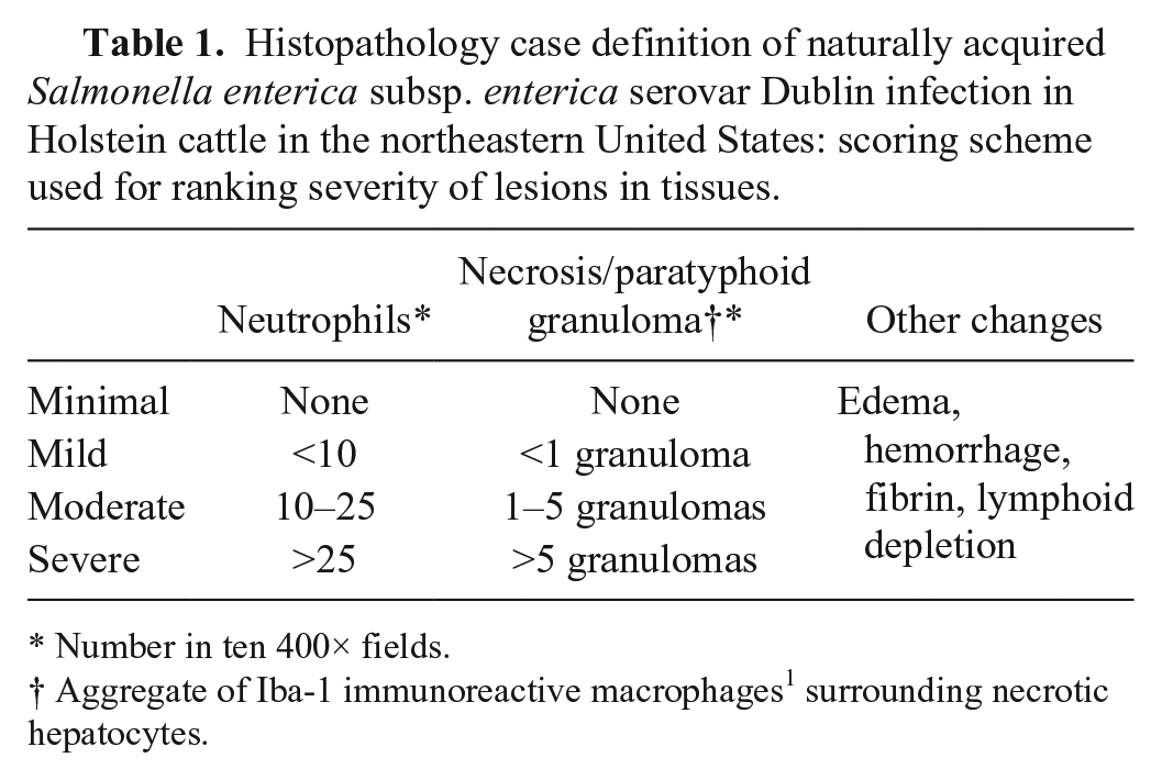
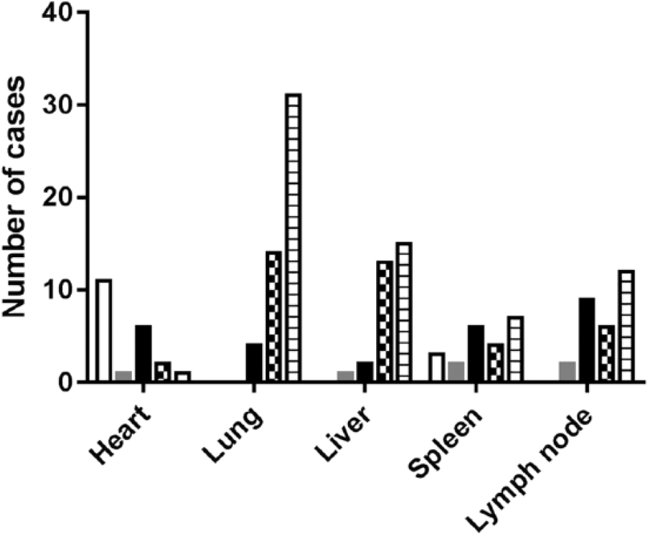
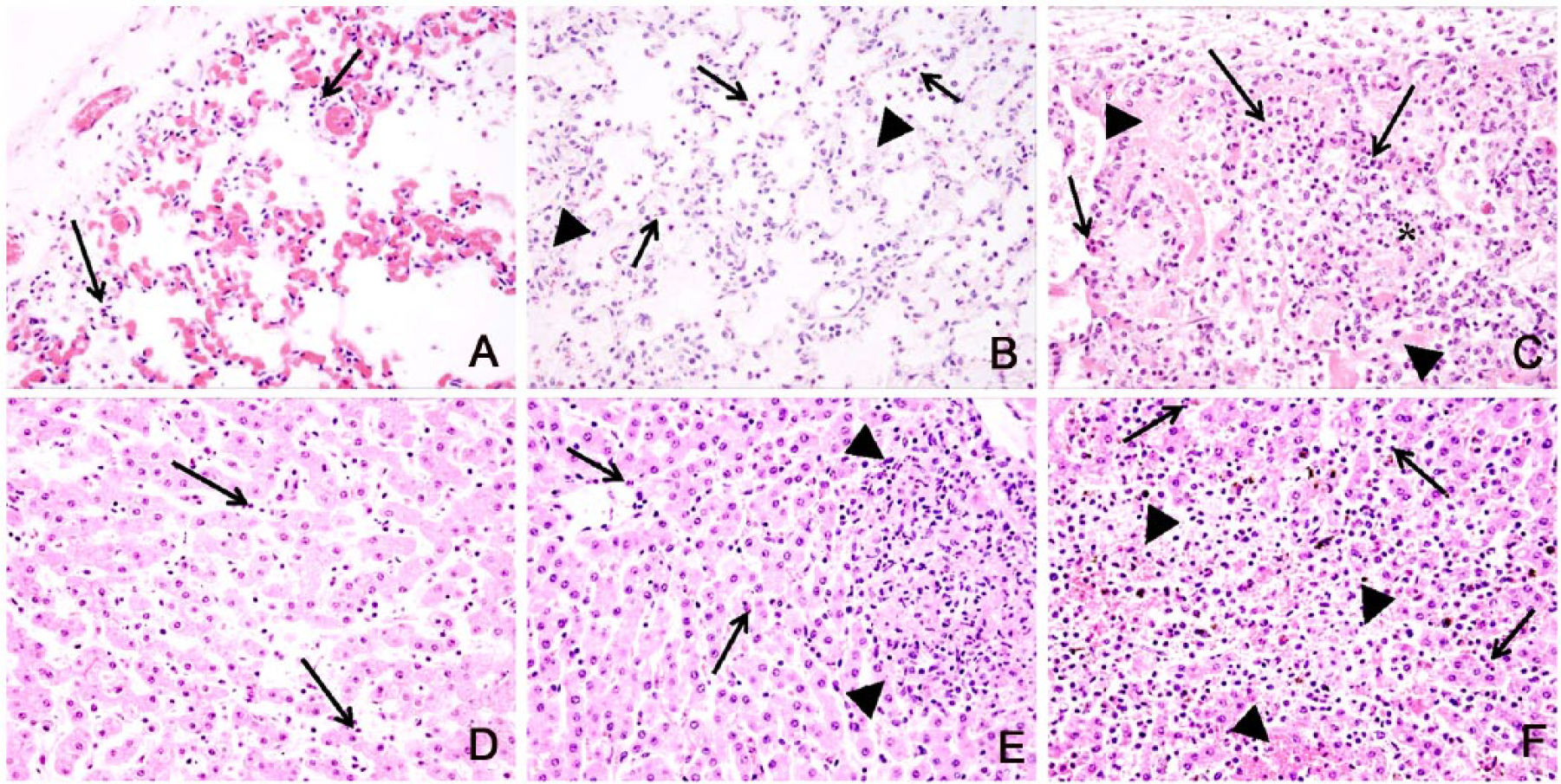
The majority of lesions in sections of lung, liver, and lymph node from calves <6 mo of age were scored as moderate-to-severe inflammation, with spleen and heart showing less consistent changes (Fig. 1). All lungs had lesions, with over 90% (45 of 49) of lesions ranging from moderate to severe. With increasing severity, lesions in sections of lung consisted of diffuse pulmonary alveolar capillary neutrophilia accompanied with neutrophilic septal infiltration, leading to multifocal-to-diffuse septal thickening or flooding of alveolar spaces by fibrin (n = 47) and mononuclear infiltrates (n = 41) with necrosis characterized by karyorrhectic debris (n = 22), and rare abscesses (n = 3; Figs. 2A–C). Similarly, 90% (28/31) of liver lesion scores ranged from moderate to severe with increasing degrees of sinusoidal neutrophilia, sinusoidal mononuclear infiltrates (n = 25), multifocal fibrin accumulation (n = 23), multifocal hepatocellular necrosis (n = 26), and paratyphoid granulomas (n = 7; Figs. 2D–F). The presence of macrophages in paratyphoid granulomas was confirmed by IHC staining with Iba-1 antibody 1 (Fig. 3), which stained Kupffer cells along sinusoids and aggregates of histiocytes forming discrete granulomas scattered within hepatic lobules. Lymph nodes had sinus neutrophilia, with 62% (18 of 29) scored as moderate to severe, most often accompanied by fibrin (n = 23) and less frequently by multifocal necrosis (n = 8). Lesions were present in 50% (11 of 22) of the spleens and ranged from moderate-to-severe marginal zone neutrophilia and/or red pulp necrosis. Lesions were present in 48% (10 of 21) of the hearts examined, and included various degrees of fibrosis, fibrin, edema, neutrophilic and/or lymphoplasmacytic infiltrates, and rare necrosis of the myocardium. Given that histopathologic changes have not been reported in the hearts of experimentally infected calves, the contribution of Salmonella Dublin to myocardial lesions seen in our calves is unknown 11 ; however, Salmonella Dublin can be isolated from broth-enriched heart tissue of infected cows as reported in a previous study. 3 Histopathologic findings in the liver of calves with naturally acquired Salmonella Dublin were similar to those reported previously in experimentally infected calves, including hepatic necrosis 2 and hepatic paratyphoid granulomas. 12 Microscopic lung lesions were generally limited to diffuse pulmonary alveolar capillary neutrophilia with multifocal interstitial neutrophilic and fibrinous infiltrates. Given that our samples were collected during field autopsies performed by referring veterinarians, not all affected lung lobes might be represented, and areas of fibrinopurulent bronchopneumonia as described in a previously reported experimental infection model were not a major finding in our study population. 11
Severity of lesions based on the scoring scheme proposed in Table 1 for Salmonella Dublin culture-positive Holstein cattle <6 mo of age. Included is a total of 49 calves with sections of heart (n = 21), lung (n = 49), liver (n = 31), spleen (n = 22), and lymph node (n = 29) tissues submitted for histologic assessment. Key: no lesion (white), minimal change (gray), mild suppurative inflammation (black), moderate suppurative inflammation (checkers), severe suppurative inflammation (stripes).
Representative photomicrographs of lung (upper panel) and liver (lower panel) from Holstein cattle <6 mo of age, infected with Salmonella Dublin. Alveolar capillaries with mild (
Paratyphoid granulomas in the liver of a Salmonella Dublin–infected Holstein calf with severe lesions. Note diffuse immunoreactivity of circulating monocytes and Kupffer cells in sinusoids (arrows) together with clusters of immunoreactive macrophages in paratyphoid granulomas (arrowheads). Immunohistochemical stain with Iba-1 antibody. 1
When compared with other calves <6 mo of age that died of non–Salmonella Dublin enteric diseases, only E. coli septicemic cattle, confirmed by culture of E. coli from lung tissue, had similar pulmonary lesions. This is not surprising, as both Salmonella spp. and E. coli are gram-negative bacteria that can disseminate in the blood stream, including to the lungs. The presence of positive bacterial culture, viral FAT, and/or fecal parasites indicative of concurrent infections were also similar among Salmonella Dublin and E. coli calves with 73% (35 of 48) and 89% (30 of 34), respectively. Indeed, bacterial cultures yielded both Salmonella Dublin and E. coli in 11 of the calves; however, postmortem E. coli bacterial proliferation cannot be ruled out in our Salmonella Dublin cases. Unlike calves infected with Salmonella Dublin, calves with systemic E. coli infection did not have hepatic paratyphoid granulomas histologically, a lesion that is characteristic of Salmonella Dublin infection in cattle. Additionally, cattle that died of E. coli systemic infection were generally <2-wk-old compared to the older 1.6-mo-old median age of Salmonella Dublin cattle. Therefore, age of cattle and presence of hepatic necrosis and paratyphoid granulomas are reliable differentiating histopathologic features of Salmonella Dublin infection in cattle.
We propose a histopathology case definition for Salmonella Dublin in <6-mo-old Holstein cattle that includes diffuse pulmonary alveolar capillary neutrophilia with variable multifocal interstitial neutrophilic and fibrinous infiltrates, necrosuppurative and histiocytic hepatitis characterized by the presence of paratyphoid granulomas, splenic marginal zone neutrophilia, and neutrophilic lymphadenitis with or without necrosis. Given that the history provided by referring veterinarians in our cases of Salmonella Dublin infection in this age group often referred to respiratory signs as the main clinical finding, histologic evaluation of multiple tissue samples in addition to the lungs, including liver, spleen, and lymph node, should increase the accuracy of diagnostic investigations of mortality in young dairy cattle.
Footnotes
Acknowledgements
We thank resident and faculty colleagues of Cornell University in the Anatomic Pathology Section for contributing cases, Isabelle Schweitzer in the Veterinary Support Services for database searches, and the staff in the histology laboratory for technical assistance. Preliminary reports of the findings were presented at the 2015 Annual American Association of Veterinary Laboratory Diagnosticians Meeting in Providence, RI and at the 2015 American College of Veterinary Pathologists, American Society for Veterinary Clinical Pathology, and Society of Toxicologic Pathology Combined Annual Meeting in Minneapolis, MN.
Declaration of conflicting interests
The authors declared no potential conflicts of interest with respect to the research, authorship, and/or publication of this article.
Funding
This work was supported in part by funds from the College of Veterinary Medicine at Cornell University.