
Abstract
The present report describes an extrathoracic bronchogenic cyst in a 30-day-old female calf. Histologically, the cyst wall was lined by a layer of ciliated pseudostratified columnar epithelium with peripheral arrangement of cartilage, glands, and smooth muscle fascicles. The mass was successfully removed by simple surgical excision.
Bronchogenic cysts are congenital cystic lesions resulting from the abnormal development of the tracheobronchial system during the embryonic period. 4 Bronchogenic cysts can be intrathoracic or extrathoracic. There are many clinical and pathologic studies of bronchogenic cysts in humans. Intrathoracic cysts usually present in the mediastinum and lung parenchyma. 4 Extrathoracic cysts characteristically present with asymptomatic neck mass, often located in the anterior neck just above the sternal notch. 6 Although various clinical and pathologic studies of bronchogenic cysts in humans exist, extrathoracic bronchogenic cysts have yet to be reported in animals. In animals, only 1 case of intrathoracic bronchogenic cyst in lung tissue of a German Shepherd Dog has been reported. 1 The purpose of the current study was to describe a case of a rare cervical cystic lesion, namely a congenital bronchogenic cyst, that presented as an asymptomatic cervical mass in a 30-day-old female calf.
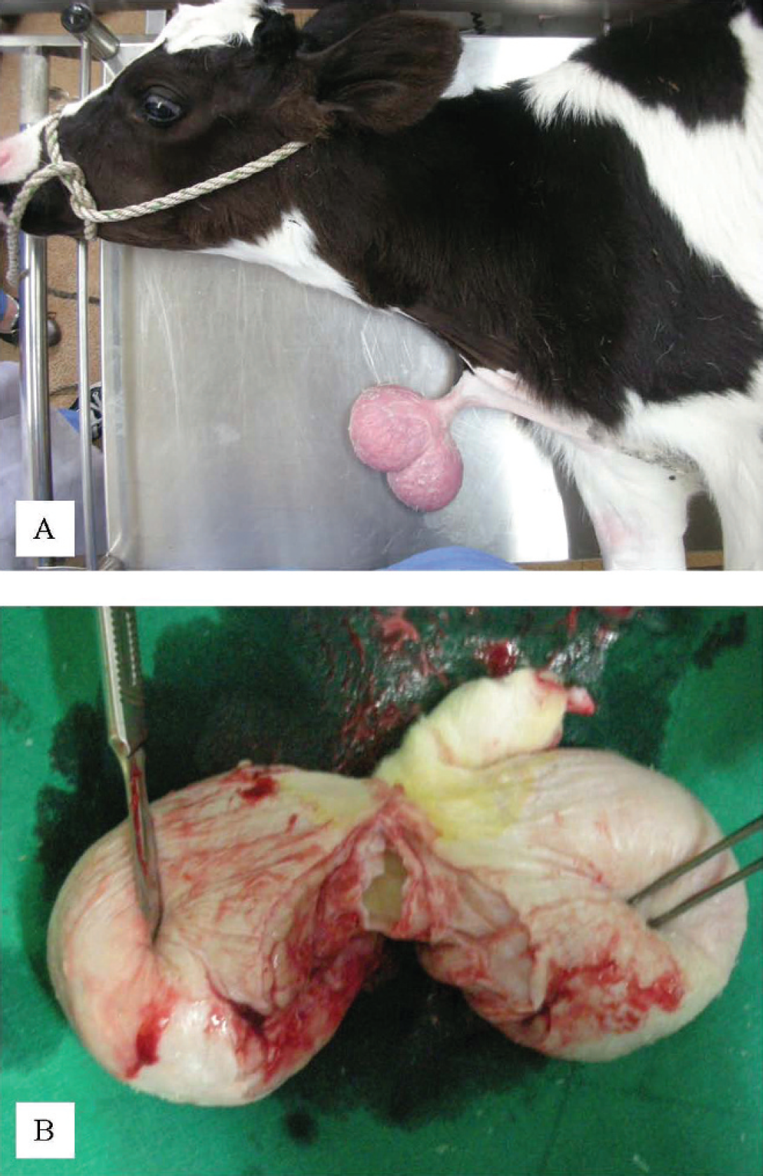
The calf was presented with a pedunculated cutaneous cervical mass, which was present at birth (Fig. 1A). Despite the progressive swelling of the mass over a period of 30 days, the calf had a good appetite and appeared healthy. There was no other relevant medical or surgical history.
On examination, a soft, nonpainful, lobulated mass 11 cm × 7 cm × 4 cm with a narrow stalk was protruding from the neck. On admission, the calf's rectal temperature, heart rate, respiratory rate, and results of hematologic examination and blood chemistry were all within reference intervals. Radiographs of the cervical regions revealed soft tissue opacity at the ventral aspect of the neck. Thoracic radiographs revealed no abnormal findings in the mediastinum and lung parenchyma. An ultrasound scan of the cervical mass demonstrated a mixed echogenic cystic mass. The initial differential diagnosis of the abnormal neck conformation was abscess or neoplasia.
Under general anesthesia, surgical excision of the cyst was performed with a skin crease incision at the peduncle of the mass. The peduncle was separated from the surrounding structures, and a fibrous band and blood vessels were traced from the ventral part of the neck to the sternal notch. The fibrous band and blood vessels were ligated, and the whole cyst was removed. The cyst did not communicate with the thoracic cavity.
Grossly, the mass consisted of a cyst filled with white mucoid material (Fig. 1B). On bacteriologic examination of the fluid, Staphylococcus spp. were isolated. Microscopically, the cyst walls were lined by a layer of ciliated pseudostratified columnar epithelium, with peripheral arrangement of cartilage, glands, and smooth muscle fascicles (Fig. 2). On the basis of the histopathologic examination, the cervical mass was diagnosed as a bronchogenic cyst.
Bronchogenic cysts are rare congenital anomalies that are usually located in the mediastinum or lung parenchyma. They are very rare in the neck and are thought to result from abnormal development of the tracheobronchial system. 8 An abnormal budding of the tracheobronchial system between the 22nd and 33rd days of gestation, and persistence of such a bud, can give rise to bronchogenic cysts in humans. 10 In addition, abnormal migration of a bud can occur during the course of development and reside in different intrathoracic or extrathoracic locations. Intrathoracic cysts are more common and can occur in the mediastinum, diaphragm, pericardium, or lung. Extrathoracic cysts are known to be present in the suprasternal notch, presternum, shoulder, neck, base of the tongue, infraclavicular region, or chin and can extend into the mediastinum. 4,5,7,9
Extrathoracic bronchogenic cysts usually present with an asymptomatic neck mass. 10 The cyst can fluctuate in size, but usually enlarges with body growth. Larger cysts can cause pressure symptoms like dyspnea, respiratory distress, cough, and dysphasia. 3 A neck abscess can also occur if the cyst becomes infected. 3 In the present case, the mass became progressively larger with body growth, but with no pressure-related signs because of the ventral position of mass.
Cervical bronchiogenic cyst in a calf.
The potential diagnoses of a neck cyst include bronchial cleft cyst, epidermal inclusion cyst, thymic cyst, thyroid cyst, cystic teratoma, and thyroglossal duct cyst. However, a definitive diagnosis of bronchogenic cyst requires histopathologic confirmation. Bronchial cleft cysts are usually lateral in position with prominent lymphoid tissues seen histologically. A thyroglossal duct cyst moves up with protrusion of the tongue and, on histologic examination, is lined by ciliated epithelia with occasional thyroid follicles. Thyroglossal duct cysts are commonly lined with squamous epithelium, respiratory epithelium, or a combination of both. Cystic teratoma can be distinguished by the complete absence of tissue components from all 3 germ layers. Bronchogenic cysts are lined by ciliated pseudostratified columnar epithelium with accessory tissues in the cyst wall, such as smooth muscle, seromucinous glands, or cartilage. 8 These histologic features were found in the current case.
Surgical excision is the best treatment option. No recurrence has been described when complete surgical excision has been performed. 4 Aspiration of the cyst is an inadequate treatment because of reoccurrence of the cyst after treatment. 2 Extrathoracic bronchogenic cysts are uncommon congenital malformations. Although cervical bronchogenic cysts are rare lesions of the neck that are difficult to diagnose preoperatively, they should nevertheless be considered in the potential diagnoses of cervical cystic lesions.
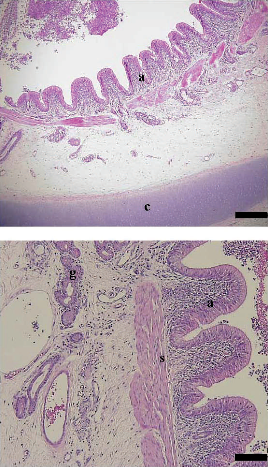
Cervical bronchiogenic cyst in a calf. Histologic findings included a cyst wall lined by pseudostratified ciliated columnar epithelium (a) that contained cartilage (c), glands (g), and smooth muscle fascicles (s). Hematoxylin and eosin. Bar = 100 μm.