
Abstract
Rapid diagnosis of tuberculosis in cattle reacting positive in antemortem assays is crucial in countries where eradication programs are operated to confirm the presence of the infection in tuberculosis-free herds. This study evaluated the accuracy of histopathologic examination by hematoxylin and eosin and Ziehl-Neelsen (ZN) staining applied in this framework, when suspected lesions are caused by low infectious doses and are detected in early stages of the disease. For this purpose, histologic methods were compared with mycobacterial culture as reference test on suspected lymph node samples from 173 cattle reacting positive in antemortem tests. Histopathology demonstrated high sensitivity (93.4%) and specificity (92.3%), while ZN sensitivity and specificity were respectively 33.9% and 100%. There was good agreement between histopathology and bacterial culture, suggesting that histopathologic examination is a reliable tool for rapid diagnosis in countries where active tuberculosis eradication programs allow the prompt identification and elimination of reactor cattle. Histopathology permits identification of typical mycobacterial lesions and its differentiation from other causes.
Introduction
Bovine tuberculosis caused by Mycobacterium bovis remains a significant disease of cattle and other species in many countries. This zoonotic disease is highly infectious in humans, and its symptoms are indistinguishable from infection caused by Mycobacterium tuberculosis. 3,10,35,38 With modern pasteurization of dairy products and widespread eradication programs, tuberculosis caused by M. bovis has become less important as a public health risk in countries with bovine tuberculosis eradication plans, 38 but it still poses one of the most important zoonotic threats in developing countries, particularly where Human immunodeficiency virus is prevalent. 25,29 Moreover, the notification system for human tuberculosis in most countries does not distinguish cases caused by nonhuman mycobacteria, so the real number of cases might be underestimated. 11
In 2005, 119 human tuberculosis cases caused by M. bovis were reported in 17 member states of the European Union (EU). This is the highest number of reported cases since 2001, 18 and it raises alarm about the potentially huge economic impact the disease could have on the international trade of animals and animal products. 3,4 Eradicating tuberculosis has long been a priority in EU policy 13 ; according to a 1990 Council Decision, the member states are responsible for having disease control and eradication programs in place, for which they receive community-funded support. 14 Nonetheless, in some countries M. bovis is persistent in the cattle population, owing in part to a large number of wildlife species (e.g., badger, wild boar) in the epidemiology of bovine tuberculosis. 3,35,38
European Union member states are classified into 2 categories based on their bovine tuberculosis status, officially tuberculosis-free (OTF) and not officially tuberculosis-free, with 11 member states declared OTF. 35 In these countries, surveillance has mainly been carried out by clinical surveillance, routine and premovement tuberculin testing, and meat inspection. In non-OTF member states, the strategies according to Community legislation are based on different systematic testing schemes depending on the local epidemiologic situation. The reactors are slaughtered and considered positive on typical tubercular findings at necropsy or following confirmation with histopathology and bacteriology. 1,11,31
Italy has an ongoing national eradication program, and several regions within Italy have set up eradication plans as well. Three regions and 15 provinces throughout Northern and Central Italy were designated bovine tuberculosis-free areas in accordance with European Commission (EC) legislation. 15–17 Other regions within Italy continue to work on obtaining this status.
In the Piedmont region (Italy), the second most important region for cattle breeding, eradication efforts have significantly reduced the prevalence of the disease over the past decade, but it remains a recalcitrant problem nevertheless. To combat bovine tuberculosis, the regional eradication program was implemented together with a dedicated one approved by the EC. 12
As a result of previous tuberculosis control, early stages of bovine tuberculosis 7,24 comprised mainly of primary complexes, represented by primary lesions with the involvement of the regional lymph node, 32 are most commonly observed, whereas advanced stages are less often reported. In this situation, rapid diagnosis of tuberculosis in cattle reacting positive to antemortem assays is pivotal to confirm the presence of infection in tuberculosis-free herds and to detect other disease causes. 8 However, mycobacterial culture and molecular diagnosis, besides being time consuming, do not permit the identification of non-mycobacterial causes of false-positive test results. The aim of the present study was to evaluate the accuracy of histopathologic examination by hematoxylin and eosin (HE) and Ziehl-Neelsen (ZN) staining applied in the framework of advanced eradication processes.
Materials and methods
Sample collection and preparation
Retropharyngeal or mediastinal lymph nodes displaying suspected lesions at abattoir postmortem inspection were collected from apparently healthy tuberculin or γ interferon-positive cattle from 2005 to 2007 in the Piedmont region during the regional eradication program. Suspected lesions were represented by granulomatous lymphadenitis, focal lymphadenitis, mineralized lesions, and lymphoid hyperplasia. Each abnormal lymph node was split into 2 halves: 1 was fixed in 10% buffered formalin for histopathology and the other was stored at −20°C in sterile plastic bags until processing for culture and identification.
Histopathologic results were compared with mycobacterial culture and M. bovis identification as a reference test. The cultivation of mycobacteria was performed according to World Organization for Animal Health (OIE) standard procedures. 41 Tissue samples, following homogenization in Stomacher and double neutralization with NaOH 2% and HPC, were centrifuged and the pellet inoculated onto Lowenstein Jensen medium and Stonebrink. After initial screening, samples that were found to be suitable for isolation of M. bovis were identified using multiplex polymerase chain reaction (PCR) for different molecular target (rRNA 16S, IS986 and mpt40 gene), followed by spoligotyping for the identification of M. bovis among those identified as Mycobacterium tuberculosis complex. 22,25,26 In 4 cases in which growth of early pigmented colonies consistent with Rhodococcus equi was observed, the colonies were identified on the basis of morphology and biochemical methods. 28
Histopathologic examination
Tissue samples were fixed in 10% neutral buffered formalin and processed by standard paraffin wax techniques. Samples displaying heavy calcification were decalcified before paraffin embedding. Samples were cut in 4 ± 2 μ serial sections at 2 different levels of the embedded sample. At each level, 2 sections were stained using the standard HE method and 2 with the ZN method to detect acid-fast bacilli. Slides were evaluated microscopically at increasing magnifications (10×, 20×, 40×).
Histopathologic classification. The histopathologic findings were evaluated microscopically and classified as:
Positive: tubercular granuloma displaying central necrosis with or without mineralization surrounded by macrophages, lymphocytes, plasma cells, neutrophils, epithelioid cells, and Langhan's giant cells, and enclosed partly or completely by a thin capsule (Fig. 1). 6
Inconclusive: lesion characterized by irregular unencapsulated clusters of epithelioid macrophages but not Langhan's-type multinucleated giant cells and necrosis, consistent with an initial stage (Fig. 2). 39
Negative: features not consistent with tubercular granuloma, including significant eosinophilic infiltrates (Fig. 3), lymphoid hyperplasia (Fig. 4), presence of bacterial colonies within necrotic area (Fig. 5). or tumors.
Acid-fast staining
Sections were stained using the ZN method. Ziehl-Neelsen carbol-fuchsin after treatment with periodic acid solution 1% was used. As a positive control for specific M. bovis staining, a bovine lymph node positive by mycobacterial culture for M. bovis was used. All slides were examined carefully, scanning the entire area of each section at 100 × magnification. They were considered positive when 1 or more acid-fast bacteria were detected in at least 1 section of the sample.
Statistical analysis
The results of histopathologic examination were compared with mycobacterial culture and M. bovis identification. The sensitivity, specificity, and positive and negative predictive values (with the 95% exact confidence intervals) were calculated by the command “diagt” implemented in STATA 9.2. a , 2,20
Results
Mycobacterial culture and identification
Of the 173 samples analyzed, 121 tested positive for mycobacterial culture and M. bovis identification, and were considered as true positives. In the remaining 52, no growth consistent with for Mycobacterium spp. was obtained and all were used as true negative samples; among these, 4 were positive for R equi.
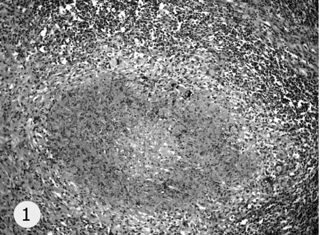
Tubercular granuloma, caseation with epithelioid and Langhan's giant cells. Hematoxylin and eosin, 10×.

Nonconclusive lesion, granulomatous lymphadenitis with macrophages and epithelioid cells. Hematoxylin and eosin, 20 ×.

Negative results, eosinophilic lymphadenitis. Hematoxylin and eosin, 10×.

Negative results, lymphoid hyperplasia. Hematoxylin and eosin, 20×.

Negative results, pyogranulomatous and necrotizing lymphadenitis. Hematoxylin and eosin, 10×.
Histopathologic examination
Histologically, 117 samples showed microscopic lesions compatible with tubercular granuloma (positive), 47 were negative, and 9 inconclusive. Of the 9 inconclusive samples, 5 were positive and 4 were negative for M. bovis culture and identification. For the purposes of statistical analysis, the inconclusive samples were considered first as positive and then as negative.
Four samples considered positive at histopathologic examination tested negative by mycobacterial culture; in these cases R equi was isolated and identified. Histopathologic features were central necrosis with mineralization delimited by degenerated neutrophils, epithelioid cells, macrophages, and multinucleated giant cells. 21
The relative sensitivity of histopathology was 93.4% (95% confidence interval [CI]: 87.4–97.1) and relative specificity was 92.3% (95% CI: 81.5–97.9) when the inconclusive lesions consistent with an early tubercle were considered positive, with a positive predictive value of 96.6% (95% CI: 91.5–99.1) and a negative predictive value of 85.7% (95% CI: 73.8–93.6). When the inconclusive results were taken as negative, the relative sensitivity rose to 97.5% (95% CI: 92.9–99.5) and the relative specificity decreased to 84.6% (95% CI: 71.9–93.1). The positive predictive value was 93.7% (95% CI: 87.9–97.2) and the negative predictive value was 93.6% (95% CI: 82.5–98.7).
Acid-fast staining
Suspected tuberculous lesions were associated with acid-fast organisms in only 31 cases. The number of acid-fast bacilli was extremely low in all cases. They were mainly observed within the cytoplasm of Langhan's-type multinucleated giant cells, less frequently within macrophage cytoplasm, and rarely free in caseous debris. The relative sensitivity and specificity of ZN staining were 33.9% (95% CI: 25.5–43.0) and 100% (95% CI: 93.2–100.0), respectively. The positive and negative predictive values were 100% (95% CI: 91.4–100) and 39.4% (95% CI: 31.0–48.3), respectively.
Discussion
By targeting the elimination of a serious zoonosis, the eradication of tuberculosis offers unquestionable benefits to society. Central to the success of eradication programs is common sense and a rational approach. 35 When disease prevalence is low, as during the later stages of an eradication campaign, the need for a definitive diagnosis becomes crucial. The key to determining the true status of tuberculin reactors is the availability of a reliable, fast, confirmatory test following positive reaction at antemortem screening. Mycobacterial culture is time consuming and, like molecular methods, does not permit identification of other causes of in vivo positive reactors.
Histopathology, on the other hand, offers the major advantage of producing results within 2 days. Furthermore, the technique has a high specificity because it can histomorphologically characterize lesions unrelated to mycobacterial agents (e.g., parasites, neoplasia). In previous studies, histologic examination was mainly applied in addition to other techniques to maximize the identification of M. bovis-infected cattle 34 and for the evaluation of lesions evolution in experimental studies of pathogenesis. 7,23
A recent study on the accuracy of histopathologic techniques was carried out on limited number of samples from cattle with classical histology features of tuberculosis. 40 The accuracy of histopathologic techniques on large number of samples are available only in cervids. 19,36
In the present study, a large number of specimens from a tuberculosis eradication campaign were collected. The major advantage to this approach is that it represents a reliable evaluation of 2 histologic techniques concerning accuracy values (small confidence intervals) and the analytical sensitivity and specificity of the techniques in field conditions. Statistical analysis of the present study's results identified that histopathologic examination by HE staining demonstrates high accuracy and good positive and negative predictive values in accordance with previous data. 19 These results suggest that the method could be a reliable tool for rapid tuberculosis diagnosis in cattle reacting positive to in vivo tests in regions with low tuberculosis prevalence, and displaying suspect lesions resembling tuberculosis prior to or after slaughter. 9,37
In the present study, all false-positive histologic diagnoses were later identified as R. equi on bacteriologic examination. This bacterium gives histologic findings similar to positive cases, with multinucleated giant cells, neutrophils infiltration, and/or extensive sheets of macrophages. 21 The similarity of the cellular response in tuberculosis and R. equi lymphadenitis is attributed to similarities in cell wall composition and antigenic structure of the causal organism. 27 In fact, the 2 bacteria belong to the same phylogenetic group. 30 The histomorphologic characteristics specific to R equi are being studied in order to highlight minor microscopic differences useful to differentiate R. equi from mycobacteria. Moreover, investigations on a larger number of histologically inconclusive cases will be needed to clarify their interpretation, and to determine whether further microscopic features can be highlighted to classify them definitively as positive because they could be consistent with an initial stage of the disease, or negative because they could be consistent with other causes.
The present study also showed that ZN staining has high specificity but low sensitivity. These data differ from that obtained in cervids, 19 but confirm data previously reported in cattle. 9,24,40 An explanation for ZN low sensitivity may be a low survival rate of mycobacteria in the environment of the central caseation 5 or loss of bacterial structure owing to immune responses operating in granulomatous inflammation in mycobacteriosis. 24 For this reason, the identification of acid-fast bacilli using the ZN method needs to be reconsidered for its application in eradication schemes with other diagnostic methods such as auramine O/rhodamine staining or immunohistochemistry. 24,40
Acknowledgements
This research was funded by the Italian Ministry of Health (Project Bovine Tuberculosis: diagnostic and epidemiologic deepening in domestic and wild animal to gain disease eradication).
Footnotes
a.
Stata Statistical Software: release 9.0 (updated to 9.2). StataCorp, College Station, TX.