
Abstract
We describe 3 outbreaks of superficial dermatitis caused by bovine herpesvirus 2 (BoHV-2) in dairy breed calves. Clinically, all of the affected calves were 12–26 d of age, had alopecia and crusts on the face and ears, and were non-pruritic and afebrile. Affected animals recovered spontaneously without any treatment within 2–4 wk after onset of clinical signs based on 1 herd with follow up. Histologic examination of all skin crust or tissue samples identified neutrophilic inflammation, mild hyperkeratosis, multinucleate syncytial cells, and intranuclear inclusion bodies in the syncytial cells. Real-time PCR testing on affected surface crusts or tissue provided evidence of BoHV-2, and testing, where performed, was negative for parapoxvirus including bovine papular stomatitis virus and the ovine form of malignant catarrhal fever tested in EDTA blood samples. Bovine viral diarrhea virus also was negative by ELISA, as well as bovine herpesvirus 1 by immunohistochemistry. Direct electron microscopy of infected tissues in the first outbreak revealed herpesvirus-like particles.
Keywords
Latent infection within specific tissues is a common feature of all herpesviruses.2,4,15 Bovine alphaherpesvirus 2 species (BoHV-2; order Herpesvirales, family Herpesviridae, subfamily Alphaherpesvirinae, genus Simplexvirus) is the cause of ulcerative mammillitis, which is usually a self-limiting cutaneous disease of the udder and teats.12,16 BoHV-2 is also responsible for pseudo-lumpy skin disease (PLSD), which causes generalized superficial cutaneous nodules with central depressions over the entire body of affected animals. 8 We describe 3 outbreaks of BoHV-2 causing head and ear skin lesions in young calves from dairy farms and calf-raising facilities located in the Tulare County region of the Central Valley of California.
The outbreaks occurred on 3 separate premises over a 10-y period. The first outbreak occurred in 2005 on a 5,000-head calf ranch with 95–100% of 2- to 3-wk-old calves affected over a 1-mo period in August. According to the referring veterinarian ~200 calves were affected on the day of sample submission. The second outbreak was on a 3,500 lactating cow dairy farm in September 2012 where nearly 100% of Jersey and Holstein calves developed ear skin lesions at ~3 wk of age, leading to alopecia; 150 calves were affected on the day of sample submission. The third outbreak occurred in July 2015 on a 5,000-head calf ranch where 50% of calves at 12–26 d of age (~100 calves affected on day of sample submission) developed face and ear lesions. Calves affected were Holsteins (premises 1 and 3) and Holstein and Jersey calves (premises 2).
Affected animals showed alopecia and surface crust formation with skin peeling from the ears. The most common sites of involvement were the dorsal portion of the head, periocular skin, and edges of the ears. Some areas demonstrated complete loss of hair (Fig. 1), whereas other calves had well-circumscribed hair loss. In all cases, calves were in good body condition. Animals were bright, alert, afebrile, non-pruritic, did not have increased sensitivity in affected areas, and ate and drank without any difficulty. It was reported that lesions spontaneously regressed within 2–4 wk after onset (premises 1).
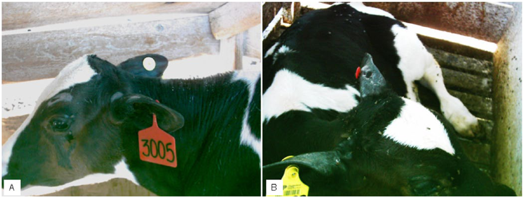
Facial skin lesions (outbreak 1). (
The submitted case material consisted of 9 EDTA blood samples (outbreak 1) and 17 skin crusts or tissue (outbreaks 1–3). No animals were submitted for postmortem examination. Samples from the skin crust or tissue were received fresh with a portion of the sample fixed in 10% neutral-buffered formalin, processed routinely, embedded in paraffin, and sectioned at 5 µm for staining with hematoxylin and eosin (H&E) for histologic examination and testing for BoHV-1 immunohistochemistry [IHC] using a specific polyclonal antibody (VMRD, Pullman, WA). The fresh skin crusts were submitted for: 1) polymerase chain reaction (PCR) testing at the California Animal Health and Food Safety (CAHFS) laboratory–Davis branch for BoHV-2 (1 sample for parapoxvirus including bovine papular stomatitis virus [BPSV]), and 2) bovine viral diarrhea virus (BVDV) antigen ELISA (BVDV PI X2, IDEXX, Westbrook, ME), following the manufacturer’s recommendation. For extraction of total nucleic acid, fresh skin samples were vortexed in viral transport medium and pulse spun. The supernatant was extracted using magnetic beads (MagMax 96 Viral RNA isolation well kit, Life Technologies, Carlsbad, CA), according to the manufacturer’s recommendation. Extracted DNA was subjected to real-time PCR (qPCR; primer and probe information for BoHV-2 targeting glycoprotein B by DNA sequencing, pan-parapox agents, and BPSV was kindly provided by Dr. K. Toohey-Kurth, WVDL, Madison, WI). All qPCR reactions utilized the Path-ID multiplex one-step RT-PCR kit (Life Technologies) and were performed with the 7500 Fast real-time PCR system (Life Technologies) under the following conditions: stage 1 at 50°C for 10 min; stage 2 at 95°C for 10 min; stage 3 at 95°C for 15 s; followed by stage 4 at 60°C for 1 min; stages 3–4 were repeated for 40 cycles. Blood samples tested for ovine herpesvirus 2 (OvHV-2) and 2 of the BoHV-2 tests were outsourced to the USDA-ARS laboratory in Pullman, Washington; all other testing was performed at CAHFS.
For direct electron microscopy via negative staining, a small section of fresh skin crust or tissue was processed in 2% phosphotungstic acid, and centrifuged at 2,300 × g for ~20 min at 30°C. The supernatant was filtered through syringe filters of descending pore size ending with 0.8 µm. Ultrafiltrate was centrifuged (Beckman 70.1 Ti rotor, Fullerton, CA) at 277,000 × g for a minimum of 45 min at 5°C. The pellet was suspended in double-distilled water and mixed with 2% neutralized phosphotungstic acid at a ratio of ~1:10 and then applied to a formvar-coated, carbon-backed grid. Grids were examined in a transmission electron microscope (Zeiss 906E, Oberkochen, Germany) at 80 kV accelerating voltage.
Given that the lesions were taken from sites with surface crusts, the skin tissue examined was usually nonviable epidermal tissue and debris. Microscopically, the epidermal crusts consisted of faintly stained epithelial cells, mixed with numerous multinucleate syncytial cells that contained lightly basophilic-to-amphophilic intranuclear inclusions filling and distending nuclei. Abundant neutrophils often infiltrated among the syncytial cells and epithelial cells, disrupting the epidermal architecture (Fig. 2). Some affected epithelial cells had undergone ballooning degeneration with irregular granularity. In some sections examined, abundant cellular debris was noted in the ostia of associated hair follicles (outbreak 1). Orthokeratotic hyperkeratosis (outbreak 1) and perivascular lymphocytic dermatitis (outbreak 3) were also noted histologically. Histologic examination of the skin lesions showed serocellular suppurative epidermitis with intranuclear inclusions within syncytia. There was no evidence of coccal bacterial colonies (except for 1 sample in outbreak 3), fungal hyphae including dermatophytes, Dermatophilus sp., or ectoparasite structures.
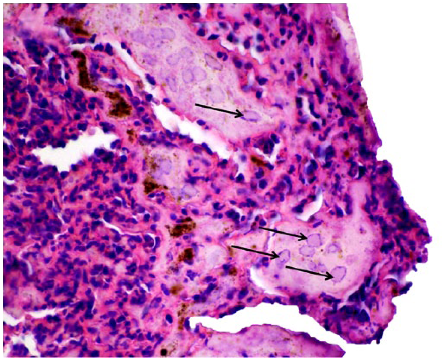
Syncytial cells in skin crust containing basophilic-to-amphophilic intranuclear inclusion bodies (arrows) are admixed with abundant neutrophilic inflammatory infiltrate and cellular debris. H&E. 600×.
Lesions noted during histologic examination were highly suggestive of a herpesvirus, and BoHV-2 was detected by molecular assays in 15 calves. In addition, direct electron microscopy of skin crusts in 2 additional calves revealed herpesviral particles (Fig. 3). No histologic lesions compatible with parapox were seen, parapox was not detected by electron microscopy, and a submitted sample from outbreak 2 had no detection by qPCR of BPSV, or of pan-parapoxvirus (Table 1). All 9 calves tested also were negative by IHC for BoHV-1. In addition, BVDV antigen was negative on ear tissue by antigen ELISA in all 3 calves tested (Table 1). The EDTA blood samples from all 5 calves tested (outbreak 1) were negative by qPCR for OvHV-2 (cause of sheep-associated malignant catarrhal fever).
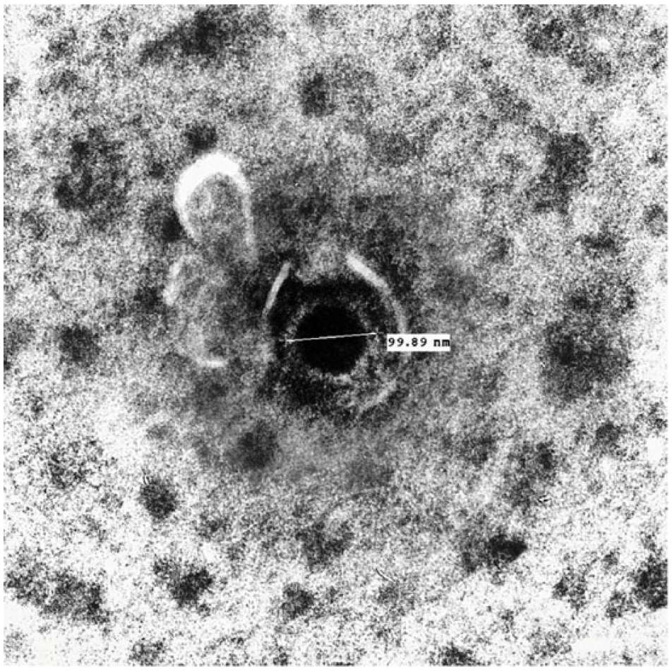
A nucleoid (round particle in the nucleus of the cell) is surrounded by a dense ring in this electron micrograph of a skin crust.
Identification of bovine herpesvirus 2 outbreaks and ancillary tests performed.
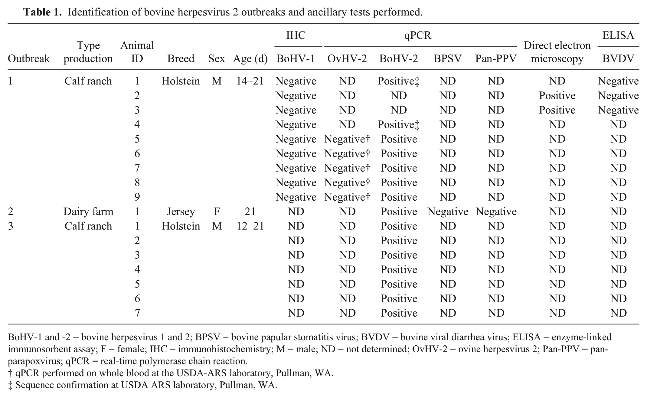
BoHV-1 and -2 = bovine herpesvirus 1 and 2; BPSV = bovine papular stomatitis virus; BVDV = bovine viral diarrhea virus; ELISA = enzyme-linked immunosorbent assay; F = female; IHC = immunohistochemistry; M = male; ND = not determined; OvHV-2 = ovine herpesvirus 2; Pan-PPV = pan-parapoxvirus; qPCR = real-time polymerase chain reaction.
qPCR performed on whole blood at the USDA-ARS laboratory, Pullman, WA.
Sequence confirmation at USDA ARS laboratory, Pullman, WA.
The gross appearance and location (around the eyes and on the ears) of these skin lesions with hair loss were the main clinical findings. Based on the estimated age of the lesions on the ear, it appeared that the lesions may have started on the dorsal pinnae along the edge and later spread over the entire ear.
BoHV-2 infection is usually associated with vesicles, crusts, and ulcers affecting 1 or more teats of lactating cows. Rarely is this virus identified in oral and muzzle lesions of nursing calves.14,19 A second syndrome associated with BoHV-2, known as PLSD or Allerton virus infection, is characterized by generalized nodular skin lesions, often accompanied by mild fever and depression.8,10 In our report, BoHV-2 cases did not show fever or depression at the time of, or prior to, the onset of the skin lesions. PCR was essential to confirm the infectious agent in these cases, given its high specificity and sensitivity in detecting and identifying viral nucleic acid.1,6,7,11
In one case, positivity for BoHV-2 virus was identified in a 3.5-y-old animal presented at a slaughterhouse. 20 The lesions characterized by lumps and scattered circular areas of alopecia were identified on the head, neck, shoulders, and on the perineum, representing the chronic pattern of PLSD.
The location of the lesions and high attack rate in the affected herds (affecting almost all calves in 2 of the outbreaks) suggest biting face flies as the mode of virus transmission to these calves. 5 At the time of these outbreaks (July–September), biting face flies were very common. In cases of PLSD, the virus is known to spread from animal to animal mechanically by biting flies.3,8,9,13
The incubation period for BoHV-2 infections is considered to be 1–2 wk. 8 Based on the age of the calves affected (12–26 d), the infection probably occurred during the first week of life. Interestingly, inclusion bodies are only observed during the exudative crust phase. 9 When the surface crust fell off, the underlying skin was alopecic. Coinfection with BoHV-2 and other bovine herpesviruses might be possible 2 ; however, in the cases described herein, BoHV-1 was not detected using IHC.
Where follow-up was available (outbreak 1), spontaneous recovery occurred in 2–4 wk after the detection of skin lesions. It is assumed that, after seroconversion, clinical signs tend to disappear, but there would be latent virus in the skin, nervous system, and lymph nodes.4,17,18
Potential differential diagnoses should include ringworm (Trichophyton verrucosum), contact dermatitis, photosensitization, poxvirus and parapoxvirus (e.g., bovine papular stomatitis virus and cowpox virus) skin diseases, and BVDV dermatitis. In the present case, ringworm was not detected. In addition, the animals had not been fed or exposed to any known photosensitizing agents or hepatotoxins.
Infection by BoHV-2 should be included in the list of differential diagnoses in cases of skin lesions in cattle. The definitive diagnosis was based on gross and histologic examinations, virus detection by qPCR, DNA sequencing, and virus particle identification by direct electronic microscopy.
Footnotes
Acknowledgements
We thank Dr. Jim Reynolds and Mr. Roger Blanchard (Visalia, CA), Dr. Ralph Walton (Tulare, CA), Drs. Hong Li and Naomi Taus (USDA-ARS Research Laboratory, Pullman, WA).
Declaration of conflicting interests
The authors declared no potential conflicts of interest with respect to the research, authorship, and/or publication of this article.
Funding
The authors received no financial support for the research, authorship, and/or publication of this article.