
Abstract
This retrospective project summarizes the types of neoplasms identified in Virginia opossums (Didelphis virginiana) presented to the University of Tennessee, College of Veterinary Medicine (UTCVM) postmortem service in 1989–2014 and serves as a review of the literature. Of the 85 Virginia opossums identified from the UTCVM case database, there were 17 diagnoses of neoplasia from 12 cases (14%). These cases included 8 females, 2 males, and 2 neutered males. All opossums with known ages (11 of 12) were >2 y old. Pulmonary tumors, specifically minimally invasive or lepidic-predominant adenocarcinomas, were the most common diagnosis and accounted for 53% (9 of 17) of the neoplasms. Additional tumors included acute myeloid leukemia with eosinophil maturation, hepatic hemangiosarcoma, sarcoma (unknown origin), squamous cell carcinoma, disseminated mast cell tumor, trichoblastoma, thyroid adenoma, and an osteoma. These findings serve as a reference for the types of spontaneous neoplasms in Virginia opossums; based on these findings, neoplasia should be considered as a differential in mature captive Virginia opossums.
Virginia opossums (syn. North American opossum; Didelphis virginiana) are the only North American marsupial. Terminology for New World species is opossum, and possum for Old World species. 13 These animals are affected by a number of infectious and neoplastic disorders. Parasitism in wild-caught opossums is common and includes lungworms (e.g., Didelphostrongylus hayesi), 1 heartworms (e.g., Dirofilaria immitis), 13 and protozoa (e.g., Besnoitia darlingi). 7 Documented bacterial infections include Streptococcus endocarditis, and bronchopneumonia from Pasteurella multocida and Bordetella bronchiseptica. 13 The most commonly documented diseases in captivity include obesity, metabolic bone disease, dental disease, chronic urogenital tract infections in unspayed females, and cardiac disease, both dilated and hypertrophic. 13
To date, there is limited information in the literature regarding neoplasia in Virginia opossums; however, in a previous report on spontaneous neoplasms in Virginia opossums, pulmonary adenomas were observed in 25% (17 of 68) of wild-caught opossums. 20 The objectives of our retrospective study were to determine the type of neoplasms in captive Virginia opossums presented to the University of Tennessee, College of Veterinary Medicine (UTCVM) in the past 25 y, review the literature on neoplasia in Virginia opossums, and compare these neoplasms to those documented in other opossum and possum species.
The UTCVM pathology database was searched for cases of Virginia opossums with a diagnosis of neoplasia from February 1989 through November 2014. Total submissions during this time period were 21,388. Autopsies were performed with conventional protocols under the direction of the duty pathologist. Slides from the identified cases were reviewed by an American College of Veterinary Pathologists board-certified pathologist (RL Donnell) and pathology resident (JP Pope) to verify the diagnosis. Signalment, presenting clinical signs, and cause of death or euthanasia were obtained from the autopsy, submission form, or case reports. To further characterize some of these neoplasms, additional stains including Giemsa, toluidine blue, and trichrome were performed on 2 cases (cases 8 and 11). Immunohistochemistry was performed on these 2 cases as well as 1 additional case (case 10).
Immunohistochemistry was performed using a conventional autostainer (Autostainer S3400, Dako North America, Carpinteria, CA) techniques per the manufacturers’ recommendations with appropriate anti-mouse– or anti-rabbit– (EnVision + System HRP anti-mouse and EnVision + System HRP anti-rabbit, Dako) labeled polymer by a horseradish peroxidase system with DAB (3,3’-diaminobenzidine tetrahydrochloride; Dako) as chromogen. For tissues stained with cluster of differentiation (CD)3, CD20, C79, and von Willebrand factor (vWF), heat-induced epitope retrieval was carried out with ethylenediamine tetra-acetic acid (EDTA) buffer at pH 9, and for tissues stained with c-kit with citrate buffer at pH 6 in a steamer at 95°C for 25 min. For tissues stained with CD18, enzyme-induced epitope retrieval (proteinase K, Dako) was carried out at room temperature for 5 min. Antibody for CD3 (monoclonal mouse CD3 antibody, Dako) was applied at a 1:200 dilution for 30 min, CD20 (polyclonal rabbit CD20 antibody, Thermo Fisher Scientific, Waltham, MA) at a 1:800 dilution for 30 min, CD79 (monoclonal mouse CD79 antibody, Thermo Fisher Scientific) at a 1:800 dilution for 30 min, CD18 (monoclonal mouse CD18 [clone Ca16.3c10] antibody, Peter Moore, University of California–Davis, Davis, CA) at 1:10 dilution for 30 min, c-kit (polyclonal rabbit c-kit antibody, Dako) at a 1:500 dilution for 60 min, and vWF (polyclonal rabbit vWF antibody, Dako) at a 1:4,000 dilution for 30 min. All immunohistochemical stains had positive external controls. vWF and c-kit were immunoreactive in normal endothelial cells and mast cells as well as in neoplasms originating from these cells. CD3 and CD18 were immunoreactive in normal T-lymphocytes and histiocytes of opossums, respectively. Normal B-lymphocytes within splenic lymphoid follicles were non-immunoreactive for both CD20 and CD79.
From the case database, the authors identified 85 Virginia opossums that had been submitted for autopsy with clinical signs and history including dead-on-arrival, dyspnea, lethargy, neurologic signs, and chronic non-healing skin wounds. Of this population, there were 17 diagnoses of neoplasia from 12 cases (14% of cases). All opossums diagnosed with neoplasia were in captivity in zoological collections, rehabilitation facilities, or as pets. Presenting clinical signs of opossums with neoplasia varied from nonspecific signs of lethargy and anorexia to skin masses and ulcers with progressive clinical deterioration. Of these opossums, 9 were euthanized either because of poor prognosis or inability to manage clinical signs, and 3 died (2 in the clinic overnight; 1 with no premonitory signs at the zoo). Signalment, types, and characteristics of neoplasia are listed in Table 1. Of the 12 cases, 8 were females, 2 were males, and 2 were neutered males. All animals identified were adults, and all those with a known age (11 of 12) were >2 y old.
Signalment, types and characteristics of neoplasia, and findings at autopsy in 12 Virginia opossums presented to the University of Tennessee, College of Veterinary Medicine from 1989–2014.
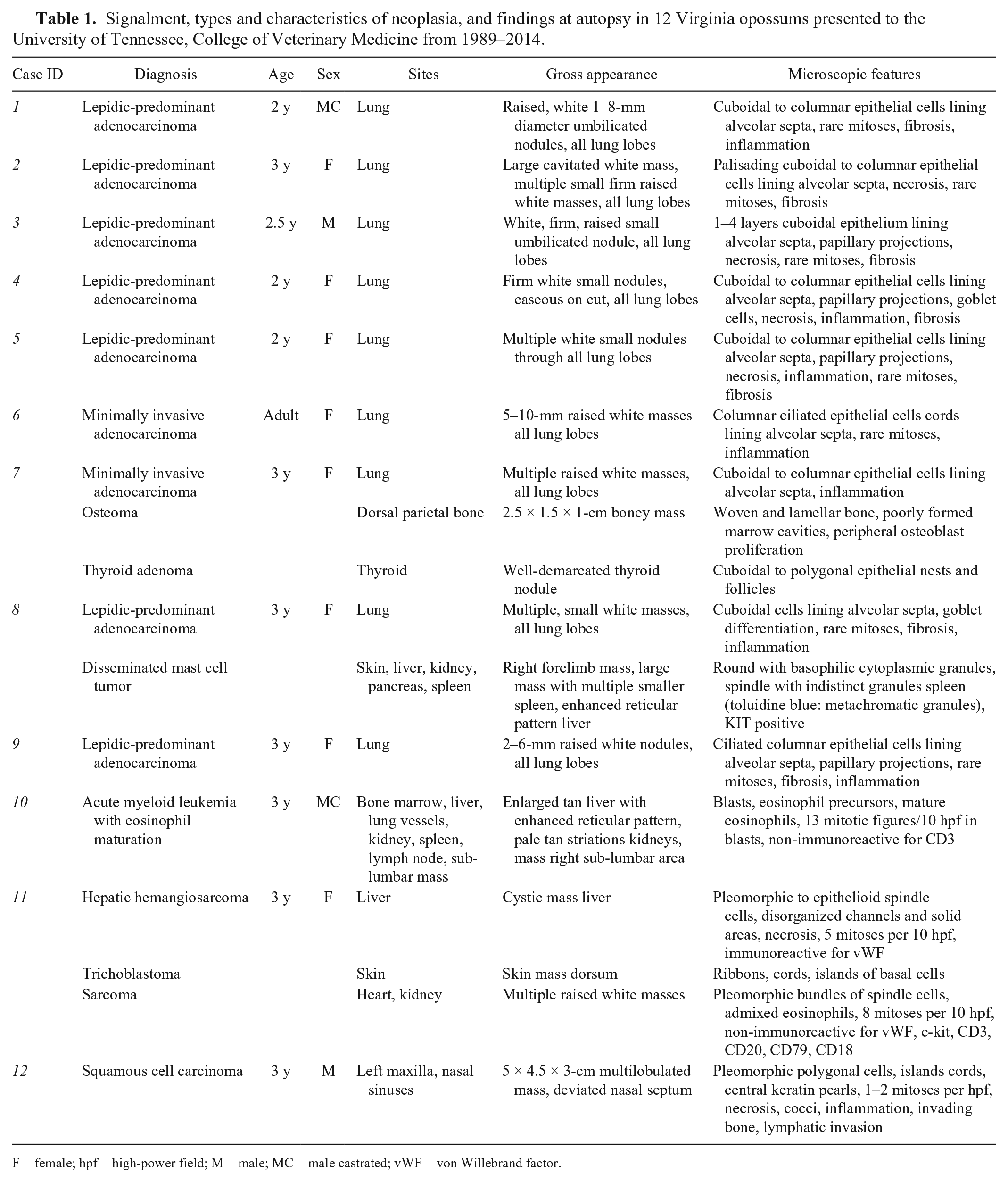
F = female; hpf = high-power field; M = male; MC = male castrated; vWF = von Willebrand factor.
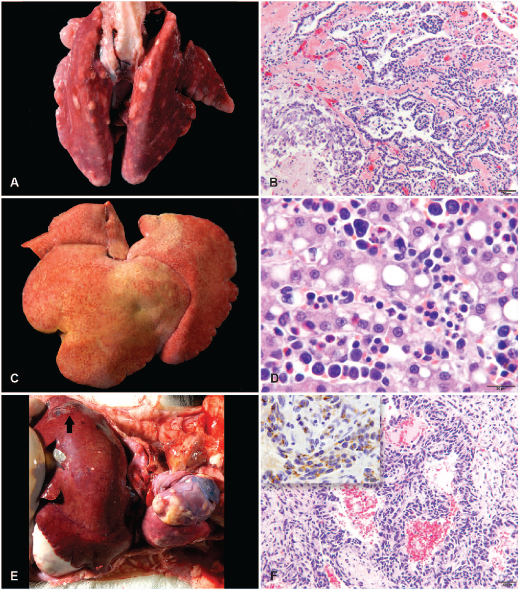
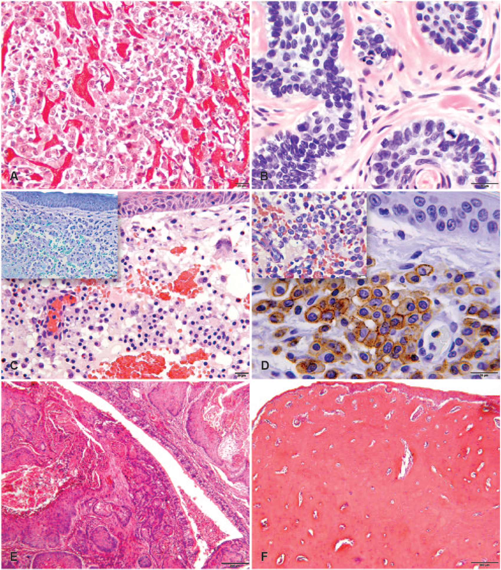
Of the 17 neoplasms identified, 14 were malignant and 3 were benign. Two animals (cases 7 and 11) were identified to have 3 concurrent neoplasms, and 1 animal (case 8) had 2 concurrent malignant neoplasms. Neoplasia was identified as the cause of death or euthanasia in 6 of 12 animals. Pulmonary tumors, specifically minimally invasive or lepidic-predominant adenocarcinomas (Fig. 1A, 1B), were the most commonly diagnosed neoplasm. Additional tumors included acute myeloid leukemia (AML) with eosinophil maturation (Fig. 1C, 1D), hemangiosarcoma (Fig. 1E, 1F), poorly differentiated sarcoma in the heart and kidney, (Fig. 2A), trichoblastoma (Fig. 2B), disseminated mast cell tumor (Fig. 2C, 2D), squamous cell carcinoma (Fig. 2E), thyroid adenoma, and an osteoma (Fig. 2F).
Neoplasms of Virginia opossums (Didelphis virginiana).
Neoplasms in Virginia opossums (Didelphis virginiana).
Approximately 53% (9 of 17) of the neoplasms diagnosed in these opossums were pulmonary tumors, comprising 11% (9 of 85) of all opossums submitted for autopsy during the study time period. In many of these cases, there were multiple small neoplastic masses with no clear primary in the lungs, consistent with spontaneous multicentric neoplasms. In 2 cases, larger masses were identified grossly, potentially consistent with a primary pulmonary carcinoma and intrapulmonary metastasis. All pulmonary neoplasms were considered malignant. Multiple pulmonary adenomas were previously reported (17 of 68) in wild-caught Virginia opossums. 20 Distinguishing between benign and malignant pulmonary tumors is inherently difficult given their growth pattern, which can lead to differences in diagnosis. 5 Additionally, the neoplasms in the previous study were often only observed microscopically and not on gross examination, which may have led to the benign diagnosis. 20 Multiple bronchioloalveolar carcinomas were previously reported in 1 opossum, 14 pulmonary adenocarcinomas were observed in 2 additional opossums,2,16 and a pulmonary squamous cell carcinoma was previously documented in 1 opossum. 21
In our study, inflammation, primarily lymphocytic, was observed in 7 of 9 cases, and fibrosis was observed in 7 of 9 cases. Interstitial pneumonia and parasites were previously reported in conjunction with pulmonary neoplasms. 20 In our study, parasites were not observed in the examined lung sections, but all animals were captive. Significant fibrosis was only reported previously in 1 case. 14 The number of neoplasms with concurrent inflammation or fibrosis both in our study and in the literature may suggest an inflammatory component to these neoplasms. It is interesting to note that opossums in captivity and wild-caught opossums14,20 both have the propensity to develop pulmonary neoplasms given that environmental conditions differ. Pulmonary neoplasms were implicated as the cause of clinical signs and resulting death or euthanasia in only 1 opossum, in our study, which is similar to previous reports.14,20 In our study, more females than males were documented with pulmonary adenocarcinomas, but this may reflect a submission bias, as, in the previous report, there was no sex bias. 20
Dogs and cats are the most common domestic animals to develop pulmonary neoplasms (~1% of dogs autopsied at veterinary schools) and these animals are typically older. 24 Pulmonary tumors are uncommonly documented in zoo and wild animals including other marsupials. Pulmonary tumors in the opossums in our study were most often minimally invasive or lepidic-predominant adenocarcinomas (previously known as bronchoalveolar/bronchioloalveolar carcinomas). These tumors most resemble those in sheep, which are also multicentric, well-differentiated pulmonary adenocarcinomas with lepidic pattern of growth, often with associated inflammation and fibrosis. 5 These tumors in sheep are caused byJaagsiekte sheep retrovirus 5 and because of noted similarities between pulmonary tumors in sheep and opossums, 20 immunohistochemistry was performed for this virus in a single case report, 14 but results were negative. 14 However, an opossum-specific retrovirus cannot be ruled out as a potential cause.
Neoplasia in our study was diagnosed in mature opossums. In the wild, the lifespan of opossums is typically 1.5 y. 13 In captivity, this lifespan can be increased to 4–7 y. 13 The mean age in this series was 2.7 y (median = 3 y). The findings in our study suggest that mature opossums are more prone to develop neoplasia and may develop multiple neoplasms: 3 of 12 (25%). Previously reported neoplasms in Virginia opossums along with signalment, captivity status, and source from the literature are documented in Table 2. As was observed in our study, 3 of the opossums from the literature had multiple neoplasms.2,14,23 In our study, the majority of the documented neoplasms, 82% (14 of 17) were malignant, which is consistent with previous reports in the literature.2,10,14,16,17,21
Previously reported neoplasms in the literature along with signalment and captivity status if available in Virginia opossums.
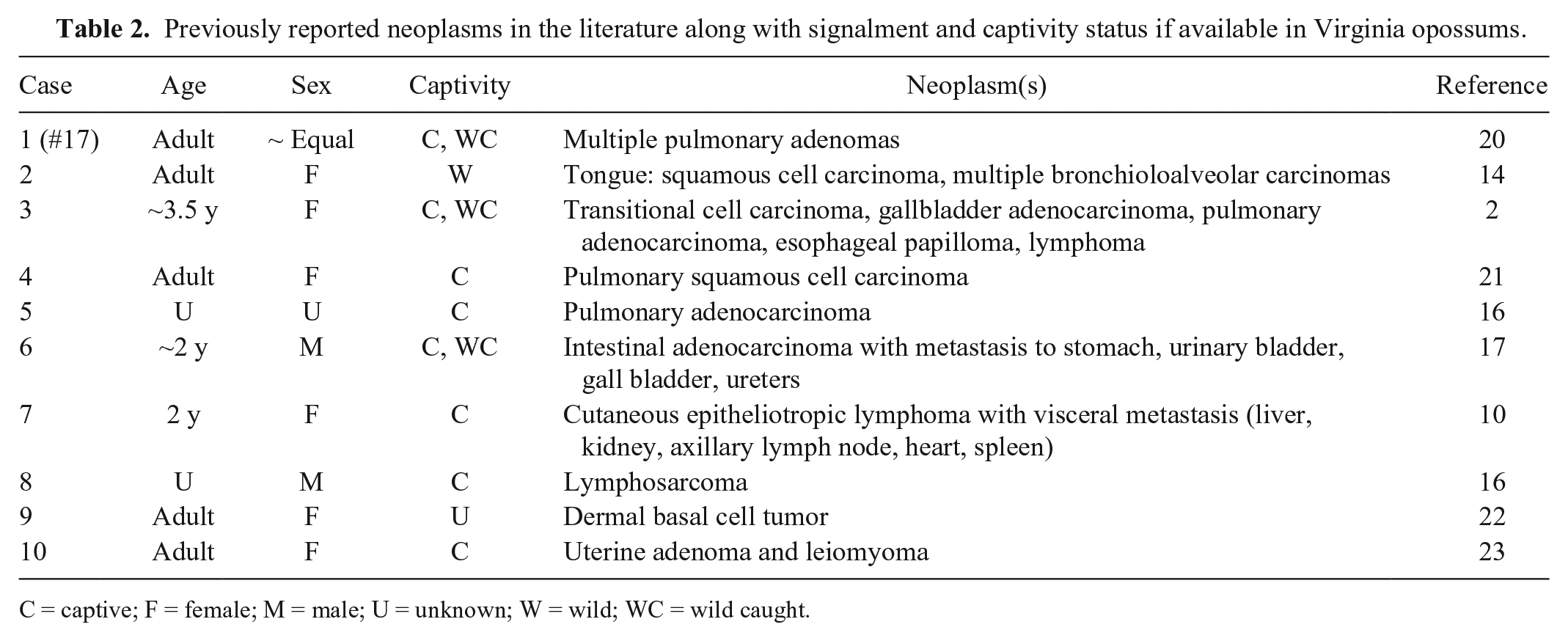
C = captive; F = female; M = male; U = unknown; W = wild; WC = wild caught.
Squamous cell carcinoma has been previously reported in the oral cavity of a Virginia opossum and was located on the tongue, 14 whereas it was located on the maxilla in case 12 in our study. The previously reported basal cell tumor 22 may have been a trichoblastoma, as in the current study, as the microscopic description was consistent and basal cell tumors have become further classified. AML has not been previously reported in Virginia opossums, to our knowledge, and is uncommonly reported in any veterinary species. Eosinophil maturation in AML is rare, and was present in only 2 of 181 (1%) cats in a previous case series. 12
Mast cell tumors have not been documented previously in Virginia opossums, to our knowledge. The spindle morphology and lack of characteristic granules on hematoxylin and eosin staining in the masses in the spleen in case 8 were similar to the described atypical mast cell tumors in cats. 18 The c-kit staining pattern was membrane-associated. Based on the knowledge of mast cell tumors in dogs and cats, it is suspected that, in case 8, the cutaneous mast cell tumor was the primary site with metastasis to other organs.
Case 11 had both a hepatic hemangiosarcoma as well as a poorly differentiated sarcoma in the heart and kidney. The liver has been documented as a primary site for visceral hemangiosarcoma in dogs, 3 cats, 6 ferrets, 8 and a rabbit, 9 and in some of these species (cats 6 and ferrets 8 ) is more common in the liver than in the spleen. Neoplastic cells in the heart and kidney were non-immunoreactive for CD3 (T-lymphocytes), CD20 (B-lymphocytes), CD79 (B-lymphocytes), CD18 (histiocytes), c-kit (mast cells), and vWF (endothelial cells). Therefore, the neoplasm was classified as a sarcoma based on cellular morphology. Because neither CD20 nor CD79 identified normal B-lymphocytes in opossums, the possibility remains that the neoplasm in the heart and kidney was a lymphosarcoma of B-cell origin. Cutaneous epitheliotropic T-cell lymphoma with visceral metastasis was previously reported in a single Virginia opossum, 10 and lymphosarcoma, unknown B- or T-cell origin, was previously diagnosed in 2 Virginia opossums.2,16
Neoplasia is common in other species of opossums and possums, as well. In gray short-tailed opossums (Monodelphis domestica) in South America, spontaneous neoplasia was reported in 39 of 150 opossums at autopsy. 11 The most commonly reported spontaneous neoplasms were pituitary adenomas (prolactinomas), followed by uterine leiomyomas, lipomas, pheochromocytomas, and hepatic carcinomas. 11 Neoplasia was reported in 8 of 48 captive mountain pygmy possums (Burramys parvus), and considered the cause of death in 5 animals. 19 The most commonly reported neoplasms were reproductive (2 mammary adenocarcinomas and 1 ovarian carcinoma) and hepatic (1 myelolipoma, 1 hepatoma, and 1 hepatic carcinoma). 19 Other documented neoplasms included a poorly differentiated sarcoma involving the lungs, and a squamous cell carcinoma, suspected to be of esophageal origin. 19
In a large study on Australian marsupials that included multiple species of possums, including brushtail possums (Trichosurus vulpecula), ring-tailed possums (Pseudocheirus peregrinus), eastern pygmy possums (Cercartetus nanus), and western pygmy possums (Cercartetus concinnus), the most commonly reported neoplasms were lymphosarcoma and hepatomas. 4 Other reported neoplasms included an adrenocortical adenoma, squamous cell carcinoma, hemangiopericytoma, metastatic adenocarcinoma, and testicular Sertoli cell tumor. 4 In the Australian Registry of Wildlife Health, possums and gliders (n = 74, 18.4%) were second only to dasyurids for neoplasms reported in terrestrial mammals. 15 In possums, epithelial neoplasms, specifically of hepatic or mammary origin, were more commonly reported than mesenchymal neoplasms. 15
Squamous cell carcinoma and lymphosarcoma seem to occur across species4,14,19; however, Virginia opossums differ with respect to the most commonly diagnosed neoplasm. Pituitary adenomas are the most frequently documented neoplasm in gray short-tailed opossums. 11 The most commonly reported neoplasm in western pygmy possums are hepatomas,4,15 and mammary adenocarcinomas are the most frequently reported neoplasm in mountain pygmy possums. 19 The most commonly reported neoplasm in Virginia opossums both in our study and in the literature are pulmonary tumors. Inflammation and fibrosis were often documented in conjunction with these neoplasms, which either suggests a role for inflammation in the development of these neoplasms or that inflammation is a sequela to these neoplasms. Further investigation into an infectious or environmental cause, mutation, or other commonality may be warranted given the propensity of both captive and wild-caught opossums to develop multiple pulmonary tumors.2,14,16,20
Footnotes
Acknowledgements
We thank Misty Bailey (paid by the University of Tennessee, College of Veterinary Medicine) for technical editing support and Joy Chambers (paid by the University of Tennessee, College of Veterinary Medicine) for graphic design support.
Declaration of conflicting interests
The authors declared no potential conflicts of interest with respect to the research, authorship, and/or publication of this article.
Funding
The authors received no financial support for the research, authorship, and/or publication of this article