
Abstract
Squamous-cell carcinoma (SCC) is the second most common tumor in horses, and 40%-50% may occur in ocular and adnexal structures. Cyclooxygenase (COX)-2 is an inducible enzyme responsible for the production of prostaglandins that control cell growth and the development and progression of cancer. Mechanisms responsible for the initial upregulation of COX-2 in neoplasia are unclear; prolonged sunlight exposure and mutations in the p53 gene may be possibilities. Because the etiopathogenesis of ocular SCC in horses may involve ultraviolet sunlight and p53 mutations, the purpose of this study was to characterize the immunoreactivity of COX-2 in these tumors. Cyclooxygenase-2 expression was found in 6 of 22 (27%) paraffin-embedded equine SCCs. Cyclooxygenase-2 immunoreactivity was associated with the mitotic index (P < 0.001). Strategies to inhibit COX-2 by the use of topical or systemic COX-2 inhibitors might prove to be a safe and economical treatment in some horses with SCC.
Cyclooxygenase (COX) is a rate-limiting enzyme that catalyzes conversion of arachidonic acid to an array of prostaglandins. There are at least 3 known COX pathways. 5 The COX-1 enzyme is constitutively present in most cells and regulates multiple physiologic processes. Cyclooxygenase-2 is an inducible enzyme that is involved in the production of prostaglandins that modulate pathologic events such as inflammation, wound healing, and neoplasia. Cyclooxygenase-2 is absent from most normal tissues, except for specific regions in the kidney, the brain, and the uterus. 6 Cyclooxygenase-3, an acetaminophen-sensitive COX isoform produced from the COX-1 gene by alternate exon usage, was recently identified. 5
Numerous experimental, clinical, and epidemiologic studies linked tumor development and progression with the presence of COX-2 in tumor cells. Stromal fibroblasts, tumor infiltrating inflammatory cells, and angiogenic endothelial cells can also express COX-2. 14 Prostaglandins, produced by COX-2 activity, increase cell proliferation; inhibit apoptosis; promote angiogenesis; alter cellular adhesion, which allows for metastasis; inhibit immune surveillance; and may activate xenobiotics to reactive substances that are carcinogenic. 16 In human patients, recent studies found that COX-2 is upregulated in pre-cancerous skin lesions, such as actinic keratosis and squamous-cell carcinoma (SCC). 3,12,19 In dogs, COX-2 was strongly expressed in neoplastic keratinocytes in all cases of SCC of the skin. 22
Piroxicam is a nonsteroidal anti-inflammatory drug that nonselectively inhibits COX. The in vivo effect of piroxicam a in a horse with metastatic SCC was previously reported. 17 A 16-year-old Morgan gelding had SCC of the lip, with spread to the submandibular lymph node. Piroxicam was administered per os, and complete clinical remission of the tumor and metastases occurred. By immunohistochemistry, the majority of neoplastic cells were positive for COX-2. 17
The hypothesis of the present study was that COX-2 would be overexpressed in SCC of the ocular and periocular structures in horses. Results of this study may help to determine if COX-2 is a potential target for therapeutic and preventative strategies of SCC in horses.
Archived biopsy samples from 1977–2004 were obtained from the Surgical Pathology and Necropsy Services of the Department of Biomedical Sciences and the Ocular Pathology Service of the Department of Clinical Science, Cornell University College of Veterinary Medicine. All samples were fixed in either 10% neutral buffered formalin, Bouin solution, or Zenker solution, and were embedded in paraffin wax. Diagnosis was confirmed by evaluation of hematoxylin and eosin (HE)-stained sections by a coauthor (B.L.N.). All tumors were classified with respect to microscopic features, including invasiveness, degree of differentiation, and mitotic index, as described by Carvalho et al. 4 Invasiveness was evaluated on a scale of 0 to 3: 0, noninvasive; 1, minimal invasion of surrounding tissues; 2, small independent islands surrounding the main tumor; and 3, deep invasion far from the main tumor. The degree of differentiation was evaluated on a scale of 0 to 2: 0, well differentiated; 1, moderately differentiated; and 2, poorly differentiated. The mitotic index was evaluated as the number of mitotic figures per high-power field (HPF) and was evaluated on a scale from 0 to 2: 0, <2; 1, 2−6; and 2, >6.
Immunoperoxidase (biotin-streptavidin) staining was performed on all sections by using a rabbit polyclonal antibody to COX-2, b as previously described. 1 To test the specificity of the immunostain, formalin-fixed, paraffin-embedded canine mandibular SCC and normal equine fetal kidney were used as positive control tissues. Positive control samples and negative control tissues were treated similarly, except that normal nonimmune rabbit serum, as a nonspecific antibody control, was substituted for the COX-2 antibody.
Immunoreactivity to assess COX-2 distribution (percentage of positive cells) and staining intensity was evaluated in a blinded fashion by one of the authors (B.L.N.), based on a previously described method. 22 Five 10 × fields from each slide were evaluated. COX-2 distribution was evaluated on a scale of 0 to 4: 0, negative; 1, <10% of cell staining positive; 2, 10%−30%;3, 31%−60%; and 4, >60%. Staining intensity was the strength of the signal and was evaluated on a scale of 0 to 3: 0, negative: 1+, weak staining; 2+, moderately intense staining; and 3+, intense staining. A final COX-2 score was determined by multiplying COX-2 distribution and intensity.
The Fisher exact test was used to analyze frequencies of COX-2 immunoreactivity (negative vs. positive) and HE features, including invasiveness (0 vs. 1–3), degree of differentiation (0 vs. 1−2), and mitotic index (0 vs. 1−2). The Wilcoxon rank sum test was used to analyze whether the COX-2 scores (the product of distribution and intensity) differed by the same HE features. P values ≤0.05 were considered statistically significant. All analyses were performed by using a commercial software package. c
Thirty-one cases were identified from the initial archive search, and the histomorphologic diagnosis of 22 was verified as SCC. Fifteen samples were fixed in formalin, 5 in Bouin solution, and the remaining 2 were fixed in Zenker solution. Anatomic origins of the 22 tumors were the cornea in 19, the conjunctiva in 2, and the nictitans in 1. Of the 22 tumors, 2 were noninvasive, 9 were minimally invasive, 8 were moderately invasive, and 3 were deeply invasive. Eight tumors were well differentiated, 12 were moderately differentiated, and 2 were poorly differentiated. The mitotic rate was 0−2/HPF in 15 cases, 2−6/HPF in 6, and >6/HPF in 1.
Six of the 22 equine ocular SCCs (27%) were immunopositive for COX-2 (Fig. 1). All positive samples arose from the cornea: 3 had been fixed in formalin, 2 in Bouin solution, and 1 in Zenker solution. Immunostaining intensity was 1+ in all cases. Positive staining was patchy and focal, and immunopositivity was confined to the cytoplasmic compartment of neoplastic cells, occasionally highlighting the perinuclear cytoplasm and, in one instance, along the intercellular junctions between adjacent neoplastic cells. The percentage of stained cells was <10% in all cases. Cyclooxygenase-2 scores ranged from 0 to 1. Positive controls (equine fetal macula densa and canine oral SCC) showed strong immunostaining for COX-2.
Cyclooxygenase-2 immunoreactivity was significantly associated with the mitotic index (P < 0.001). The mitotic index of 5 COX-2 positive tumors was 2−6/HPF, and the mitotic index of the remaining COX-2 positive tumor was >6/HPF. Fifteen COX-2-negative tumors had a mitotic index of <2/HPF, and 1 had a mitotic index of 2−6/HPF. There was no association between COX-2 immunoreactivity and other HE features, such as the degree of invasiveness or the degree of differentiation.
Squamous-cell carcinoma is the second most common tumor in horses, accounting for 20% of all neoplasms in the species. Common anatomical locations include the skin, the external genitalia, and the stomach, but involvement of ocular and adnexal structures account for 40%-50% of SCC. In a study of 157 cases of equine SCC, 55% occurred on the eyelid, 25% occurred on the conjunctiva, and 19% occurred on the cornea. Approximately 20% of horses had bilateral disease. 18
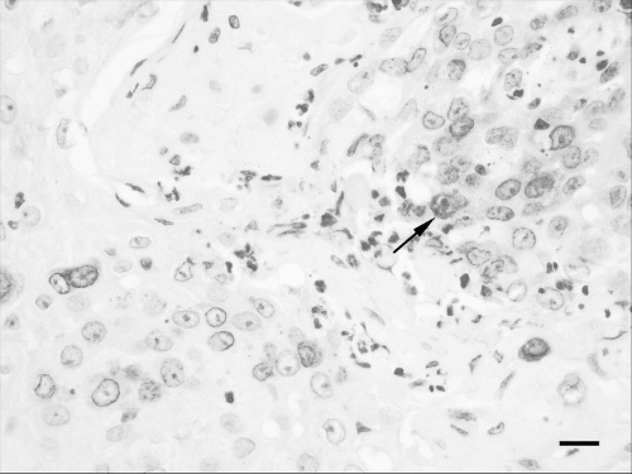
Squamous-cell carcinoma of the cornea, cyclooxygenase (COX)-2 immunoreactivity. There is weakly intense cytoplasmic and perinuclear staining for COX-2 in neoplastic epithelial cells. Chromagen 3,3'-diaminobenzidine tetrachloride (DAB), Gill hematoxylin counterstain, 400× magnification. Bar = 20 μm.
The etiopathogenesis of SCC involving ocular and periocular structures in horses is not known. The ultraviolet component of sunlight is the most plausible carcinogenic agent, because many lesions occur in the nonpigmented adnexa. Pazzi et al 20 described p53 point mutations consistent with ultraviolet radiation carcinogenesis in 25% (2 of 8) equine SCCs. Other factors implicated include viral agents and genetic and immunologic factors.
The mechanisms responsible for the initial upregulation of COX-2 in neoplastic tissues are still unclear. In vitro ultraviolet irradiation of human keratinocytes induced upregulation of COX-2 expression, suggesting prolonged sunlight exposure as an underlying mechanism in human SCC of the skin. 3,11 In a further study, the administration of a COX-2 inhibitor, celecoxib, d to mice irradiated with ultraviolet light reduced tumor development if the COX-2 inhibitor was given during the irradiation period or prevented new tumor formation if the compound was administered after the onset of photocarcinogenesis. 8,21 It is possible that ultraviolet irradiation contributes to increased COX-2 expression through its action on p53; p53 has been shown to cause a decrease in the expression of COX-2 in mouse fibroblasts, 23 and, as previously mentioned, mutation of the p53 gene has been reported in natural cases of equine SCC. 20 In some horses, mutations of p53 might contribute to high levels of COX-2. A correlation between COX-2 and p53 has been suggested for human SCC. 9 However, this has not been substantiated in all studies. 13
Based on the likely association between ultraviolet radiation and the development of ocular SCC in horses and laboratory studies to evaluate the role of COX-2 in SCC of mice and people, this study was undertaken to evaluate COX-2 immunoreactivity in 22 equine SCCs. Cyclooxygenase-2 expression was present in 6 tumors (27%) arising from the cornea. These results are in striking contrast to those in dogs, where COX-2 immunostaining was demonstrated in 100% of neoplastic keratinocytes of 7 tumors originating from the skin. 22 Recently, 5 corneal and 10 eyelid SCC lesions in horses were evaluated for COX-2 immunoreactivity. 15 Tumors were noted to be positive, and positivity was quantified by the use of computer analysis; however, the proportions of negative and positive tumors were not reported. 15 In 2 combined human studies, 16 of 25 cutaneous SCCs (64%) had a strong expression of COX-2. 12,13 Finally, no COX-2 immunoreactivity was detected in cutaneous SCC of 6 cats. 1
Results of the present study suggest that COX-2 is not expressed in all cases of naturally occurring equine ocular SCC. However, investigators recently harvested 14 equine SCC lesions in liquid nitrogen and by Western blot analysis detected COX-2 enzyme in all samples. 7 The lack of detectable COX-2 expression in the majority (>60%)of equine ocular SCC lesions in the study reported here may have been because of sample size; failure of the antigen retrieval method used 1 ; or other confounding variables, such as the type of fixative or prolonged fixation. Tissue fixation (i.e., formalin, Bouin solution) and/or tissue processing destroys or denatures several proteins, thereby altering antigenic recognition by immunohistochemistry. It is possible that the polyclonal antibody used may not be a sensitive indicator of COX-2 antigenic epitopes present in the horse. Potentially, other polyclonal COX-2 antibodies or a monoclonal antibody could be used to more fully characterize the presence of COX-2 in equine tumors. An alternative explanation is that COX-2 concentrations in equine ocular SCC are below the level of immunohistochemical detection. Although not attempted in the present study, the use of frozen sections for these studies may yield results that are more reliable. Also, immunohistochemistry is a semiquantitative method, and Western blotting might be a better method to quantify the level of COX-2 expression. Finally, determination of prostaglandin E2 concentration is commonly used as a marker for COX-2 activity. Future studies to evaluate a larger number of equine tumors with these techniques and other methods to assess functional COX-2 activity would be useful to confirm the findings of the present study.
Data reported herein showed that COX-2 immunoreactivity was significantly associated with the mitotic index. The proportion of COX-2 positive tumors with a mitotic index >2/HPF was significantly higher than that among tumors that were COX-2 negative. Because of the retrospective nature of the study, patient follow-up was not available; attempts to associate these findings with an outcome such as metastases or survival could not be done. No association was found between COX-2 immunoreactivity and the degree of invasiveness or the degree of differentiation. Kagoura et al 12 investigated COX-2 expression in various human skin tumors, and their results also indicated no correlation with the extent of malignancy; COX-2 was more strongly expressed in differentiated cells than immature cells.
Numerous treatment modalities are described for equine ocular SCC, including surgical resection, strontium-90 beta irradiation, interstitial radiotherapy, external beam radiotherapy, radiofrequency hyperthermia, intratumoral or topical chemotherapy, CO2 laser ablation, and cryotherapy. 18 The treatment modality chosen is influenced by tumor size, tumor location, availability of equipment, clinical expertise, and, often, cost of treatment. Even if treatment of SCC is undertaken, therapy fails to be curative for many horses. After treatment, local recurrence may occur in up to 68% of horses. 18
New and more effective treatment modalities are needed for some horses with ocular SCC. As in other species with SCC, the inhibition of COX-2 might prove to be a valuable alternative therapy. Drugs that appear to inhibit COX-2 in vitro in horses have been identified, 2 and COX inhibitors have demonstrated antiproliferative effects that are independent of COX-2 expression. 10 However, routine treatment of equine ocular SCC with COX-2 inhibitors is not supported by the results of the present study. Further investigations into the relations between COX-2 and p53 during tumorigenesis of equine ocular SCC should be pursued.
acknowledgements. This work was funded by a grant from the Harry M. Zweig Memorial Fund for Equine Research. The authors thank Drs. Nita Irby, Thomas Kern, and Ronald Riis of the Section of Ophthalmology, Cornell University College of Veterinary Medicine, for the contribution of case material to this study.
Footnotes
a.
Feldine, Pfizer, New York, NY
b.
PG27B, Oxford Biomedical Research, Oxford, MI
c.
SPSS version 10, SPSS, Chicago, IL
d.
Celebrex, Pfizer, New York, NY