
Abstract
A 9-year-old, intact female alpaca (Vicugna pacos) was presented for a second opinion with a 1-year history of nonpruritic, multifocal scaling and crusted cutaneous lesions, mainly involving skin on the face, axillae, and ventral abdomen. Clinical abnormalities were limited to the skin, and the alpaca was otherwise healthy. The initial veterinarian had examined the alpaca, found no evidence of ectoparasites with laboratory testing, and had tried several trial therapies including oral antibiotics, ivermectin, and topical use of betadine solution. At the time of presentation, the lesions had neither improved nor worsened with any attempted therapy, and multiple skin biopsies were collected. Histopathology and immunohistochemical staining findings were consistent with the pagetoid reticulosis type of cutaneous epitheliotropic T-cell lymphoma. Our report describes the clinical, histopathologic, and immunophenotypic features of pagetoid reticulosis epitheliotropic cutaneous T-cell lymphoma in an alpaca.
Lymphoma is recognized as one of the most common malignant neoplasms affecting South American camelids (SACs).11,20,22 Numerous publications have demonstrated the reliability of immunophenotyping alpaca and llama lymphocytes using anti-human cluster of differentiation (CD)3 and CD79α antibodies to confirm the presence of T and B lymphocytes, respectively, in both healthy2,4 and diseased skin.1,8,11,16,18,20 The vast majority of cases of lymphoma reported in SACs have involved malignant T- or B-lymphocyte neoplasms with dissemination to various organs.1,11,18,20 In contrast, cutaneous lymphoma (both nonepitheliotropic and epitheliotropic) appears to be quite rare in SACs. A retrospective study describing the prevalence and types of neoplasia diagnosed in SACs for the Pacific Northwest observed that cutaneous and mucocutaneous neoplasms were the most common types of tumors diagnosed in the llama and alpaca; however, none of those cases were cutaneous lymphoma. 22 A 2010 review of skin disease in the alpaca reported a single case of subcutaneous lymphoma in a young alpaca. 19 Additionally, a larger study describing the immunophenotypic characterizations of 26 camelid malignant round cell tumors identified a single case of epitheliotropic cutaneous T-cell lymphoma (CTCL) in an aged llama. 1
Epitheliotropic CTCL has been described extensively in humans, more recently in dogs,5,13 and in horses. 3 In these species, epitheliotropic CTCL is a heterogeneous group of neoplasms, which are often subclassified based on their various clinical patterns and histologic features. In all cases, epitheliotropic lymphoma is characterized by a cutaneous T-cell infiltrate with tropism for the epidermal, follicular, and/or mucosal epithelium. 21 The 3 primary classifications are mycosis fungoides, Sézary syndrome, or pagetoid reticulosis. 6 Mycosis fungoides is well recognized as the most common form of CTCL in humans and domestic animals,5,13,15,21 whereas both Sézary syndrome and pagetoid reticulosis are considered to be rare.6,13 In humans, CTCL is clonal; however, the cells have a heterogeneous immunophenotypic profile.7,10,17 Our report describes the clinical, histopathologic, and immunophenotypic features of pagetoid reticulosis epitheliotropic CTCL in an alpaca (Vicugna pacos).
A 9-year-old, intact female alpaca was presented with a 1-year history of cutaneous lesions characterized by nonpruritic, multifocal thickening of the skin with associated scaling and crusts. The lesions predominantly affected the perioral, periocular, and periauricular tissue, as well as skin in the axillae and on the ventral abdomen near the udder. Differential diagnoses for this clinical presentation include dermatophytosis, chorioptic mange, zinc-responsive dermatosis, cutaneous neoplasia, idiopathic necrolytic neutrophilic hyperkeratotic dermatosis, and less likely dermatophilosis, bacterial folliculitis, as well as other ectoparasitic infestations. A previous veterinarian had examined the alpaca, found no evidence of ectoparasites with laboratory testing, and had tried several trial therapies including oral antibiotics (unknown types and doses), ivermectin (unknown dose), and topical use of betadine solution. All therapeutic trials failed to improve her condition after 5 months, and the therapies were discontinued without progression or improvement of the skin lesions.
Two months later, the alpaca was presented for a second opinion evaluation to a veterinarian who collected several skin biopsies and submitted them to the Veterinary Diagnostic Laboratory at the University of Illinois (Urbana, Illinois). The histopathology revealed abundant orthokeratotic hyperkeratosis with several small aggregates of degenerative leukocytes within the keratin, and a moderately hyperplastic epidermis. The epidermis and follicular epithelium were being infiltrated by numerous individual or aggregates of neoplastic rounds cells (Fig. 1A). The neoplastic cells had distinct cell margins, a large nuclear-to-cytoplasmic ratio, round to ovoid to indented nucleus, vesiculate chromatin, and a single prominent nucleolus. There was moderate to marked anisocytosis and anisokaryosis with an average of 2 mitotic figures within 10 random 400× fields. Accompanying the neoplastic round cells were intermediate numbers of eosinophils within the superficial dermis with minimal numbers infiltrating the epidermis. Dermal and subcutaneous neoplastic round cells were not observed. Immunohistochemical staining demonstrated positive membranous immunoreactivity for CD3 in the neoplastic population (Fig. 1B) and no immunoreactivity for CD79α. The histopathologic and immunohistochemical findings were consistent with pagetoid reticulosis epitheliotropic CTCL. Attempts to contact the owners for follow-up after the diagnosis were unsuccessful, and there is no further information on the progression of lesions or the clinical state of the alpaca. To our knowledge, blood work was not performed at any time during the diagnostic evaluation of this patient.
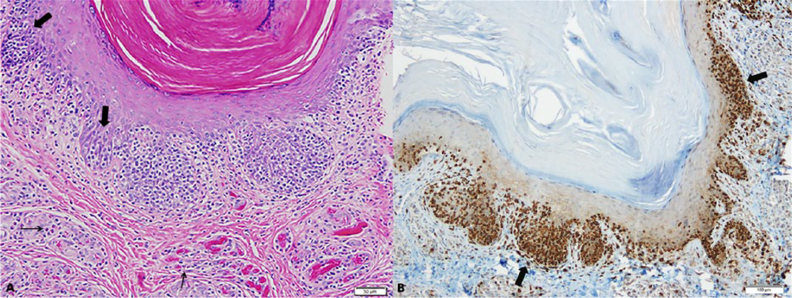
Epitheliotropic T-cell lymphoma in a 9-year-old female alpaca (Vicugna pacos).
Epitheliotropic cutaneous T-cell lymphoma is a disease characterized by proliferating T-lymphocyte infiltration of the epidermis and epithelium of adnexal structures. 6 Mucosal surfaces can be affected, and extracutaneous involvement can occur with spread to regional lymph nodes, spleen, liver, and bone marrow. 21 Although no specific etiology for clonal expansion has been identified, there is some speculation that chronic environmental allergen stimulation or defects in the cutaneous antigen-presenting cells may lead to progression from chronic T-cell activation to clonal expansion of memory T cells. 6 Several classification systems have been used to distinguish between different types of epitheliotropic CTCL; however, the classification system derived from human medicine is the most useful from the standpoint of histopathologic differentiation. This scheme recognizes 3 distinct subtypes: mycosis fungoides (“classic” form and “d’emblée” variant), Sézary syndrome, and pagetoid reticulosis.
Classical mycosis fungoides typically progresses through several distinct or overlapping clinical stages consisting of patch and plaque formation, followed by subsequent progression to the tumor stage with potential for metastasis to regional lymph nodes and visceral organs. 14 Histologically, mycosis fungoides is characterized by epitheliotropism of neoplastic T lymphocytes, formation of Pautrier microabscesses, and pleomorphic lymphoid cells (larger mycoses cells and smaller Sézary cells) that often form a lichenoid band that can extend into the superficial dermis and surrounding adnexal structures.6,12 Although histopathologically indistinguishable from mycosis fungoides, Sézary syndrome is a generalized epitheliotropic T-cell lymphoma characterized by peripheral lymphadenomegaly and the presence of neoplastic T cells (Sézary cells) within the cutaneous tissue, lymph nodes, and peripheral circulation. 21
Pagetoid reticulosis can either be localized (Woringer–Kolopp form) with a relatively benign clinical course or generalized (Ketron–Goodman form) with a typically more progressive nature. 12 In dogs and cats, lesions are characterized by exfoliative erythroderma, scale formation, alopecia, and erosions or ulcerations, without the presence of distinct masses. 6 Histologically, pagetoid reticulosis can resemble mycosis fungoides and Sézary syndrome due to the monomorphic lymphocytic infiltrate within the epidermis; however, the lesions are distinct from the other forms of epitheliotropic CTCL in that the neoplastic cells are located nearly exclusively within the epidermis and associated appendages, without dermal or subcutaneous involvement.6,15
Lymphoma is one of the most common malignant neoplasms affecting SACs. Most cases of SAC lymphoma described in the literature are multicentric, with formed masses within the abdominal and/or thoracic cavities, and multifocal to locally extensive masses in visceral organs, including the liver, stomach, kidneys, heart, lungs, and lymph nodes.8,9,11,16,18,20 In SACs, cutaneous lymphoma has only previously been reported in 2 animals. The first case involved a 1.5-year-old female alpaca with multifocal subcutaneous lymphoma widely distributed over the body. That alpaca was euthanized, and no postmortem examination was performed to assess the presence of further organ involvement. 19 The second case involved a 20-year-old female llama with a single subcutaneous eyelid mass. That eyelid mass demonstrated histologic tropism for the epidermis, formation of Pautrier abscesses, and prominent CD3 immunoreactivity, classifying the mass as an epitheliotropic CTCL of the classic mycosis fungoides form. 1 Our report documents a third reported case of CTCL in a 9-year-old female alpaca. The alpaca’s lesions consisted of chronic and multifocal skin thickening, scaling, and crusting predominantly affecting the tissue around the eyes, mouth, ears, axillae, and ventral abdomen. The diagnosis of epitheliotropic CTCL was based on the histopathologic and immunohistochemical identification of CD3+ T-lymphocyte infiltration of the epidermis. The absence of dermal or subcutaneous involvement seen in this case is consistent with the pagetoid reticulosis form of epitheliotropic CTCL.
Lymphoma in SAC does not appear to demonstrate any age or sex predilection, with multicentric forms having been described in animals ranging from late gestation to 23 years of age. 11 It has been shown that lymphoma occurs at a younger age in alpacas when compared with llamas.1,11,22 One case of subcutaneous lymphoma affected a young animal (1.5-year-old alpaca), whereas the other 2 cases of cutaneous lymphoma affected adult animals (9-year-old alpaca and 20-year-old llama). In other species, the clinical course of epitheliotropic CTCL can range from months to a few years 6 ; however, the small number of cases that have been reported in SACs restricts any ability to draw reliable conclusions with regards to the clinical course of this disease in camelids. As more cases are described, we will likely develop a better understanding of the natural course of progression for the various forms of epitheliotropic CTCL in SACs.
Footnotes
Authors’ contributions
AE Hasbach contributed to analysis and interpretation of data, and drafted the manuscript. AW Stern contributed to conception and design of the study, and critically revised the manuscript. All authors gave final approval and agreed to be accountable for all aspects of the work in ensuring that questions relating to the accuracy or integrity of any part of the work are appropriately investigated and resolved.
Declaration of conflicting interests
The author(s) declared no potential conflicts of interest with respect to the research, authorship, and/or publication of this article.
Funding
The author(s) declared that they received no financial support for their research and/or authorship of this article.