
Abstract
A large breeding kennel of Bulldogs (n = 57) experienced several Canid herpesvirus 1 (CHV-1)–related diseases in older puppies (9 weeks of age) in Arkansas. CHV-1 has been repeatedly confirmed in the kennel in several animals for 3 years (January 2012–February 2015) using various virology tests. I was able to detect a partial sequence of CHV DNA (~120 bp) in archived formalin-fixed, paraffin-embedded tissue blocks after 3 years of storage. CHV-1 is persistently circulating in this kennel in spite of high serum antibody titers in the adult dogs. The dogs were negative for canine brucellosis antibodies based on Brucella canis rapid card test.
Canid herpesvirus 1 (CHV-1; order Herpesvirales, family Herpesviridae, subfamily Alphaherpesvirinae, genus Varicellovirus) is well documented to cause disease in newborn puppies <3 weeks of age. The infection is transmitted by contact with secretions and excretions of the carrier dogs housed in the groups. 8 CHV-1 is widespread in canine populations based on serum antibody prevalence (36–94%) in the Western hemisphere. 8 The role of CHV-1 in canine disease is still not completely understood. CHV-1 circulates in animal shelters as a result of exposure of naive animals to dogs shedding CHV-1 following close contact between animals kept in groups and comingled from various sources. 5 Several forms of diseases caused by CHV-1 have been documented. 6 Various CHV syndromes are caused by a virus that has 1 serotype and is closely related genetically, worldwide. 12 Diagnostic methods such as histology for CHV-1 detection lack sensitivity and specificity. In this study, I validated and applied diagnostic methods for rapid (within 3 hr) detection of CHV-1 in antemortem vaginal epithelial specimens.
For rapid confirmation of CHV-1 infection in fresh tissues collected at autopsy of the puppies, I performed direct fluorescent antibody test (FAT) with canine origin, anti–CHV-1 polyclonal fluorescein isothiocyanate (FITC) conjugate. a Briefly, sections of fresh tissues collected at autopsy were fixed with acetone–methanol at room temperature for 15 min. The sections were incubated with anti–CHV-1 FITC conjugate for 30 min. After rinsing the unbound conjugate, the sections were counterstained with Evans blue.
To prepare the antemortem spot slides from external epithelial surfaces, swabs were collected from conjunctiva, nasal cavity, and vagina or prepuce of dogs. The swabs were vortexed to release the epithelial cells, lymphocytes, and other cells. After centrifugation, the cell pellet was deposited on charged slides. b After settling of cells by gravity, the epithelial cells were fixed and stained by CHV-1 FITC conjugate.
For isolating CHV-1, ~10% (w/v) suspension of puppy tissues was prepared in virus transport medium. The fresh chilled tissues were homogenized, chopped into smaller pieces with scissors, and then placed into a 50-mL tube. After vortexing for 2 min, the suspension was centrifuged at 1,000 rcf for 15 min to pellet the tissue debris. The clear supernatant was syringe-filtered through a 0.22-μm filter. c Approximately 1 mL of tissue suspension was inoculated on Madin–Darby canine kidney (MDCK) cells that were plated to form a semiconfluent monolayer (80%) on day 2. The cells were observed daily for 1 week for herpesvirus cytopathology (rounding and detachment of the cells). Cytopathic effect was confirmed by direct CHV-1 FAT using a polyclonal CHV FITC conjugate.
For detection of CHV DNA in formalin-fixed, paraffin-embedded (FFPE) sections from archived cases, 4-μm thick sections were used. The sections were treated with a FFPE tissue DNA isolation kit d (1 mL) to remove paraffin. After washing with ethanol, the tissue section material was air-dried. The section material total DNA was extracted using a commercial kit. e
Canid herpesvirus glycoprotein B–specific polymerase chain reaction (CHV-gB PCR), designed using the gB gene of CHV as the target for PCR, was performed using MgCl2 (1.5 mM), DNA polymerase 10× buffer, f CHV gB forward and reverse primers (15 μM), deoxyribonucleotide triphosphates (10 mM each), and DNA polymerase f (5 U/μL). The identity of PCR amplicon was confirmed by sequencing and BLASTn analysis (http://www.ncbi.nlm.nih.gov/blast/Blast.cgi). The primers for CHV detection have been described previously. 2 The details of PCR conditions were not described in the previous study and were optimized for this investigation on FFPE sections. The following primers were used for amplifying the CHV gB partial sequence: forward primer, 5′-CAGGACTATTGGACTATAGT-3′; and reverse primer, 5′-TTGCAATGCCCCTCATAATT-3′. To optimize the amplification conditions, a CHV-1 isolate and tissue culture medium were used as CHV-1–positive and –negative controls, respectively. The PCR program was designed as follows: denaturation at 94°C for 10 min; and 35 cycles of 94°C for 30 sec, 50°C for 1 min, and extension of 72°C for 1 min. Final PCR extension was 72°C for 10 min, and reaction was held at 4°C. The PCR products were electrophoresed on 2% agarose gel; the resultant amplicon was 120 bp. The PCR amplicons were gel-purified and sequenced. The PCR amplicon was found to have maximum homology with the gB gene of CHV on BLASTn analysis. All 9 out of 9 known positive CHV-1 FFPE tissues from 3 CHV-1 puppies were PCR positive after 3 years of storage as paraffin blocks at room temperature. All the 9 tissues were CHV-1 positive based on direct FAT on fresh tissues examined immediately after submission to the Oklahoma Animal Disease Diagnostic Laboratory (OADDL; Stillwater, Oklahoma).
Canid herpesvirus 1 was persistently detected in a large kennel of Bulldogs (57 females and 4 males) in Arkansas. A total of 6 whole body autopsies were performed over a period of 3 years at OADDL from this kennel (Table 1). A total of 30 antemortem swabs (vaginal swabs) from prewhelping female dogs (in the last week of pregnancy) were examined at OADDL for CHV-1. A total of 17 sera were examined by CHV serum neutralization (SN) assay at the Cornell Animal Diagnostic Laboratory (CADL; Ithaca, New York).
Summary of Canid herpesvirus 1 (CHV-1) findings in autopsied puppies from a breeding kennel over a 3-year period (2012–2015).
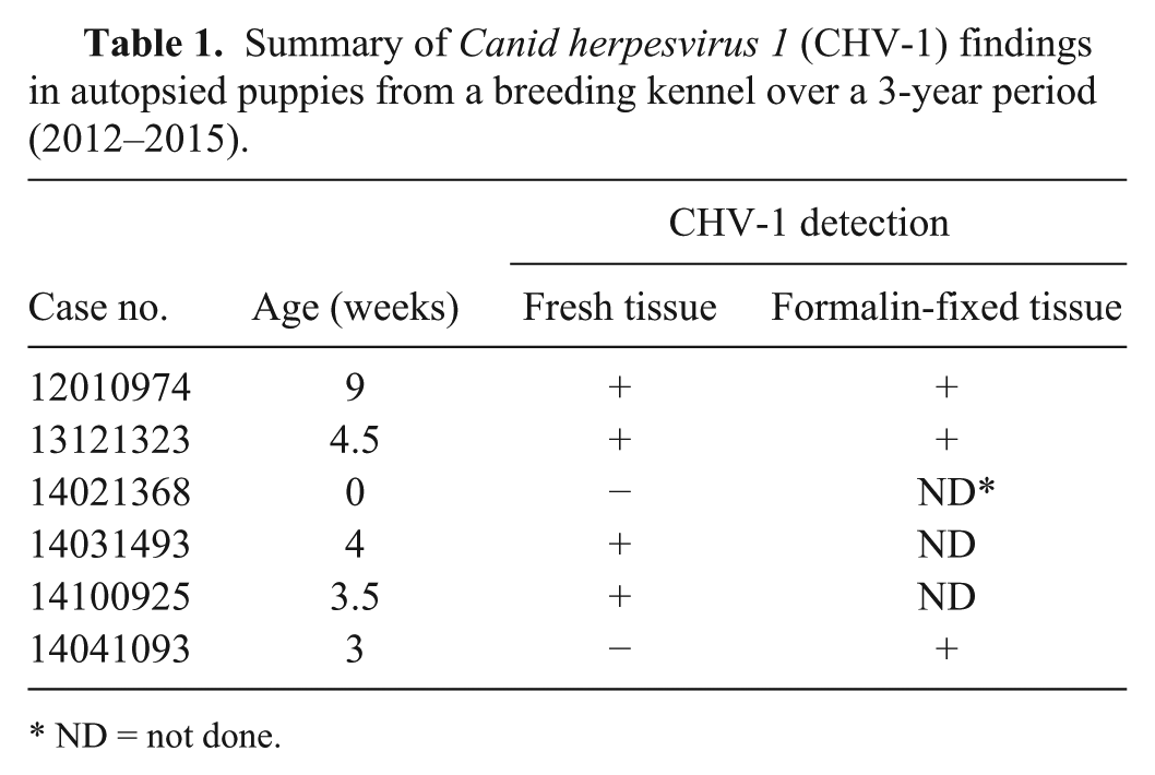
ND = not done.
Brucella canis antibodies were repeatedly negative by B. canis card test on 81 animals in this kennel (February 2008–March 2015). Dogs were screened for B. canis by serology tests using a B. canis rapid slide agglutination test (card test) h at Arkansas Livestock (Little Rock, Arkansas) and the Poultry Commission and Animal Health Laboratory (PC-AHL; Jefferson, Missouri). Dogs have been found to be negative for B. canis by culture at PC-AHL (81 tests performed since August 6, 2008). A few dogs were also checked by Brucella PCR on vaginal discharge and found to be negative (n = 2). Thus, B. canis antibodies and B. canis antigen were not detected in this kennel of Bulldogs. A semen culture was submitted by the kennel, and no significant bacterial pathogen and no mycoplasma was detected (case 15020359). Thus, there is no evidence of bacterial pathogens as cause of reproductive problems in this kennel.
The index case of CHV-1 (12010974) was submitted in January 2012. The CHV-1–infected dog was a 9-week old female Bulldog. During postmortem examination, petechial hemorrhages were observed on the cortical surfaces of both kidneys. The lungs were edematous and contained petechial hemorrhages. Hemorrhage in the kidney and lung parenchyma was centered on discrete foci of necrosis, occasionally accompanied by small deposits of fibrin. Necrotic foci were scattered throughout the liver and spleen. Rare intranuclear inclusion bodies consistent with herpesvirus were detected in the necrotic areas in the kidney, lung, and liver. Only virology testing was performed on this puppy because CHV-1 was suspected by the submitting veterinarian.
Based on direct FAT for CHV-1, the liver was found to be negative and the kidney sections were recorded as suspect for CHV-1 as a result of weak-positive fluorescence in the interstitial cells of the kidney. The presence of CHV-1 was confirmed by virus isolation.
In July 2014, 5 archived FFPE tissue blocks from the puppy submitted as OADDL case 12010974 were examined using the CHV-gB PCR. All tissues (block 1: liver; block 2: lung and spleen tested together; blocks 3 and 4: kidneys; block 5: small intestine, pancreas, and colon) examined were found to be positive for CHV by the CHV-gB PCR. Presence of CHV-1 DNA in multiple tissues from all puppies supported the diagnosis of systemic CHV infection.
In December 2013 (13121323), in a second submission, a 4.5-week-old puppy in the same kennel developed respiratory disease and died within 48 hr. On gross examination, necrotic foci were observed in the liver, lung, and kidneys. Based on the lesions, the pathologist concluded that necrotic foci were pathognomonic of CHV-1 disease. On histology, discrete foci of necrosis were scattered in the liver, lung, and renal cortex. In the lungs, the necrotic foci with small deposits of fibrin, edema, and alveolar histocytic infiltrates. On direct FAT, the liver was negative for CHV-1 and the lung was found to be suspect for CHV-1 based on detection of weak fluorescence. The presence of CHV-1 DNA was confirmed by CHV-gB PCR in 4 stored FFPE tissue blocks (block 1: small intestine, mesenteric lymph node, and colon; block 2: small intestine, mesenteric lymph node, and colon; block 3: liver, lung, and kidney; block 4: heart, spleen, thymus, and urinary bladder). The PCR amplicon was sequenced and confirmed as gB of CHV-1.
In the third case submitted (14021368), 2 mummified fetuses were submitted. The lung, liver, and kidney of the fetuses were negative for CHV-1 by direct FAT due to autolysis.
In the fourth case (14031493), a 4-week-old puppy was submitted. On gross examination, the internal tissues had pinpoint hemorrhages on the kidney. On direct FAT, the tissues were weakly positive for CHV-1. Presence of bacteria (Streptococcus and Klebsiella) was an incidental finding.
In the fifth case (14100925), a 3.5-week-old puppy exhibited mild respiratory signs followed by anorexia and death over a 1-week period. On histology, moderate amounts of pulmonary edema and vacuolar degeneration of liver were observed. CHV FAT was suspect and Canine distemper virus (CDV) was negative by CDV FAT.
In the sixth case (14041093), 1 of the 4 dead puppies was submitted for autopsy. The puppy was 3 weeks old. The cause of death was severe, interstitial pneumonia. Common viruses for canine infectious respiratory syndrome were negative by canine respiratory complex FAT profile (canine adenovirus 2, CDV, canine parainfluenza 2, and CHV). However, CHV-1 was still suspected as the cause of death in this puppy based on histological examination.
The client submitted several sera (n = 17) for CHV SN to the CADL. The CHV SN titers on these individual canine sera were 1:24, 1:24, 1:16, 1:48, 1:96, 1:128, 1:128, 1:128, 1:48,1:128, 1:96, 1:64, 1:64, 1:48, 1:24, 1:3, and <1:2. Based on high CHV-1 titers, it was concluded that all adult dogs tested, except 1, in this kennel were exposed to CHV-1. To date, there is no approved vaccine for CHV-1 in the United States. In spite of widespread detection of high titers of serum antibodies in adult dogs, clinical problems have persisted in newborn puppies that are immunologically naive. Thirty-six vaginal swabs were examined for CHV-1 by direct FAT. Five pregnant animals were shedding CHV-1 one week before whelping. Thirty animals were negative. One specimen had insufficient cells.
If a prewhelping vaginal swab is found to be positive, the owner should take additional precautions for preventing the transmission of CHV and perform passive immune therapy from CHV seropositive adults by plasma therapy. If a puppy gets sick with respiratory symptoms caused by CHV-1, plasma and oxygen therapy should be immediately administered. There is a commercial CHV-1 vaccine g in Europe but it is not licensed in the United States. 11
Canid herpesvirus 1 can be difficult to confirm because of the lack of stability of the virus in stored tissues caused by the fragility of the virus. 4 PCR on FFPE tissues is the most sensitive technique compared to immunohistochemistry for herpesviruses. 10
In this study, a CHV-1–specific PCR test was optimized to detect CHV-1 DNA in FFPE tissues. The CHV-1 PCR showed good correlation with lesions (9 out of 9 FFPE tissues from known CHV-1–positive cases were positive by CHV-1 PCR). Although the stability of the CHV-1 is short term in fresh tissues, the stability of the CHV DNA was found to be long term in FFPE tissues. Glycoprotein B (UL27) was used as a target for the CHV PCR. The gB gene offered a suitable PCR target for CHV-1 detection in FFPE samples. CHV was not detected in sick and healthy dogs with respiratory disease using CHV-1 polymerase gene as the target for CHV PCR. 13 Thus, selection of gene target for PCR can make a difference in the sensitivity of the PCR.
There are only a few published reports of fatal systemic infections with CHV-1 in older dogs. In a serious outbreak in Japan with fatalities, the dogs were predisposed by immunosuppressive therapy for cancer. 7 CHV-1 was the only pathogen identified in this fatal infectious tracheobronchitis outbreak in dogs stressed in a shelter environment. 5 In a case report from the United States, disseminated CHV-1 infection was diagnosed in an immunocompromised adult dog. 9 The problems related to CHV-1 have persisted in the Arkansas kennel in the current study. Dog breeding is done year-long; however, more problems occurred in this kennel during the winter months (December–February). On inquiry, the kennel ambient temperature is set at 21°C. The floors are concrete with wood shavings as padding. Puppies may have been predisposed to CHV-1 as a result of hypothermia and the effects of cooler ambient temperature on the immune system. 3 In this kennel, CHV-1 was associated with fatal systemic disease with lesions, respiratory symptoms, and mummification. The Arkansas kennel breeding stock is comprised of Bulldogs from the European Union and the United States. I could not demonstrate transplacental transfer of CHV-1 in this kennel (n = 1, placenta from a female animal that was positive in vagina was negative by CHV-1 FAT on the placenta sample). There are no previous reports of long-term longitudinal studies (~3-year follow-up) on effects of CHV-1 after introduction in a breeding kennel. In humans, vertical transmission of herpesvirus is reported. 1 However, I detected shedding of CHV-1 in the vaginal canal during whelping as a risk factor for puppies.
Footnotes
Author’s contributions
S Kapil contributed to conception and design of the study; contributed to acquisition, analysis, and interpretation of data; drafted the manuscript; critically revised the manuscript; gave final approval; and agrees to be accountable for all aspects of the work in ensuring that questions relating to the accuracy or integrity of any part of the work are appropriately investigated and resolved.
a.
VMRD Inc., Pullman, WA.
b.
HTC super-cured, charged 8-spot slides, Cel-Line/Erie Scientific, Thermo Fisher Scientific Inc., Waltham, MA.
c.
GE Healthcare UK, Little Chalfont, Buckinghamshire, United Kingdom.
d.
MO BIO Laboratories Inc., Carlsbad, CA.
e.
D-40724, Qiagen GmbH, Hilden, Germany.
f.
AmpliTaq Gold, Applied Biosystems, Foster City, CA.
g.
Merial, Lyon, France.
h.
Canine brucellosis antibody test kit, Synbiotics Corp., Kansas City, MO.
Declaration of conflicting interests
The author(s) declared no potential conflicts of interest with respect to the research, authorship, and/or publication of this article.
Funding
The author(s) disclosed receipt of the following financial support for the research, authorship, and/or publication of this article: This work was supported by user fees, and was partially supported by a grant from Merck Animal Health, USA on epidemiology of CHV in the United States.