
Abstract
Cats are infected by Anaplasma phagocytophilum and Borrelia burgdorferi when exposed to infected Ixodes scapularis (black-legged ticks). The purpose of our study was to allow wild-caught I. scapularis to feed on healthy research cats (n = 4) and temporally evaluate for A. phagocytophilum DNA in blood by a polymerase chain reaction (PCR) assay as well as for antibody responses to the B. burgdorferi C6 peptide, to the A. phagocytophilum P44 peptide, and to a novel A. phagocytophilum peptide (P44-4). Prior to I. scapularis infestation, all cats were negative for antibodies against both organisms based on a kit optimized for dog serum, and negative for A. phagocytophilum DNA in blood using a conventional PCR assay. Using the pre-infestation samples, an enzyme-linked immunosorbent assay for detecting antibodies against the P44-4 peptide was optimized. Cats were infested with wild-caught I. scapularis for 7 days. Genomic DNA of A. phagocytophilum was amplified from the blood before antibodies were detected in all 4 cats. Antibodies against the C6 peptide, P44 peptide, and P44-4 peptide were detected in the sera of all 4 cats. Antibodies against P44-4 were detected prior to those against P44 in 3 out of 4 cats. The results suggest that a PCR assay should be considered in acutely ill cats with suspected anaplasmosis that are seronegative.
Ixodes scapularis, the black-legged tick, is the vector for Anaplasma phagocytophilum and Borrelia burgdorferi in the eastern and midwestern United States. 5 While little is known about these infections in cats, some clinically ill, client-owned cats have had A. phagocytophilum DNA amplified from blood and have apparent clinical responses to tetracycline administration.1,2,6,14 Antibodies against B. burgdorferi have been detected in serum from some client-owned cats.7,9 In one study, 15 of 93 cat sera (16%) were positive for antibodies against both B. burgdorferi and A. phagocytophilum. 9 Some temporal information regarding infection exists after inoculation of cats with A. phagocytophilum–infected blood,4,8 but minimal information exists tracking the clinical and laboratory changes associated with A. phagocytophilum or B. burgdorferi infections induced by experimental exposure to I. scapularis.
Cats naturally infected with A. phagocytophilum have occasionally been positive for A. phagocytophilum DNA in blood prior to detection of A. phagocytophilum antibodies in serum. 6 The P44 peptide of A. phagocytophilum is immunodominant and is used as the target in some assays for detecting A. phagocytophilum antibodies in dogs. 3 There also are novel peptides related to P44 that may be associated with more recent infection. 15 In our pilot study, it was hypothesized that cats experimentally exposed to wild-caught I. scapularis carrying B. burgdorferi and A. phagocytophilum would have variable clinical signs of disease, would become positive in a polymerase chain reaction (PCR) assay for A. phagocytophilum DNA in blood, would develop antibodies against the A. phagocytophilum P44 peptide and the B. burgdorferi C6 peptide, and a novel A. phagocytophilum peptide (A. phagocytophilum P44-4) would detect antibodies more quickly than A. phagocytophilum P44.
Our pilot study was approved by the Institutional Animal Care and Use Committee at a contract research facility. The specific pathogen–free cats were approximately 1 year of age and were neutered males (2 cats) or spayed females (2 cats). Each of the cats was shown to be negative for Feline leukemia virus antigen, a negative for antibodies against Feline immunodeficiency virus, a and negative for B. burgdorferi and A. phagocytophilum antibodies by a commercially available kit used to test dog serum. b In addition, blood from each cat was shown to be negative for the DNA of A. phagocytophilum when tested by a commercial service c using a published conventional PCR assay. 6 Ixodes scapularis (13 females, 12 males) collected in Rhode Island were placed on each cat under a tick chamber made of adhesive bandage materials for 7 days. d The prevalence rate of A. phagocytophilum DNA in a representative aliquot of adult female ticks (n = 30) from the capture area that year was shown to be approximately 15% using previously described methods. 10 While DNA of Ehrlichia canis, Ehrlichia ewingii, Ehrlichia chaffeensis, or Anaplasma platys were not amplified, approximately 50% of the ticks contained B. burgdorferi DNA. The cats were assessed for clinical signs of depression or inappetence daily. A temperature-sensing microchip was used to monitor body temperature daily. 11
Blood for a complete blood cell count, for A. phagocytophilum PCR assay, and to obtain serum for assessing B. burgdorferi and A. phagocytophilum antibodies was collected prior to placing I. scapularis on the cats, every week after tick infestation for 10 weeks, and then weekly from 13 to 18 weeks after tick infestation. Doxycycline was orally administered at 10 mg/kg daily for 14 days starting 13 weeks after tick infestation. Sera from each collection date was assayed for B. burgdorferi and A. phagocytophilum P44 antibodies using a commercially available kit and A. phagocytophilum P44-4 using an enzyme-linked immunosorbent assay (ELISA) titrated at the research laboratory but not available commercially. Western blot immunoassay was used to aid in optimizing the A. phagocytophilum P44-4 ELISA and to confirm that positive results for P44 and C6 in the commercial kit were true. 9
Multiple I. scapularis attached and had fed on each of the 4 cats by day 7. Clinical signs of illness were not recognized in any of the cats over the course of the study. When compared with the week 0 group mean, results for significant anemia, neutrophilia, thrombocytopenia, and monocytosis were not noted. Significant (P < 0.05) lymphopenia was detected on weeks 2 and 5 using a Student t-test (http://graphpad.com/quickcalcs/contingency1.cfm).
Each cat had A. phagocytophilum DNA amplified from blood and 1 positive sample from each cat was shown to be most homologous with A. phagocytophilum by genetic sequencing (Fig. 1). Over the first 10 weeks of the study, A. phagocytophilum morulae were noted in neutrophils of all 4 cats at least once: week 3 (1 cat), week 5 (3 cats), and week 8 (1 cat), with an overall prevalence rate of 12.5% (5/40 samples). All of the morulae-positive samples also were positive for A. phagocytophilum DNA. During and after doxycycline administration, all samples were negative for A. phagocytophilum morulae and A. phagocytophilum DNA.
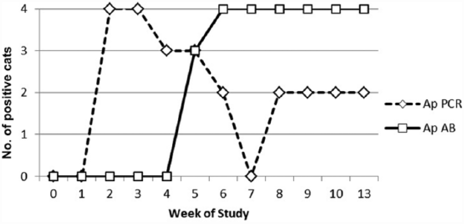
Temporal appearance of Anaplasma phagocytophilum DNA and antibodies in cats following exposure to Ixodes scapularis ticks. Ap = A. phagocytophilum; PCR = polymerase chain reaction; AB = antibodies.
Each cat developed detectable A. phagocytophilum and B. burgdorferi antibodies in serum. However, in all 4 cats, DNA of A. phagocytophilum was amplified from blood prior to detecting A. phagocytophilum antibodies (Table 1). Antibodies against A. phagocytophilum P44-4 were detected 2–3 weeks prior to those detected by the commercially available kit in 3 out of 4 cats (Table 1). Antibodies against A. phagocytophilum and B. burgdorferi persisted during and after doxycycline administration.
Duration of Anaplasma phagocytophilum (AP)- and Borrelia burgdorferi (BB)-positive test results in cats exposed to Ixodes scapularis.*
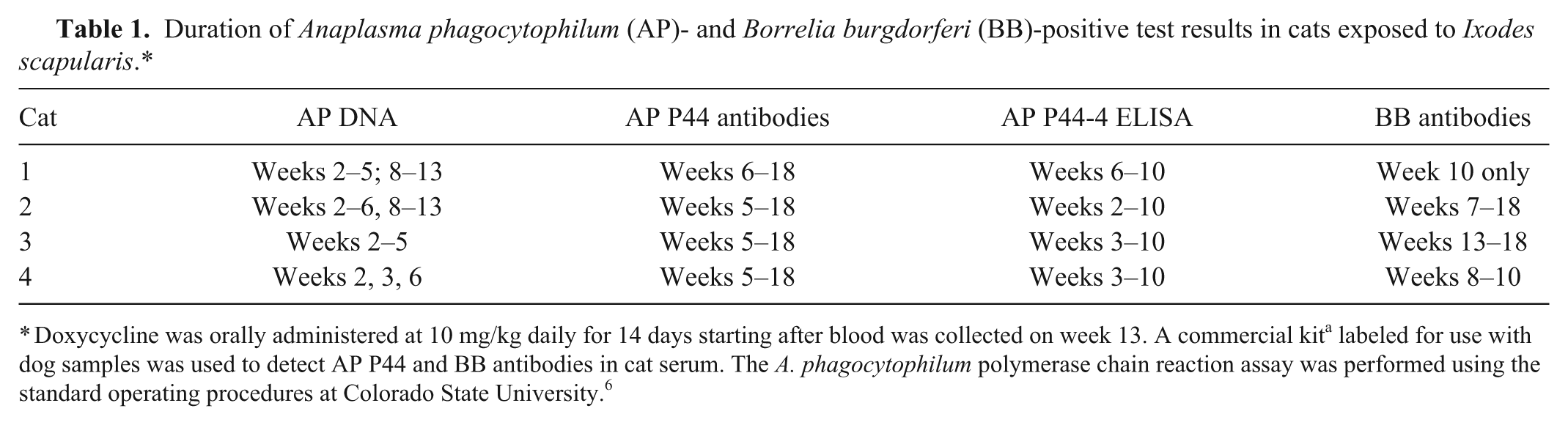
Doxycycline was orally administered at 10 mg/kg daily for 14 days starting after blood was collected on week 13. A commercial kit a labeled for use with dog samples was used to detect AP P44 and BB antibodies in cat serum. The A. phagocytophilum polymerase chain reaction assay was performed using the standard operating procedures at Colorado State University. 6
The results of our study document that cats can be infected with A. phagocytophilum and B. burgdorferi after being fed upon by infected I. scapularis. However, clinical abnormalities were not recognized in this small group of cats, and significant laboratory abnormalities were minimal, even though evidence of dual infection with A. phagocytophilum and B. burgdorferi existed. To our knowledge, evidence documenting B. burgdorferi–associated disease in cats has not been published, suggesting that the agent is not a primary pathogen in cats. 5 In contrast, several publications have suggested clinical disease associated with A. phagocytophilum infection in cats.1,2,6 The failure of the cats in the current study to develop clinical anaplasmosis may have been related to the immune status of the cats (immunocompetent), the dose of the organism, or the strain of A. phagocytophilum.4,13
As with most rickettsial infections, A. phagocytophilum rickettsemia in cats can be detected by amplification of specific DNA from blood prior to detecting antibodies in serum. Thus, if a cat with clinical findings of anaplasmosis is seronegative for A. phagocytophilum antibodies, submission of blood for PCR amplification of Anaplasma spp. DNA is indicated. While all cats were negative for B. burgdorferi and A. phagocytophilum antibodies before I. scapularis infestation and then positive for antibodies against both organisms on multiple dates after tick infestation, further research will be needed to validate the use of this assay b with samples from cats, as the kit is not currently approved for this use. A cat with suspected anaplasmosis that is negative for A. phagocytophilum antibodies can have a second sample tested within 2–4 weeks to evaluate for seroconversion. Our study also suggests that the A. phagocytophilum P44-4 peptide might be a better target than the A. phagocytophilum P44 peptide when attempting to diagnose recent A. phagocytophilum infection in cats.
In our study, the Anaplasma species infecting the cats was shown to be A. phagocytophilum by genetic sequencing. However, it has been shown that A. platys also can infect naturally exposed cats. 12 In dogs, antibodies against both A. phagocytophilum and A. platys bind to the A. phagocytophilum P44 peptide. e Further work will be needed to determine whether this also is true in cats. As for dogs, genetic sequencing or use of specific PCR assays should be used to determine whether the Anaplasma spp. causing infections in cats is A. phagocytophilum or A. platys.
Anaplasma phagocytophilum DNA was not amplified from blood during or after doxycycline administration to experimentally infected cats in our study. Most naturally infected cats become PCR negative after treatment as well. 6 There appear to be no reports of a cat with clinical anaplasmosis having recurrent clinical signs of disease or evidence of chronic illness following doxycycline treatment. However, it would be prudent to maintain tick control, as repeat infections could occur. In the experimentally exposed cats in our study, serum antibodies against A. phagocytophilum and B. burgdorferi persisted after the short course of doxycycline used. Further research will be required to determine whether there is clinical benefit to repeat serological testing of cats with A. phagocytophilum or B. burgdorferi infection.
Footnotes
Authors’ contributions
MR Lappin and R Chandrashekar contributed to conception and design of study. B Stillman and J Liu contributed to design of study. MR Lappin, R Chandrashekar, and TN Mather contributed to acquisition, analysis, and interpretation of data, and drafted the manuscript. B Stillman contributed to acquisition and interpretation of data. J Liu contributed to analysis and interpretation of data. All authors critically revised the manuscript, gave final approval, and agree to be accountable for all aspects of the work in ensuring that questions relating to the accuracy or integrity of any part of the work are appropriately investigated and resolved.
a.
SNAP FeLV/FIV, IDEXX Laboratories Inc., Portland, ME.
b.
SNAP 4Dx, IDEXX Laboratories Inc., Portland, ME.
c.
Ehrlichia/Anaplasma PCR assay, Veterinary Diagnostic Laboratory, Colorado State University, Fort Collins, CO.
d.
Dr. T Mather, University of Rhode Island, Kingston, RI.
e.
SNAP 4DxPlus, IDEXX Laboratories Inc., Portland, ME.
Declaration of conflicting interests
The author(s) declared the following potential conflicts of interest with respect to the research, authorship, and/or publication of this article: R Chandrashekar, B Stillman, and J Liu are employees of IDEXX Laboratories.
Funding
The author(s) disclosed receipt of the following financial support for the research, authorship, and/or publication of this article: The study was funded by IDEXX Laboratories.