
Abstract
An adult Domestic Longhair cat developed a subcutaneous mass in its tail. Histologically, this mass consisted of ill-defined pyogranulomas centered around aggregates of gram-positive, acid-fast filamentous bacteria, consistent with Nocardia. Due to the lack of fresh samples, DNA was extracted from formalin-fixed paraffin-embedded tissue sections and subjected to polymerase chain reaction amplification and DNA sequencing of 16S ribosomal RNA gene encompassing Nocardia sp.-specific sequences. Sequences analyzed using the GenBank database revealed 99.5% homology with Nocardia spp. and had the highest sequence homology of 98.2% with Nocardia tenerifensis among Nocardia spp. To the authors’ knowledge, this is the first report of detection of N. tenerifensis genome associated with cutaneous nocardiosis in an animal.
Nocardiosis, infection with aerobic actinomycetes of the family Nocardiaceae, results in suppurative to pyogranu-lomatous inflammation. 5 Most domestic animals can be infected with this organism. 4,5 Nocardia asteroides is the most commonly isolated species in cats, but Nocardia brasiliensis, Nocardia otitidiscaviarum, Nocardia Africana, and Nocardia nova infections also have been documented in this species. 1,4,13,14,17 The microscopic findings of cutaneous nocardiosis in a cat are reported. Based on molecular methods on formalin-fixed, paraffin-embedded tissues, it was considered to be Nocardia tenerifensis.
An adult, neutered male Domestic Longhair cat developed a 1.5 − 1.5 − 2.5 cm subcutaneous mass at the mid portion of the tail (Fig. 1). Other clinical signs were not apparent. Because neoplasia was suspected, the tail was amputated. The mass was located in the subcutis involving mostly the dorsal aspect of the tail and extending slightly on to its lateral aspect (Fig. 2). This mass was tan and firm and did not appear to affect adjacent coccygeal bone. The dorsal skin was extensively ulcerated. The referring veterinarian submitted the entire mass fixed in neutral buffered 10% formaldehyde. This sample was trimmed and routinely processed for histopathology. Histologically, indistinct and confluent granulomas, mostly in the subcutis and deep dermis, made up the mass (Fig. 3). The center of these granulomas contained numerous, faintly basophilic, filamentous organisms that were surrounded by variable numbers of neutrophils and foreign body-type multinucleated giant cells (Figs. 4, 5). Filamentous organisms were rarely seen within multinucleated giant cells. Epithelioid macrophages with foamy cytoplasm and some neutrophils formed an ill-defined outer layer that merged with adjacent granulomas. Individual granulomas were not surrounded by a fibrous capsule. However, dense trabeculae of collagen separated groups of 30 or more granulomas. The coccygeal vertebrae and surrounding skeletal muscle and tendons were not affected by this inflammation. Filamentous organisms were gram positive and strongly acid fast (Fig. 6). Their filamentous morphology was best depicted with Gomori methenamine silver stain. The microscopic features of this lesion along with the tinctorial characteristics of these organisms warranted a diagnosis of cutaneous nocardiosis.
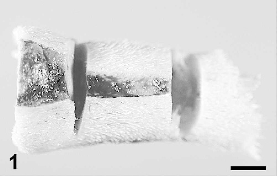
Tail; cat. A linear ulcer is present in the dorsal aspect of the midsection of the tail. Bar = 5 mm.

Tail; cat. Cross-section of the subcutaneous mass. The mass occupies the dorsal and lateral aspects of the tail. Arrows indicate the margins of the lesion. V = vertebral body. Bar = 5 mm.

Skin; cat. Confluent pyogranulomas in the subcutis (S). The center of pyogranulomas (asterisk) contains bacterial colonies. D = dermis. Hematoxylin and eosin stain. Bar =1.2 mm.

Skin; cat. Bacterial colonies occupy the center of granulomas (asterisk) and are surrounded by neutrophils, epithelioid macrophages, and multinucleated giant cells. Hematoxylin and eosin stain. Bar = 240 μm.

Skin; cat. Multinucleated giant cells (arrow) are in close contact with filamentous bacteria (asterisk), but bacteria seldom are phagocytized. Hematoxylin and eosin stain. Bar = 50 μm.

Skin; cat. Filamentous bacteria are strongly gram positive. Gram stain. Bar =120 μm. Inset: Bacterial colonies are also strongly acid fast. Fite stain. Bar = 240 μm.
Because no specimen was available for bacterial cultures, bacteria were characterized by polymerase chain reaction (PCR) using formalin-fixed, paraffin-embedded tissue sections according to a published protocol. 6,7 Briefly, 5 10-μm-thick paraffin sections were cut from a paraffin block containing the granulomatous mass that had been fixed in formalin. The paraffin was removed from these sections using xylene. The DNA in the deparaffmized tissue sections was extracted using a DNeasy Kit for animal cells or bacteria a following the procedures recommended by the manufacturer. Five microliters of the extracted DNA was used as the template in a 50-μl PCR mixture (1 − PCR buffer, 1.5 mM MgCl2, 0.2 mM each deoxynucleoside triphosphate, 0.25 μM each primer, and 1.5 U of Taq polymerase b ) to amplify a 999 base-pair 16S ribosomal RNA (rRNA) gene sequence. The sequences for the 2 primers used were 5‘-cga acg ctg gcg tgc tta ac-3’ (sense primer) and DN (5‘-cct gta cac cga cca caa ggg gg-3’ (antisense primer). 7 The PCR conditions entailed 5 minutes of denaturation at 94°C, followed by 40 of amplification cycles of 94°C for 60 seconds, 68°C for 45 seconds, and 72°C for 60 seconds, and ended with a final extension at 72°C for 10 minutes. The PCR amplicon was visualized on a 1.5% agarose gel containing ethidium bromide (data not shown), excised using a sterile scalpel blade under ultraviolet illumination, and submitted for direct sequencing of both strands of DNA. c orward and reverse sequences were subjected to GenBank search for similarities with other Nocardia spp. using the online Basic Local Alignment Search Tool program provided by National Center for Biotechnology Information (http://www.ncbi.nlm.nih.gov/BLAST). Sequencing of amplified 16S rRNA gene (999 base pairs encompassing Nocardia-specific sequences) from nucleic acid extracted from paraffin sections yielded 99.5% homology with Nocardia sp. The highest sequence homology of the index case was with N. tenerifensis (98.2%) followed by N. brasiliensis (97.4%), N. asteroides (96.8%), and N. nova (95.9%). Based on the amplified nucleotide sequence of 16S rRNA, N. brasiliensis, N. beijingensis, N. transvalensis, and N. asteroides had 98.0%, 97.3%, 97.5%, and 97.3%, respectively, homology with N. tenerifensis. 15
Nocardia spp. are gram-positive, weakly acid-fast, strictly aerobic bacteria that form filamentous branched cells that may fragment into pleomorphic rod-shaped or coccoid elements. 19 Nocardia are saprophytes involved in the decomposition of plant material, but some species can infect humans and animals regardless of their immune status. 19 Nocardial infections produce suppurative to pyogranulomatous inflammation that, in dogs and cats, typically has 1 of 3 clinical presentations: pulmonary, systemic or disseminated, or cutaneous. 2,9 Nocardial peritonitis also has been reported in a cat. 22 Cutaneous nocardiosis is the most common form in cats and usually involves the extremities, inguinal area, and neck. 9 The mass in the cat of this report is a typical presentation of cutaneous nocardiosis. Other cutaneous forms include abscesses, cellulitis, and draining fistulous tracts. 1,11,23
The diagnosis of cutaneous nocardiosis reported in this cat was based on microscopic morphology and staining features (gram positive and acid fast) of organisms present in granulomas, whereas species identification was made by PCR and sequencing of the amplified 16S rRNA gene product. Nocardia are considered partially acid fast, 9,11 and the use of a modified acid-fast stain is recommended. 11 In this cat, the staining was intense and most organisms were positive by the Fite-Faraco modification of acid-fast stain. Another actinomycete, Actinomyces, is also a filamentous, gram-positive organism, but it is not acid fast, which permits distinction with Nocardia. 9 Nocardia identification requires laborious, time-consuming phenotypic and chemotaxonomic methods. Molecular methods offer an alternative, particularly when bacterial cultures are not possible. The advantages of molecular identification of bacteria using 16S rRNA sequencing over phenotypic identification are rapid turnaround time, reliable identification, and taxonomic meaningfulness. 12 The 16S rRNA gene of Nocardia is highly conserved with constant regions that are identical for all Nocardia spp. and variable regions that are species specific. 6 The first 500 base-pair genes in the variable region of 16S rRNA, included in the 999 base-pair genes amplified in the present case, have been reliable for the identification of Nocardia spp. 6 Molecular methods have been used in the identification and characterization of more than 40 Nocardia spp. 7,18 20,24 Because formaldehyde fixation and paraffin embedding produces nucleic acid fragmentation, a large number of organisms present in this sample was important to produce successful DNA recovery and PCR amplification. 10,16 N. tenerifensis has been recently isolated from soil of the Spanish island of Tenerife, 15 and to the authors’ knowledge it has not been reported as a cause of nocardiosis of animals or humans. 6
Based on clinical examination, no evidence of dissemination was present 18 months after the diagnosis of this mass. The most common concurrent noncutaneous nocardial infection is pulmonary infection and osteomyelitis. 9 Sulfur granules (macroscopic aggregates of bacterial colonies and inflammatory cells) were not observed in the present study, although they have been reported in feline cutaneous nocardiosis. 8 The number and size of multinucleated giant cells within pyogranulomas was remarkable in the present study. Nocardia can be phagocytized by macrophages, but the efficacy of this process depends in part on bacterial virulence. 5 Nocardia spp. in this cat's lesion were only occasionally observed intracellularly, indicating resistance to phagocytosis by macrophages, as others have reported. 1 The origin of this infection was not determined, but because Nocardia is a saprophite present in soil, contamination of a pre-existing wound or trauma by an animal harboring this organism is likely. 3,11,21 In summary, this study demonstrates the usefulness of molecular methods by performing PCR amplification and DNA sequencing of 16S rRNA gene on formalin-fixed, paraffin-embedded tissues to detect and speciate microorganisms, particularly when other procedures are not available. To the authors’ knowledge, this is the first report of detection of N. tenerifensis genome associated with cutaneous nocardiosis in humans or animals. In addition, this paper illustrates the usefulness of molecular methods to detect microorganisms in formalin-fixed, paraffin-embedded tissues when no other sample is available.
Footnotes
a.
Qiagen, Valencia, CA.
b.
Perkin-Elmer, Shelton, CT.
c.
Purdue Genomics Center, West Lafayette, IN.