
Abstract
Hermann’s tortoise (Testudo hermanni) is considered near threatened in the wild but, by contrast, it is one of the most popular pet tortoises in Europe. Scant data is reported in the veterinary literature on hematological values for T. hermanni and, to our knowledge, none focused on the subspecies boettgeri (eastern Hermann’s tortoise). Published reports are based on small sample populations, and confusion arises when comparing the sampling sites, the anticoagulants, and the counting methods used. The purpose of the current study was to establish the normal mean values and reference intervals for the main hematological parameters for captive adult T. hermanni boettgeri and to evaluate the reliability of a semiautomated blood analyzer for red blood cell count and hematocrit determination. Blood values were determined in 23 adult tortoises using a Neubauer chamber with Natt and Herrick solution; red blood cells and hematocrit were also measured using a semiautomated blood analyzer. Statistical analysis included descriptive statistics, differences between sexes, and agreement between the counting methods. Reference intervals were calculated with the robust method. Wilcoxon signed rank test with continuity correction was used to investigate differences between sexes, and Bland–Altman analysis was performed to compare manual versus semiautomated values. Red blood cells, hematocrit, and hemoglobin determinations were significantly higher in males than in females. White blood cell counts did not show any sex variability. The agreement of manual versus semiautomated determination was considered acceptable for clinical use.
Among the 323 known species of tortoises, 132 are listed on the International Union for Conservation of Nature (IUCN) red list as critically endangered, endangered, or vulnerable. Testudo hermanni (Hermann’s tortoise) is considered “near threatened” by the IUCN (http://www.iucnredlist.org/details/21648/0), is listed in appendix II of the Convention on International Trade of Endangered Species, and in Annex A of the European Council Regulation No. 338/79 on the protection of species of wild fauna and flora by regulating their trade. Testudo hermanni occurs in Mediterranean Europe, from coastal northeastern Spain, through southeastern France, Mallorca (Spain), Menorca (Spain), Corsica (France), Sardinia (including Asinara Island), and Sicily (Italy), the coastal plains of peninsular Italy, coastal Croatia, coastal Bosnia-Herzegovina, coastal Montenegro, central and southern Serbia, inland to southwestern Romania, much of Bulgaria, Macedonia, nearly all of Albania, the Greek mainland plus islands from Corfu to Zakynthos, and European Turkey. 6 The subspecies T. h. hermanni, is present in northeastern Spain, southeastern France, the Balearic Islands (Spain), Corsica (France), and Sardinia (Italy). Testudo hermanni boettgeri is the eastern subspecies distributed in the Balkan region. 6 Despite the threatened status of this tortoise in its natural habitats, T. hermanni is very popular as a pet animal and is kept in many gardens and households mainly in Europe.
Hematological parameters of reptiles vary depending on many factors, including physiology of the species and techniques employed for analysis. The purpose of the current study was to standardize sampling and counting procedures in this species, to provide reference intervals for the main hematological variables, and to describe agreement of a semiautomated blood cell counter with manual count.
All 23 tortoises (15 males and 8 females) were adult reproductive animals belonging to the species T. hermanni subspecies boettgeri, originating from 2 different locations: location A with 13 tortoises (8 males and 5 females) and location B with 10 tortoises (7 males and 3 females). The 15 males had a carapace straight line length ranging from 16 to 23 cm and a weight ranging from 735 to 1,726 g. The 8 females had a carapace straight line length ranging from 19 to 32 cm and a weight ranging from 801 to 2,300 g. In both locations, the tortoises were kept outdoors in northeastern Italy under semi-natural conditions. They were housed in pens with other T. h. boettgeri and allowed to spontaneously hibernate, usually from the beginning of October until March. Their usual diet was based on native wild grasses (e.g., Plantago lanceolata, Plantago major, Taraxacum officinale, Rumex acetosella, Eruca vesicaria subsp. sativa, Sonchus oleraceus, Malva sylvestris, and Trifolium repens) occasionally supplemented with cultivated leafy vegetables (romaine lettuce, red radicchio, endive, and Belgian and catalogna chicory). Sporadically, other vegetables and fruit (Indian fig [Opuntia ficus-indica], tomatoes, cucumber, and watermelon) were offered.
At clinical examination, all tortoises showed a good body condition, were alert and responsive, and none of them presented signs of illness. All the animals previously tested negative by serology for Chelonid herpesvirus 1 and 2 as well as for Virus X. Fecal examinations revealed a mild presence of oxyurids in all of the tortoises. The samples were taken in September when the tortoises were temporarily housed indoors for use in a future pharmacokinetic study. The tortoises were housed indoors in a heated room (24°C from 8:00
Normality of variables was assessed by use of the Kolmogorov–Smirnov test. Median and nonparametric statistical tests would be performed to account for non-normality. Tukey method for outlier detection was employed. Reference intervals were calculated using the robust method, with 90% confidence interval (CI) of upper and lower limit. 9 To study possible variations between sexes, a Wilcoxon signed rank test with continuity correction was used considering the following hematological parameters: RBC, WBC, PCV, and the WBC differential counts as measured by manual count. To evaluate whether the automated analyzer agreed with the manual count, Bland–Altman analysis and Passing–Bablok regression were performed. Mean difference and limits of agreement (LOA) were determined. 2 Statistical analysis was performed using commercial software.j,k
The results for the whole population are summarized in Tables 1 and 2. Blood sampling was easily accomplished by the chosen technique. The size, anatomy, and temperament of the study animals allowed proper restraint and a precise visualization of the jugular vessels for an effective sampling.
Mean values and reference intervals for hematological parameters in Testudo hermanni boettgeri (tortoise no. 23) obtained by manual method.*
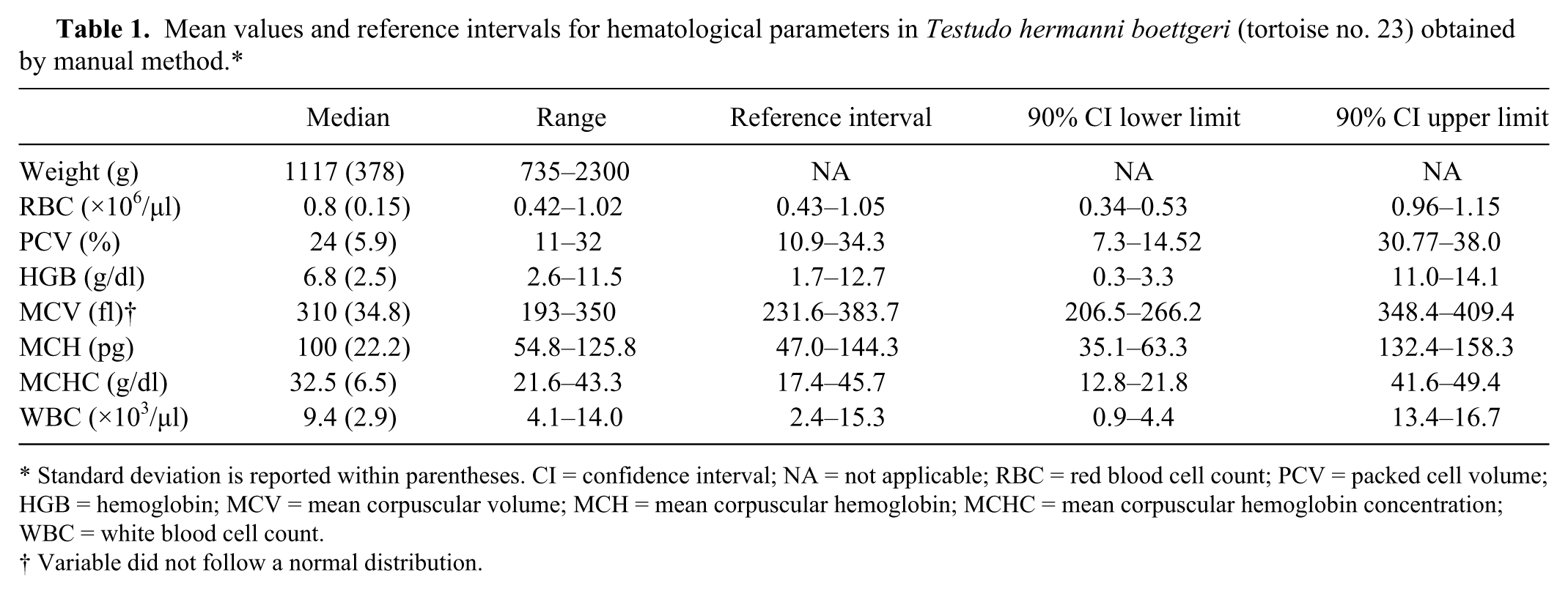
Standard deviation is reported within parentheses. CI = confidence interval; NA = not applicable; RBC = red blood cell count; PCV = packed cell volume; HGB = hemoglobin; MCV = mean corpuscular volume; MCH = mean corpuscular hemoglobin; MCHC = mean corpuscular hemoglobin concentration; WBC = white blood cell count.
Variable did not follow a normal distribution.
Normal mean values and ranges for differential white blood cell counts in Testudo hermanni boettgeri (tortoise no. 23).*
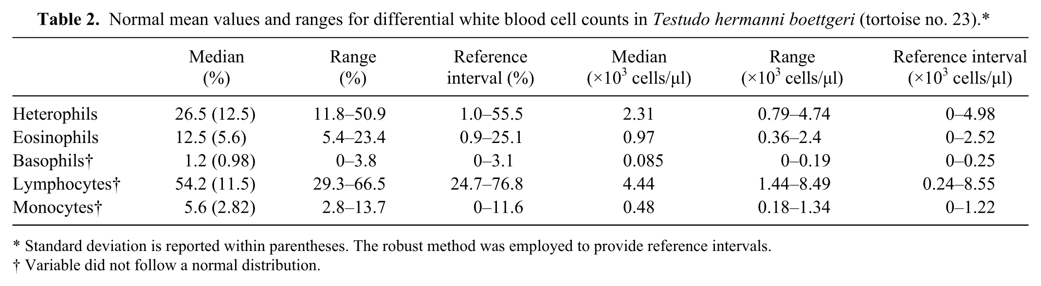
Standard deviation is reported within parentheses. The robust method was employed to provide reference intervals.
Variable did not follow a normal distribution.
The anticoagulant and the counting procedure chosen did not present any challenges, and WBC clumps were rarely seen in the hemocytometer; the procedure to repeat the reading solution was requested in 3 cases only. The rapid staining method was adequate for the differential count and, at 1,000× magnification, there were no impediments to cell type identification.
Most variables were normally distributed, with the exception of mean corpuscular volume (MCV; manual and automatic), monocytes (percentage), lymphocytes (percentage), and basophils (absolute count) that were nonnormally distributed (Fig. 1). No outliers were detected. A significant statistical difference was found between sexes in RBC counts (P = 0.04), in PCV (P < 0.001), and in HGB values (P < 0.001; Fig. 2). No differences were evident in the total WBC count (P = 0.6) nor in the WBC differential counts between sexes.
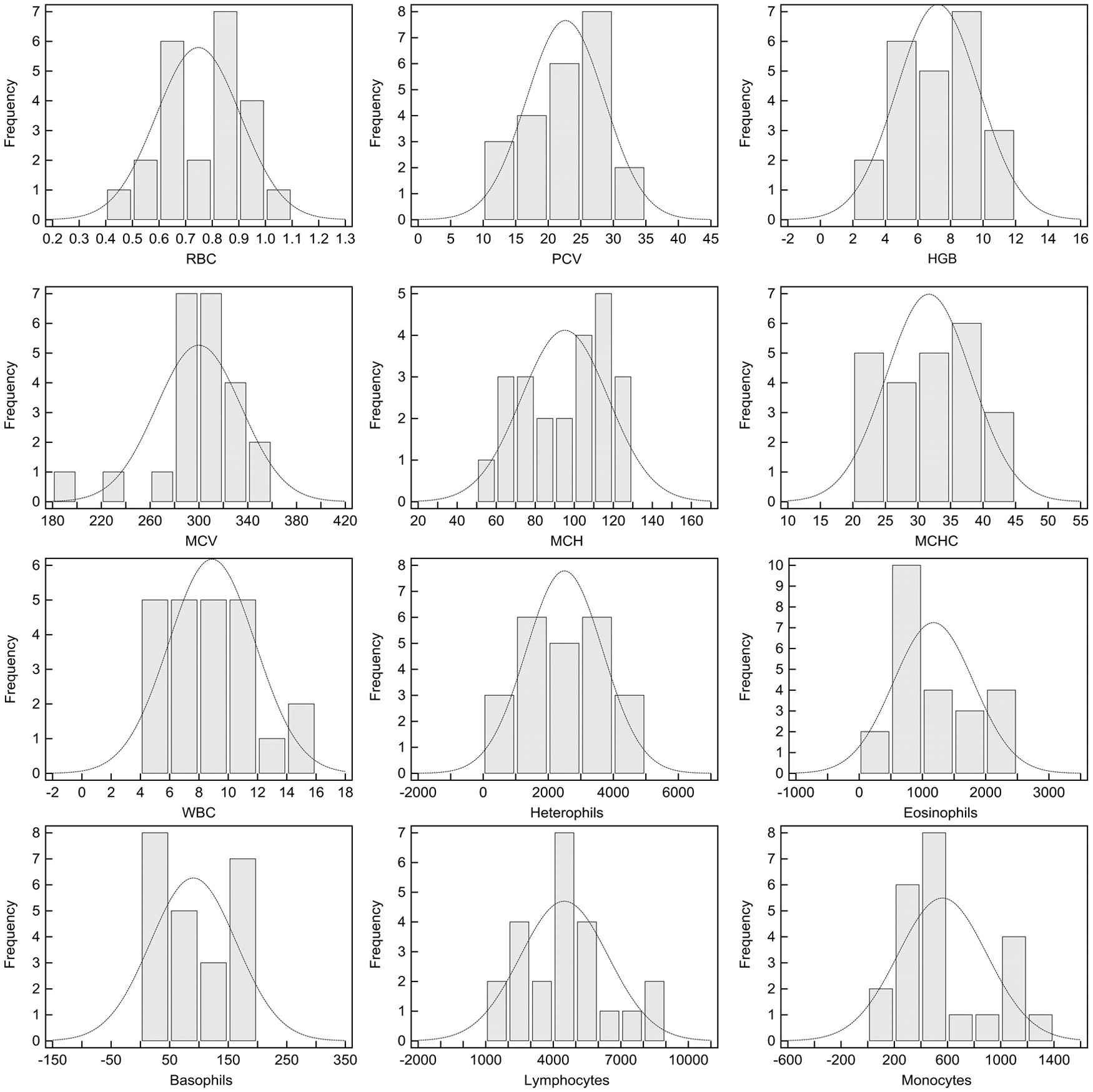
Histograms depicting the distribution of hematological values measured in a sample of 23 clinically healthy eastern Hermann’s tortoises (Testudo hermanni boettgeri). RBC = red blood cell count; PCV = packed cell volume; HGB = hemoglobin; MCV = mean corpuscular volume; MCH = mean corpuscular hemoglobin; MCHC = mean corpuscular hemoglobin concentration; WBC = white blood cell count.
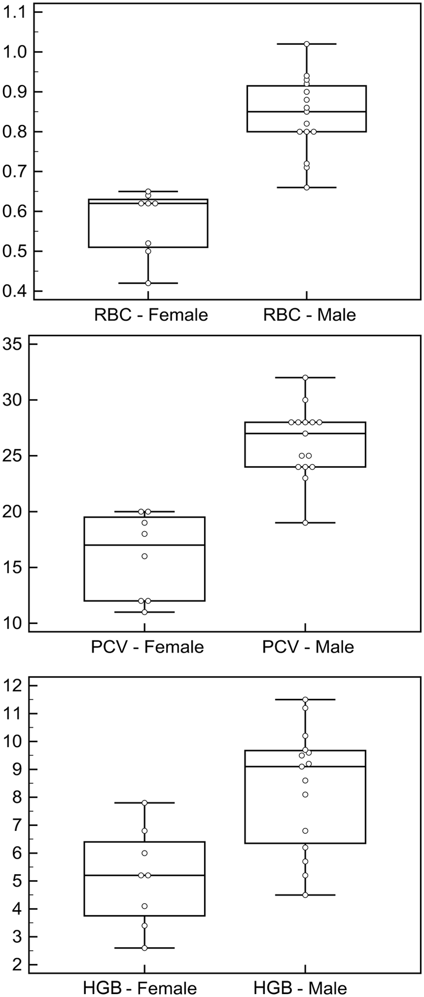
Combined box-and-whisker and dot plots showing the differences in red blood cell (RBC) count, packed cell volume (PCV), and hemoglobin (HGB) between female and male eastern Hermann’s tortoises (Testudo hermanni boettgeri). The boxes represent the values from the first to the third quartile (25–75th percentile). The horizontal line in each box represents the median. The whiskers include values of 1.5 times the interquartile range.
Mean difference between manual and semiautomated RBC count was 0.07 × 10⁶/µl (LOA: −0.03 to 0.17; Fig. 3). Mean difference between manual PCV and semiautomated HCT was 3.2% (LOA: 1.5–4.9%). Mean difference between manual and semiautomated MCV was 15.4 fl (LOA: −26.5 to 57.5 fl). Mean difference between manual and semiautomated mean corpuscular hemoglobin (MCH) was −11.4 pg (LOA: −31.0 to 8.2 pg). Mean difference between manual and semiautomated MCH concentration (MCHC) was 5.9 g/dl (LOA: −0.12 to 11.9 g/dl). Based on Passing–Bablok regression, at-least-constant difference (y-intercept ≠ 0) was present between manual and automatic count of RBC, PCV, and MCV. An at-least-proportional difference (slope ≠ 1) was present between manual and automatic count of MCV and MCHC.
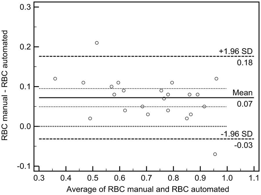
Representative Bland–Altman plot, showing agreement between automated and manual count of red blood cells (RBCs) in blood samples of eastern Hermann’s tortoises (Testudo hermanni boettgeri). Mean difference (solid line) with its 95% confidence interval (dotted and dashed lines) and 2 standard deviation (SD) limits of agreement (dashed lines) are provided. The slope regression line is depicted.
The range values in our study of manually counted RBC, WBC, and HGB are wider than those previously reported in the literature for T. hermanni.13,15,20 Possible explanations for the differences can be attributed to the size of sample population, to seasonal variability, to the venipuncture site, to the anticoagulant used, or to the counting method.7,12–15,19,20 In fact, some sampling sites are more prone to lead to lymph contamination in chelonians, causing spurious low values of blood cell count. In our experience, lymph contamination is less likely to occur when sampling from the jugular vein.
The stress induced by keeping the tortoises indoors does not seem to have influenced WBC count, as our values are not dissimilar to others reported in the literature. The mild parasite load of oxyurids found in some individuals is a typical finding in asymptomatic Testudo species.
Differences in RBC count between males and females are commonly reported in reptiles. 11 Among chelonians, sex-based differences in RBC, HTC, and HGB have been previously reported for Astrochelys radiata, 21 Geochelone gigantea (syn. Aldabrachelys gigantea), 12 Gopherus agassizii, 10 Ocadia sinensis (syn. Mauremys sinensis), 8 and Terrapene carolina. 1 Sex-based differences have not been found in Chelonia mydas. 3 Because erythrocytic indexes are correlated to RBC, HTC, and HGB, the same sex-based differences are likely to be found in MCV, MCH, and MCHC. The finding of a sex-based difference in selected parameters in T. h. boettgeri may have a clinical relevance and suggests the need for sex-specific reference intervals. Unfortunately, our results are based on a small population. For this reason, data is summarized in Table 3 and graphically represented in Figure 2 as suggested by the American Society for Veterinary Clinical Pathology quality assurance and laboratory standards committee guidelines (http://www.asvcp.org/pubs/pdf/RI%20Guidelines%20For%20ASVCP%20website.pdf).
Red blood cell (RBC), packed cell volume (PCV), and hemoglobin (HGB) values in ascending order for Testudo hermanni boettgeri sorted by sex.*
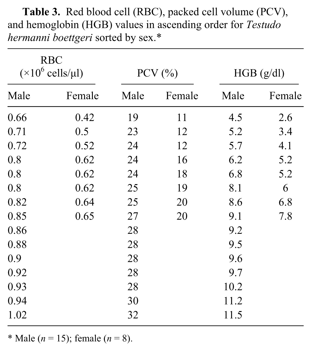
Male (n = 15); female (n = 8).
In the current study, we employed an impedance blood cells counter that yielded results comparable to the manual count for RBC, HTC, and erythrocytic indexes. The automated count seemed to overestimate the RBC count and to underestimate the HTC determination in a way not likely to affect clinical judgment. A possible explanation is that some small WBCs or thrombocytes are counted as RBCs and that the spun PCV overestimates the actual HTC. 4 Even though some authors recommend the use of an automated or semiautomated counting machine for reptile RBC and HTC determinations,16–18 according to our findings, the use of such equipment should be previously validated in order to obtain reliable results.
Clinical evaluation of reptilian species is always challenging and, though more extensive studies are needed, clinical pathology can aid in chelonian health monitoring. 22 Validation of automated and semiautomated blood counters for reptilian blood count may make clinical pathology more feasible.
Footnotes
a
Repti Bark no. 8, Zoo Med Laboratories Inc., San Luis Obispo, CA.
b.
Coco Earth, Mondialfauna, Monopoli, Italy.
c.
Powersun, Zoo Med Laboratories Inc., San Luis Obispo, CA.
d.
Micro tube, Sarstedt AG & Co., Numbrecht, Germany.
e.
Spencer Bright Line, American Optical Co., Buffalo, NY.
f.
NRIS, Vitrex Medical A/S, Herlev, Denmark.
g.
Mod. 4223, ALC International Srl, Cologno Monzese, Italy.
h.
Mod.S-8300, Semar Srl, Sesto Fiorentino, Italy.
i.
Hemacolor, Merck KGaA, Darmstadt, Germany.
j.
SPSS version 22.0, IBM North America, New York, NY.
k.
MedCalc 12.1, MedCalc Software, Ostend, Belgium.
Declaration of conflicting interests
The author(s) declared no potential conflicts of interest with respect to the research, authorship, and/or publication of this article.