
Abstract
The effect of fixation and storage conditions on the performance of polymerase chain reaction (PCR) assays for Babesia odocoilei were examined using 3 different primer sets targeting the eukaryotic 18S ribosomal RNA gene, with variably sized products of 1,723 base pairs (bp), 483 bp, and 306 bp. All primer sets performed well on fresh-frozen tissue, and storage for 1 year at −20°C did not affect PCR performance. Formalin fixation markedly affected the amplicon length that could be amplified. However, DNA was successfully amplified after storage in formalin for 2 months using the primer set with a 483-bp product, and up to 6 months using the primer set with a 306-bp product. The latter primer set successfully differentiated B. odocoilei and Babesia microti DNA; however, further evaluation is required to confirm its specificity. Treatment of tissues with formic acid, at concentrations typically used to denature prions, degraded the DNA and made it unsuitable for PCR testing.
Babesia odocoilei is a tick-borne intraerythrocytic protozoal parasite of cervids, which can manifest as a subclinical infection, a chronic debilitating disease, or an acute fatal hemolytic anemia.3,6,14 Common methods of diagnosis include visualization of organisms on peripheral blood smears, serology, and polymerase chain reaction (PCR) testing. There are several advantages in the diagnosis of babesiosis via PCR, including high sensitivity and specificity, with detection of parasitemias as low as 0.0001−0.00001% (based on serial dilutions of blood with 1 infected red blood cell [RBC] per 100 RBCs),1,2 differentiation from morphologically similar organisms such as Theileria spp., 15 and the ability to precisely identify different Babesia species. 21 As well, PCR offers more sample options for the detection of B. odocoilei in cervids, including fresh or frozen ethylenediamine tetra-acetic acid (EDTA)−treated blood, as well as spleen and velvet antler. 13 At necropsy, gross and histologic changes are nonspecific and therefore the diagnosis may be missed in areas where the disease is not considered endemic. 8 In these cases, frozen tissues may no longer be available, and the investigation of babesiosis may be retrospective using formalin-fixed tissues. The use of fixed, paraffin-embedded tissues has been extensively investigated for PCR in general.4,5,9 Incorporation of fixed tissues into the sample repertoire for B. odocoilei testing would further facilitate retrospective diagnosis, as well as enable the use of large archived tissue banks for retrospective research studies.
The method and duration of fixation appear to be critical factors in PCR performance.4,5,9 It has been well established that formalin, the most common method of fixation for pathologic specimens, results in chemical modification and crosslinking of proteins thereby reducing amplicon length or rendering samples unsuitable for PCR amplification. 12 An inverse relationship between the amplicon length and concentration of formic acid has also been shown, and appears to be due to degradation of DNA.9,17 In contrast, when the duration of storage after paraffin embedding was examined, there was no effect on the amplification efficiency. 9 These factors are particularly relevant in areas where chronic wasting disease (CWD) is endemic in cervids as tissues from animals of unknown CWD status are typically treated with formic acid in order to denature prions and reduce the potential risk of transmission to people involved in processing histological sections. 11 As well, tissues may remain immersed in formalin fixative for prolonged periods while waiting on CWD test results. Similarly, in field investigations of disease in wildlife, samples may be collected and stored in formalin for varying lengths of time before they are transported to a diagnostic laboratory for histological evaluation. The purpose of the current study was to evaluate the performance of 3 sets of primers targeting the eukaryotic 18S ribosomal RNA (rRNA) gene of B. odocoilei in archived tissue stored under variable conditions.
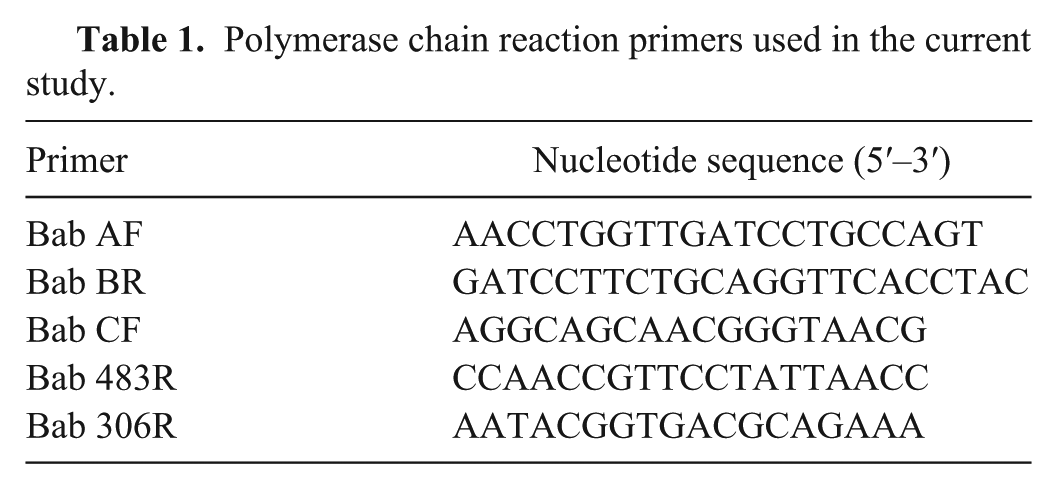
The primers used in the current study are listed in Table 1. The first primer set, Bab A–Bab B, a has been widely used in the veterinary literature,7,8,18 and are broad-range primers targeting the eukaryotic 18S rRNA gene. These primers produce a large product of 1,723 base pairs (bp). Two additional sets of primers were designed, based on the B. odocoilei DNA sequence (GenBank accession no. U16369), to produce smaller products more suitable for the reduced amplicon length often associated with formalin fixation. The forward primer (Bab CF) a was identical for both primer sets, starting at position 318 of the U16369 sequence. When used with the reverse primer Bab 483R, a ending at position 800 of the U16369 sequence, a 483-bp product was produced. This primer combination was within a region of the B. odocoilei DNA sequence where it differs from that of B. divergens by 32 positions. 7 The size of the product would be sufficient for DNA sequencing, and its position would allow species identification based on 17 of these 32 positions. Alternately, when the reverse primer Bab 306R a was used, ending at position 623 of the U16369 sequence, a 306-bp product would result. The annealing site of this reverse primer was in the region of the DNA sequence where the 32 position difference between B. odocoilei and B. divergens was located. 7 Therefore, based on the current knowledge of cervid Babesia spp. and available 18S rRNA gene sequences, Bab 306R was designed so that it could only anneal with the B. odocoilei sequence, making the primer specific to this species of Babesia. However, in making this reverse primer species specific, and making the product size small enough to accommodate formalin-associated protein modifications, the ability to confirm Babesia species through sequencing was lost, as the annealing site, providing specificity in this case, is typically removed during trimming and editing.
Polymerase chain reaction primers used in the current study.
The extraction of DNA from tissues was performed as previously described, as was the PCR procedure when using Bab A–Bab B primer set. 13 The newly designed primers were optimized for temperature and magnesium concentration at 58.8°C and 4 mM, respectively, but the PCR procedure was otherwise unaltered. Prior to each PCR, adequate DNA concentration (50–1,000 ng) was confirmed using a spectrophotometer. b
In order to confirm that the newly designed primers would amplify target DNA, PCR was performed on a B. odocoilei–positive elk spleen and a negative fresh elk spleen, stored frozen at −20°C, using all 3 primer sets. The positive status of the above sample was assigned through identification of intraerythrocytic parasites, morphologically consistent with B. odocoilei, in 1.5% of the erythrocytes on a direct smear of whole blood, and confirmed by PCR using the widely used Bab A–Bab B primer set and DNA sequencing. 13 Negative status was assigned based on the rare occurrence of cervid babesiosis in Canada, as well as a negative result on PCR using the Bab A–Bab B primer set. Specificity of the Bab306 R was also evaluated by comparing amplification of purified B. microti DNA c using the Bab A–Bab B and Bab CF–Bab 306R primer sets. In order to investigate the influence of fixation on PCR performance, a comparison between the 3 primers was made using the B. odocoilei–positive spleen that was stored frozen, fixed in 10% neutral buffered formalin for 2 days, or fixed in 10% neutral buffered formalin for 2 days followed by a 1-hr treatment with 96% formic acid (25 M). The influence that duration of storage had on performance was also evaluated. The PCR assays were performed on fresh-frozen B. odocoilei–positive EDTA blood and spleen, from the same B. odocoilei–positive animal described above, after storage at −20°C for 1 year using all 3 primer sets. In addition, 9 small sections of the B. odocoilei–positive spleen, ranging in weight from 0.03 to 0.13 g, were placed in separate containers of 10% neutral buffered formalin for storage. One of the 9 sections was removed from formalin for DNA extraction and PCR using all 3 primer sets, as above, weekly for the first 4 weeks, then monthly up to a final storage time of 6 months. The PCR was repeated for any of the time points where there was a negative result, after heating the samples to 70°C for 1 hr; as heat has been previously shown to enhance PCR amplification of DNA extracted from formalin-fixed, paraffin-embedded tissues. 19 The effect of paraffin embedding was not evaluated, as this was previously determined to be of minor influence by other groups. 9
When PCR was performed on a B. odocoilei–positive fresh-frozen spleen using the 3 different primer sets, bands of the expected size (1,723 bp, 483 bp, and 306 bp, respectively) were produced, while no bands were produced in the B. odocoilei–negative sample. The Bab A–Bab B primer set was also able to produce a band of the expected size when purified B. microti DNA was tested; however, only nonspecific binding occurred with the Bab CF–Bab 306R primer set with no band present at the 306-bp mark. The fixation method was shown to influence PCR performance when the 3 primer sets were compared. While all 3 primer sets successfully amplified DNA from fresh-frozen tissue, only the Bab 483R and Bab 306R primer sets produced bands of the expected size in formalin-fixed tissues. The PCR for all primer sets was uniformly negative when samples were treated with formic acid. Storage for 1 year at −20°C did not influence PCR performance, as bands of the expected size were produced with all primer sets. In contrast, fixation time in formalin did influence PCR performance. Positive results were consistently obtained using the Bab 306R primer set on samples that were stored in formalin for up to 4 months. The PCR was negative when performed with this primer set on a sample stored in formalin for 5 months; however, after a 2.5× increase in the amount of template, a band of the expected size was produced. The PCR was also positive after 6 months of storage in formalin, with a 2.5× increase in template concentration. Positive results were obtained in samples stored in formalin for up to 2 months using the Bab 483R primer set; however, samples stored for 3–6 months were consistently negative despite adjustments to the template concentration. The PCR remained negative after incubating samples at 70°C for 1 hr, and adjusting the amount of template as in the preincubation samples. Similarly, the PCR remained negative in all formalin-fixed samples incubated at 70°C for 1 hr when using the Bab A–Bab B primer set.
Although PCR provides numerous advantages in the diagnosis of infectious disease, sample storage conditions can limit performance. In the current study, primers were designed in the hope of expanding the repertoire of sample types available for B. odocoilei testing. The primers were designed bearing in mind both the size of product, as reduced amplicon length is common with formalin fixation, and the ability to identify species either by DNA sequencing or as a result of the specificity of the primer itself. Both of the newly designed primer sets were successful in amplification of B. odocoilei DNA in fresh-frozen tissue, with similar performance to the more traditional Bab A–Bab B primer set. Although the specificity of the Bab 306R primer would have ideally been evaluated using several different species of Babesia, access was limited to B. microti DNA at this time. Results of the current study demonstrate the ability of the Bab 306R primer to differentiate these 2 Babesia species, but further work will be necessary to confirm these primers are specific for B. odocoilei.
A significant decrease in amplicon length with formalin fixation was demonstrated in the current report, as only the primers with the smallest products (483 bp and 306 bp) successfully amplified the target DNA. This was consistent with previous reports.5,9 Amplification was not possible using any of the primer sets when tissues were treated with formic acid. There is limited information in the literature regarding the effects of formic acid on PCR performance. When described in the literature, it is generally in the context of increasing formic acid content during the acidification of nonbuffered formalin or the use of formic acid in decalcification processes.9,17 Concentrations of formic acid in these applications are much lower than what was used in the current study, and while an inverse relationship was found between the sensitivity of PCR and formic acid concentration, amplification was possible.
The presence of CWD in Saskatchewan wild and domestic cervid populations has necessitated the use of high concentration formic acid on cases with unknown prion status, as a biosafety precaution. 10 Because of the shared host species, fixed samples from many cases where B. odocoilei testing may be relevant have also been treated with formic acid. The findings of the current study indicate that under these formic acid concentrations, samples are not suitable for PCR testing. If tissue processing can be delayed until CWD status is known, the use of formic acid can be avoided. However, this delay would prolong exposure to formalin, which has also been shown to have an inverse relationship with PCR performance and amplicon length.5,9
As numerous other factors can also delay tissue processing in wildlife species, particularly in field study scenarios, the effect of formalin fixation for up to 6 months was evaluated. Extended exposure to formalin did not influence the PCR performance for the first 2 months using the Bab 483R primer set, and up to 6 months for the Bab 306R primer set with minimal adjustments to the PCR protocol (specifically, the template concentration). The evaluation of fixation time on PCR performance has often been limited to 7 days duration, with only rare studies examining samples stored in fixative for up to 1 month.4,5,9,16 In these reports, the size of the PCR product after 1 month of storage in formalin was limited to 268 bp and 135 bp, respectively, which is comparable to the size of the PCR product maintained through 6 months of storage in the current study, but smaller than that achieved after 2 months of storage in formalin.5,16 In the current study, heat treatment of previously extracted DNA was not successful in restoring PCR amplification, in contrast to that demonstrated by others. 19 It has been suggested that heat treatment may improve PCR amplification due to the partial elimination of chemical modification of nucleic acids generated by formalin fixation. 19 The current study was likely unable to reproduce these results due to the size of the largest PCR product (1,723 bp), and prolonged fixation times evaluated. 19 Confirmation that amplification of B. odocoilei DNA is still possible after extended fixation times greatly increases flexibility when short storage times are not possible, or when longer storage times may be preferable to formic acid treatment and the associated degradation of DNA. In cases where babesiosis is suspected and formic acid treatment is required, retention of fresh-frozen spleen, blood, or velvet antler for PCR testing is recommended. It has been previously reported that the sensitivity of PCR for Babesia gibsoni decreased after whole blood was stored at −20°C for 2 months in cases with percent parasitemias of 10−5 or less. 20 In the current study, PCR using all 3 primer sets effectively amplified the target DNA in EDTA blood and spleen after storage at −20°C for 1 year. The difference likely reflects the higher levels of parasitemia (1.5%) in the current report.
Although 3 primer sets were evaluated in the current study, the Bab CF–Bab 306R primer set provided the largest advantage in expanding the sample repertoire available for B. odocoilei testing. In addition to amplification of the target DNA in fresh-frozen tissues stored up to 1 year, this primer set was also effective in tissues stored in 10% neutral buffered formalin for up to 6 months. Specificity of the primer was demonstrated when challenged with B. microti DNA, but more extensive testing of various Babesia spp. will be required to confirm the degree of specificity.
Footnotes
Acknowledgements
The authors wish to thank Dr. Robbin Lindsay and Antonia Dibernardo from the National Microbiology Laboratory in Winnipeg, Manitoba, for the generous donation of B. microti DNA for this study.
a.
Sigma-Aldrich Canada Ltd., Oakville, Ontario, Canada.
b.
NanoDrop spectrophotometer, Thermo Fisher Scientific, Wilmington, DE.
c.
Supplied by the National Microbiology Laboratory, Winnipeg, Manitoba, Canada.
Declaration of conflicting interests
The author(s) declared no potential conflicts of interest with respect to the research, authorship, and/or publication of this article.
Funding
Funding for this study was provided by the Saskatchewan Ministry of Agriculture, Disease Investigation Unit.