
Abstract
Little is known about the presence of mycoplasmas in the genital tracts of domestic and stray bitches or in the vaginas of ovariohysterectomized (OHE) bitches. Moreover, to our knowledge, there has been no research to investigate the presence of canine vaginal mycoplasmas during the different stages of the reproductive cycle. We investigated the occurrence of mycoplasmas in the vaginas of healthy domestic and stray intact bitches, to correlate their presence with specific stages of the reproductive cycle, and to compare them with those in OHE bitches. We also investigated the presence of uterine mycoplasmas. Mycoplasmas were isolated from 41 of 122 vaginal swabs (34%) from domestic (27%) and stray (39%) bitches. Mycoplasma canis was the most commonly identified species (n = 26; 63%), and was detected in both intact (60%) and OHE (73%) bitches. Mycoplasma isolates from the vaginas of healthy bitches did not vary during the various stages of the estrous cycle. Mycoplasmas were not detected in uterine samples.
Escherichia coli, Pasteurella multocida, Staphylococcus aureus, S. pseudintermedius, and hemolytic Streptococcus spp. are frequently detected in the vaginas of healthy bitches.2,11,14 Most of these species have also been isolated from the vaginas of bitches with various reproductive disorders.1,3 Information regarding the frequency of isolation of canine genital mycoplasmas and their pathogenicity is limited. 5 Furthermore, the results of the few available studies describing the presence of canine vaginal and uterine mycoplasmas are inconsistent. Some earlier papers report the absence of mycoplasmas in vaginas and uteri of healthy bitches,8,16,20 whereas others report frequent isolation of mycoplasmas.2,7 In addition, the most commonly identified canine genital mycoplasma, Mycoplasma canis, is often isolated from clinically healthy dogs, infertile dogs, and dogs with urogenital disease.5,7 Other mycoplasmas routinely recovered from the vaginas of bitches with or without signs of genital disorders include M. cynos, M. edwardii, M. maculosum, M. molare, and M. spumans.7,17 In contrast to M. canis, which is an opportunistic pathogen isolated from urogenital and respiratory tracts, M. cynos is a proven pathogen of the respiratory tract in dogs.5,12 M. edwardii is associated with pneumonia, and M. spumans with arthritis; M. molare has been isolated from the canine oral cavity and reproductive tract, and M. maculosum from canine respiratory and reproductive tracts. 5
Little is known about the presence of mycoplasmas in the genital tracts of domestic and stray bitches, as well as in the vaginas of ovariohysterectomized (OHE) bitches. Moreover, there has been no research reported with respect to the presence of canine vaginal mycoplasmas during various stages of the reproductive cycle, to our knowledge. We investigated the occurrence of mycoplasmas in the vaginas of healthy domestic and stray intact bitches and correlated this with specific stages of the reproductive cycle. We compared these findings with those in OHE bitches. We also investigated the possible presence of uterine mycoplasmas.
A total of 122 vaginal swabs from domestic (n = 56) and stray (n = 66) bitches of different breeds and ages from different regions in Bosnia and Herzegovina were collected for isolation of mycoplasmas. Of these 122 bitches sampled, 95 swabs were also obtained for vaginal cytology. Sampling from the vagina and the determination of the stage of the reproductive cycle were conducted as described previously.9,11 In addition, a swab of the mucosa from the uterine horns of 35 bitches was aseptically obtained immediately following ovariohysterectomy. All samples were cultured in Thiaucourt (TH) liquid medium 18 and incubated at 37°C for up to 7 d. Prior to identification, all mycoplasma isolates were cloned using TH liquid and solid media. All incubations were in a 95% N2 and 5% CO2 atmosphere at 37°C.
Genomic DNA was extracted from mycoplasma broth cultures (QIAamp DNA extraction kit, Qiagen, Hilden, Germany) according to the manufacturer’s instructions. PCR assays for the Mycoplasma genus, 19 M. canis, 13 M. cynos, M. edwardii, M. maculosum, and M. spumans 6 were used for identification.
Statistical evaluations were performed using a chi-squared test and Fisher exact test; p ≤ 0.05 was considered statistically significant. The calculations were conducted utilizing GraphPad (GraphPad Software, San Diego, CA) and Social Science Statistics (http://www.socscistatistics.com/).
Mycoplasmas were isolated from 41 vaginal swabs (34%) from domestic (15 of 56; 27%) and stray (26 of 66; 39%) bitches. These mycoplasmas were found in the vaginas of OHE (11 of 27; 41%) and intact (30 of 95; 32%) bitches. M. canis was the most commonly identified species (n = 26; 63%), detected in domestic (53%) and stray (69%), intact (60%) and OHE (73%) bitches (Table 1). Significant differences were not found in the numbers of vaginal samples from which mycoplasmas were isolated among the observed groups of animals. Mycoplasma isolates did not vary significantly during the different stages of the reproductive cycle (Table 2).
Mycoplasma isolates from the vaginas of domestic and stray bitches.
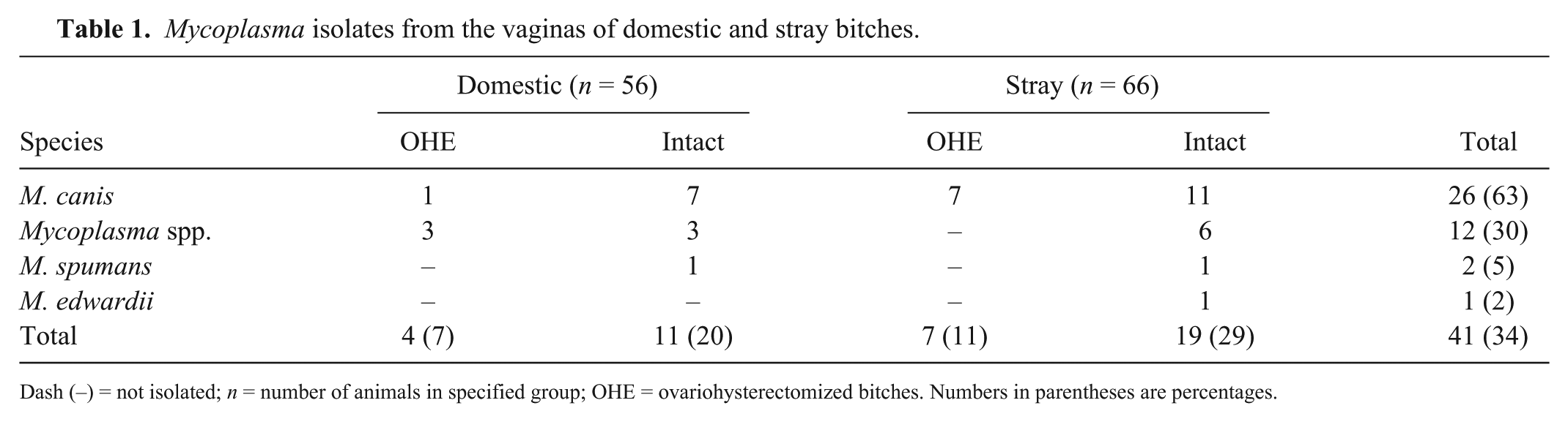
Dash (–) = not isolated; n = number of animals in specified group; OHE = ovariohysterectomized bitches. Numbers in parentheses are percentages.
Mycoplasma isolates from the vaginas of intact bitches during different stages of the reproductive cycle.
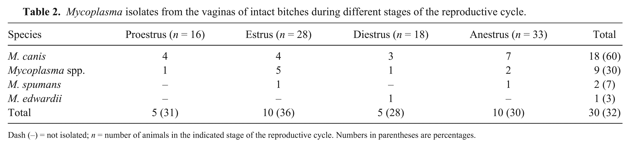
Dash (–) = not isolated; n = number of animals in the indicated stage of the reproductive cycle. Numbers in parentheses are percentages.
All uterine samples tested (n = 35) were negative for mycoplasmas. Similarly, other researchers8,14,16 found no mycoplasmas in uterine samples collected at ovariohysterectomy. However, some studies2,15 reported the frequent isolation of mycoplasmas from uterine contents obtained postmortem. Based on the results of our study, mycoplasmas do not seem to be a part of the normal uterine bacterial flora of clinically healthy bitches.
Our finding of mycoplasmas in the vaginas of healthy bitches is in agreement with previous findings.4,7,10,15 The frequency of isolation (34% of total samples) was lower than in previous studies, which recovered mycoplasmas from 81%, 7 59%, 4 46%, 15 and 42% 10 of clinically healthy bitches. Similar to previous research, 4 all of our isolates were obtained from the cranial vagina. In contrast with our findings, mycoplasmas were not recovered from this region in a previous study. 20 Other researchers7,10,15 did not specify the regions of the vagina from which they obtained samples. Negative results reported in some studies8,15 could be the result of the lack of specific and suitable techniques for the growth and detection of mycoplasmas, including the choice of media and the sensitivity of detection and identification methods used. 10
In our study, the isolation of mycoplasmas did not vary during the various stages of the estrous cycle. This finding is supported by previous studies on general vaginal bacteria.2,11 M. canis was the predominant species we isolated, which is in agreement with previous studies.7,10,15 M. spumans and M. edwardii were also isolated from canine vaginas in earlier studies, 7 as found in our study. Not all mycoplasma isolates could be identified by the several PCR assays used in our study. Therefore, future studies should consider investigating less common mycoplasma species of the canine reproductive tract.
The presence of M. canis in the reproductive tracts of both healthy and diseased bitches is inconclusive, and the reason could possibly be found in differences between strains, and differences in virulence. Future studies should be focused on phenotypic and genotypic characterization of M. canis strains isolated from bitches, with and without urogenital disorders. Furthermore, such studies should include a comparison of phenotypic and genotypic profiles among the strains isolated from the vaginas of domestic and stray bitches, as well as between intact and OHE bitches.
Footnotes
Acknowledgements
We thank M. Scott Cornwell for the English language revision of this article.
Declaration of conflicting interests
The authors declared no potential conflicts of interest with respect to the research, authorship, and/or publication of this article.
Funding
The authors received no financial support for the research, authorship, and/or publication of this article.