
Abstract
A 24-year-old, spayed female Malayan sun bear (Helarctos malayanus) in the Taipei Zoo (Taipei, Taiwan) showed clinical signs of slowly progressive anorexia, dullness, compulsive pacing, and circling. The animal subsequently developed acute severe stupor and persistent recumbency. Postcontrast study of computed tomography revealed a spheroid, extra-axial mass with strong but heterogeneous hyperattenuation in the left temporal lobe of the cerebrum. At necropsy, a solitary, well-circumscribed intracranial mass measuring 3 cm × 2.5 cm × 2 cm was attached to the left pyriform lobe with compression of the adjacent neuroparenchyma. Cytological examination obtained from the mass revealed large clumps and sheets of cohesive polyhedral cells with round nuclei, wispy cytoplasm, and indistinct cell borders. Microscopically, the mass was composed of densely packed round to polygonal cells arranged in lobules and small nests. Psammoma bodies, xanthomatous change, and cholesterol deposition were also noted. Immunohistochemical staining of the tumor was positive for vimentin, pancytokeratin, cytokeratin (CK)34BE12, neuron-specific enolase, and epithelial membrane antigen, but negative for glial fibrillary acidic protein and S100 protein. The cytological, histological, and immunohistochemical features were compatible with a meningothelial meningioma.
Keywords
Meningiomas are common central nervous system neoplasms found in human beings, dogs, and cats but are rarely documented in wild animals. In human beings, intracranial meningiomas have a predilection for cerebral convexities and parasagittal regions. 16 Most canine meningiomas are solitary intracranial neoplasms that mainly involve the olfactory and/or frontal region, the floor of the cranial cavity, the optic chiasm, or the suprasellar and parasellar regions. In cats, multiple tumors are not uncommon and frequently involve the supratentorial meninges or the third ventricle. 13 Women are more likely to develop a meningioma, whereas there is no significant sex predilection found in animals.3,13 To date, 4 meningiomas have been identified in wild animals including a microcystic meningioma in a dolphin (Delphinus delphis), 9 a meningioma in a baboon (Papio spp.), 15 a meningioma in a rhesus macaque (Macaca mulatta), 21 and a fibroblastic meningioma in a Bactrian camel (Camelus bactrianus). 10 Of these 4 meningiomas, all were intracranial, 3 arose from the base of the cranial vault, and 3 of the animals were female.
A 24-year-old, spayed female Malayan sun bear (Helarctos malayanus) housed at the Taipei Zoo (Taipei, Taiwan), with a previous history of tubulopapillary mammary adenocarcinoma with regional nodal metastasis treated by mastectomy, lymphadenectomy, and ovariohysterectomy 5 years before, developed slowly progressive anorexia, dullness, compulsive pacing, and circling to the left over 3 months from January 2012. The animal further deteriorated and acutely developed severe stupor and persistent recumbency. Physical examination revealed an abrasion wound on the left forepaw. The complete blood count and serum biochemistry analyses were unremarkable. Computed tomography (CT) scan of the head was planned. Because of the unstable anesthetic status, only the postcontrast imaging was performed. A single, well-demarcated, spheroid, extra-axial mass with strong but heterogeneous hyperattenuation was revealed (Fig. 1). The mass was located in the left temporal lobe, at the level of caudate nucleus and thalamus. Midline shift, perimass edema, and compression of the left lateral ventricle were also noted. Based on the imaging findings and the progressive clinical signs, intracranial neoplasia was the main differential diagnosis, and meningioma was highly suspected. The animal failed to recover from anesthesia and expired after CT examination.
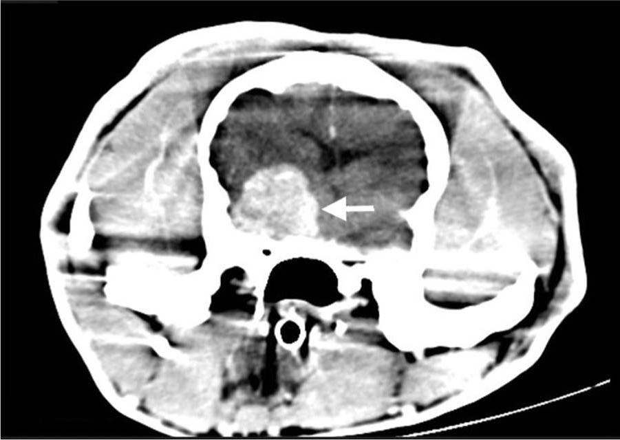
Postcontrast image of computed tomography; Malayan sun bear (Helarctos malayanus). There is an extra-axial, demarcated mass with heterogeneous hyperattenuation in the left temporal lobe (arrow).
At necropsy, a solitary, well-circumscribed, rubbery to firm, red mass measuring 3 cm × 2.5 cm × 2 cm was tightly attached to the left pyriform lobe of the cerebrum and adhered to the dura of the cranial base (Fig. 2). The surface of the mass was granular and flat to mildly irregular. Serial sections of the cerebrum revealed compression of the adjacent neuroparenchyma concomitant with moderate asymmetry of the lateral ventricles. The right thoracic cavity contained approximately 22 ml of serosanguinous fluid. The lungs were diffusely red, heavy, and wet. Within the trachea and bronchi was yellow to brown semisolid material resembling ingesta. The serial cut sections of the kidney revealed a 3-mm diameter white nodule within the cortex. Other lesions included focal ulceration of the left metatarsal pad, and multiple areas of congestion in the stomach, small intestines, pancreas, and urinary bladder.
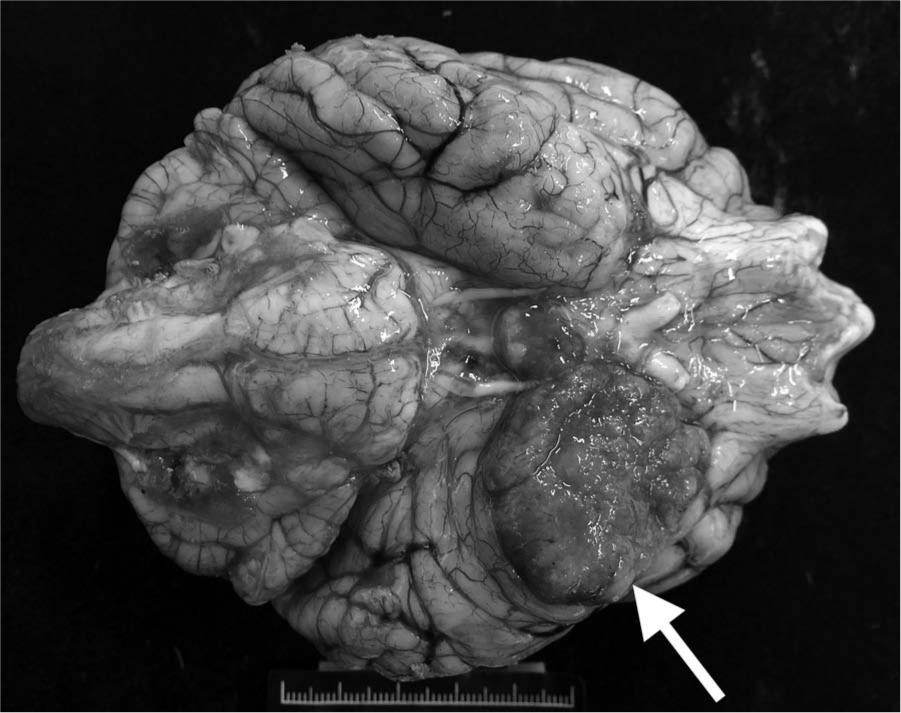
Ventral brain; Malayan sun bear (Helarctos malayanus). A well-defined, rubbery to firm, red mass is attached to the left pyriform lobe (arrow) with compression of the surrounding cerebrum.
Imprints of the cerebral mass were made at necropsy for cytological examination. The slides were air-dried and stained with Liu stain (a modified Romanowsky stain). Large clumps and sheets of cohesive polyhedral cells with a moderate to high nuclear-to-cytoplasmic ratio were noted (Fig. 3). Nuclei were uniformly round to oval, slightly eccentric, and sharply delineated with a coarse chromatin pattern and 1 or 2 eccentric nucleoli. The cytoplasm was wispy, and the cell borders were indistinct. Naked nuclei were occasionally identified. Mitotic figures and inflammatory cells were absent.
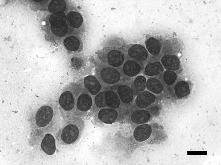
Imprints of the tumor; Malayan sun bear (Helarctos malayanus). Clumps of cohesive polygonal cells with round to oval nuclei, wispy cytoplasm, and indistinct cell boundaries. Liu stain. Bar = 10 µm.
Tissue samples were fixed in 10% neutral buffered formalin and routinely processed for histology. Sections were stained with hematoxylin and eosin while selected sections were stained with periodic acid–Schiff and Alcian blue at pH 2.5. Immunohistochemical staining was performed with a non-avidin–biotin immunoperoxidase and diaminobenzidine detection method a for the following markers: antibodies against vimentin, b pancytokeratin, b S100 protein, b neuron-specific enolase (NSE), b glial fibrillary acidic protein (GFAP), b and cytokeratin (CK)34BE12. c Staining of epithelial membrane antigen (EMA) c was accomplished by an automated immunohistochemistry system. d Appropriate tissues as positive controls of aforementioned markers were included in the staining procedure.
Microscopically, the tumor arising from the left pyriform lobe of the cerebrum was circumscribed, unencapsulated, and consisted of lobules and small nests of densely packed, round to polygonal cells separated by fine fibrovascular septa. Neoplastic cells had round to ovoid nuclei with conspicuous nucleoli, ample eosinophilic cytoplasm, and indistinct cell borders. The chromatin pattern ranged from evenly distributed to coarsely granular. Mitotic figures were rarely seen, with less than 1 per high-power field. Within the neoplasm were a small number of scattered whorls of elongate cells with amorphous to concentrically laminated central calcification (psammoma bodies). There were multifocal areas of xanthomatous change, evidenced by cells distended with abundant, coarsely vacuolated cytoplasm (Fig. 4). Cholesterol deposition in clefts and cystic formation were occasionally seen. The adjacent neuroparenchyma compressed by the tumor contained extensive areas of spongy vacuolation with dispersed gemistocytes. Histochemical staining demonstrated there was no periodic acid–Schiff- or Alcian blue–positive substance within the neoplasm. Immunohistochemically, the neoplasm showed strong, diffuse, cytoplasmic labeling for vimentin, pancytokeratin, and CK34BE12 (Fig. 5A–5C) as well as diffuse, low-intensity cytoplasmic labeling for NSE. Approximately half of neoplastic cells showed immunoreactivity for EMA with a perinuclear dot-like pattern (Fig. 5D). Neoplastic cells did not express GFAP or S100. In the kidney, a sharply defined, unencapsulated mass in the deep cortex consisted of projecting papillae lined by a single layer of well-differentiated columnar epithelial cells. The morphologic features of the mass were compatible with renal papillary adenoma. Other histologic findings included mild suppurative pneumonia with edema, ulcerative dermatitis of the metatarsal pad of left forepaw, and mild lymphocytic hepatitis and cystitis.
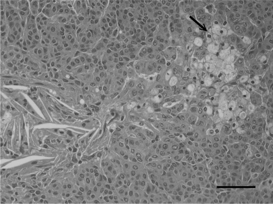
Meninges; Malayan sun bear (Helarctos malayanus). Neoplastic cells with xanthomatous change (arrow) characterized by a gradual cellular transition from finely to coarsely vacuolated cytoplasm. Cholesterol deposition in clefts is also noted. Hematoxylin and eosin. Bar = 50 μm.
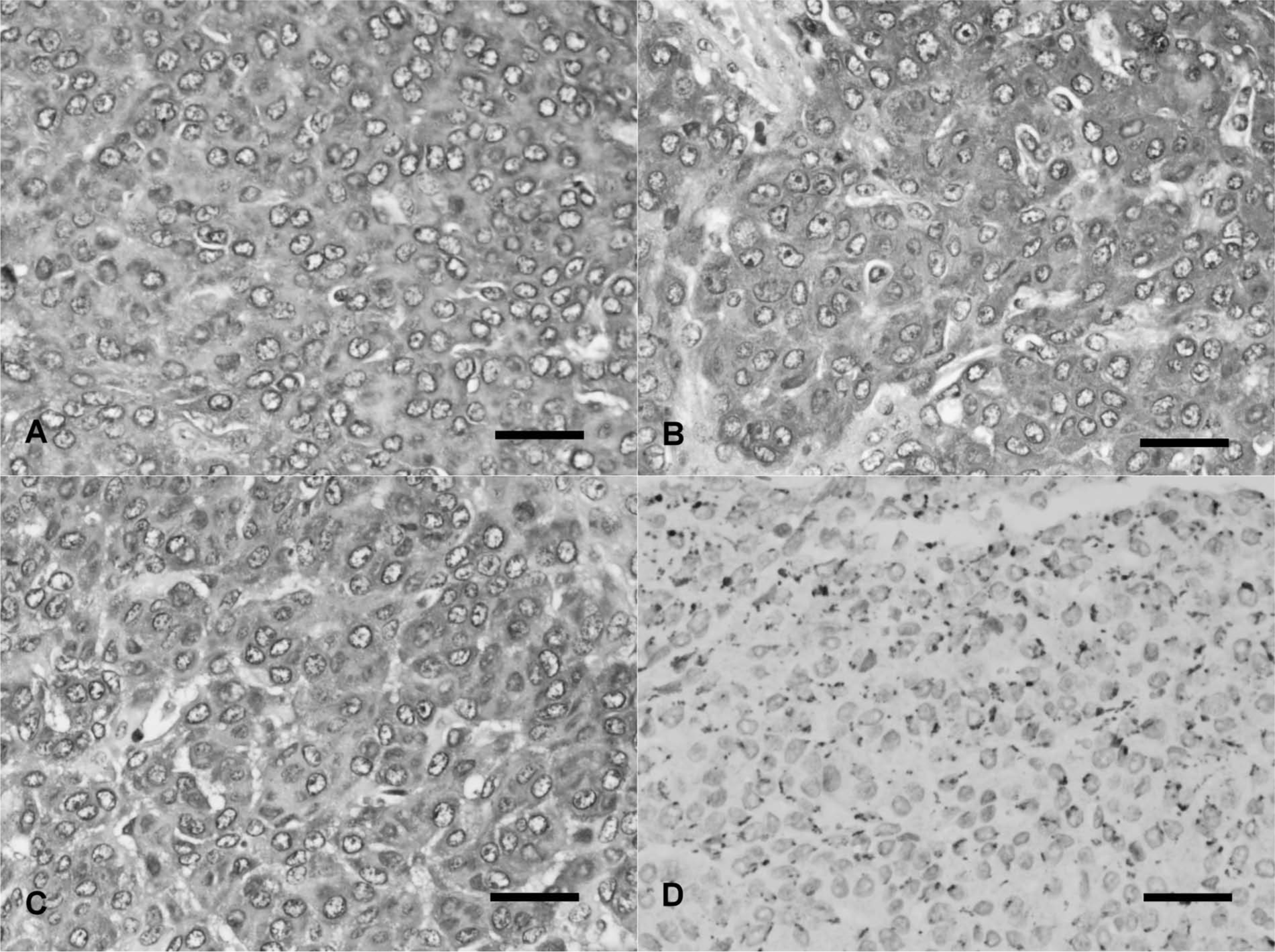
Meninges; Malayan sun bear (Helarctos malayanus). Diffuse intense cytoplasmic immunolabeling for vimentin (
Meningioma is characterized by a histologically benign, slow-growing central nervous system neoplasm that produces clinical signs by local compression of the surrounding nervous parenchyma. 6 Usually the clinical signs develop slowly but progressively, and the type of clinical signs depends on the tumor location, tumor size, and its secondary pathologic changes. 25 In the current case, the tumor was located in the ventral aspect of the left pyriform lobe. The altered mentation was presumably associated with the disturbance of the ascending reticular activating system, owing to tumor compression of the nearby diencephalon and diffuse perimass edema in the surrounding cerebrum. Circling and compulsive pacing are common signs suggestive of a forebrain lesion. Furthermore, circling toward the left side typically reflects a left-sided lesion of the forebrain.
Meningiomas are most likely derived from arachnoid cap cells that form the outer layer of the arachnoid. Arachnoid cap cells are capable of developing both mesenchymal- and epithelial-like functions and morphology. 17 The current neoplasm was histologically consistent with meningothelial meningioma, characterized by lobules or nests of polygonal cells with a moderate to large amount of eosinophilic cytoplasm, ill-defined cell borders imparting a syncytial appearance, and low mitotic activity.6,16 Xanthomatous change is well described in human meningiomas and typified by a gradual transition of meningothelial cells to cells with vacuolated cytoplasm owing to lysosome or fat accumulation.5,16 Xanthomatous cells resemble macrophages both morphologically and immunohistochemically, as indicated by immunoreactivity for a series of histiocytic markers, while their meningothelial origin can be demonstrated by immunoreactivity for EMA or by the presence of plasmalemmal interdigitations and desmosomal junctions ultrastructurally.5,22
Because of lack of a sensitive and specific marker for meningiomas, a broad panel of immunohistochemical stains is required to assist in diagnosis. Vimentin and EMA are consistently expressed in human meningiomas, whereas the application of antibodies against EMA in animals has not been established.12,23 Human meningiomas exhibit variable immunoreactivity for cytokeratin and S100. 18 Immunohistochemical analysis of canine meningiomas has demonstrated vimentin expression in all reviewed cases.1,12 In dogs, most tumors were immunolabeled by S100 and NSE antibodies with an inconsistent intensity and percentage of positive cells. Cytokeratin immunoreactivity was detected in a small number of canine cases in which limited positive cells were observed. Moreover, rare canine tumors revealed positive GFAP staining. In the current case, the tumor displayed a diffuse, strong, and cytoplasmic immunolabeling for vimentin and pancytokeratin, indicative of both mesenchymal and epithelial differentiation; the latter was substantiated by CK34BE12 labeling. The NSE-positive and GFAP-negative staining results were also similar to most canine meningiomas. 12 Regarding EMA staining, the staining pattern was inconsistent with that of human meningothelial meningiomas, in which tumor cells reveal both cytoplasmic and membranous immunoreactivity. 23
Cytological features of meningioma cells are highly variable. Generally there is a mixed pattern of sheets, clusters, or whorls of cells with both mesenchymal and epithelial morphology.24,25 The exception is the meningothelial type, which is, like the human counterpart, characterized by a homogenous population of polygonal to spindle cells with eosinophilic wispy cytoplasm and ill-defined cytoplasmic borders with a syncytial appearance.20,25 The nuclei are round to oval and slightly eccentric with sharply delineated nuclear membrane, finely dispersed chromatin, and prominent nucleoli. Intranuclear cytoplasmic invaginations (pseudoinclusions) and nuclear grooves are rarely observed in animals but relatively common in human meningiomas.20,24 In the present case, both cytological and histological characteristics were compatible with meningothelial meningioma. Clinically, cytological assessment of brain tumor cells in smear or crush preparations in combination with frozen sections provides an immediate and accurate diagnosis intraoperatively in human medicine, 20 and this technique is also applicable to canine and feline nervous system neoplasms, with a satisfying diagnostic accuracy of 80%. 24
Computed tomography is a valuable diagnostic technique to evaluate intracranial lesions in veterinary medicine. Imaging characteristics of meningiomas include a lenticular, broad-based or plaque-like mass, a well-defined margin with tissue displacement, and an extra-axial location with strong and homogenous contrast enhancement, sometimes concurrent with a dural tail sign.7,14 More often, meningiomas appear isoattenuating to hyperattenuating on precontrast CT images. 14 Meningiomas usually display a space-occupying effect with associated pathological changes, such as edema or necrosis with a hypodense appearance or hemorrhage with hyperdensity. 7 In the current case, without comparing to the precontrast images, the differential diagnosis of a hyperattenuated mass on the postcontrast CT image included hematoma and mass with contrast enhancement. Given the progressive clinical signs and extra-axial location with strong hyperattenuated pattern on the postcontrast images, meningioma was considered to be the most likely diagnosis during antemortem examination.
Malayan sun bears are in the family Ursidae, which includes all bear species. The most commonly reported neoplasm in elderly bears is hepatobiliary tumors.2,4,11,19 The reason why bears are highly vulnerable to hepatobiliary neoplasms remains unclear; it has been postulated that genetic predilection and dietary and environmental factors may contribute to the oncogenesis. Renal adenoma is an uncommon neoplasm in domestic animals and is reported most commonly in dogs, cats, horses, and cattle. 8 In the present case, renal papillary adenoma is considered to be an incidental finding.
Footnotes
Acknowledgements
The authors thank Dr. Pei-Yi Chu, Head of Department of Pathology, St. Martin De Porres Hospital for assistance with epithelial membrane antigen staining.
a.
Super Sensitive Polymer-HRP detection system, BioGenex Laboratories Inc., San Ramon, CA.
b.
Dako North America Inc., Carpinteria, CA.
c.
Novocastra Laboratories Ltd., Newcastle, UK.
d.
BOND-MAX autostainer, Leica Biosystems, Newcastle, UK.
Declaration of conflicting interests
The author(s) declared no potential conflicts of interest with respect to the research, authorship, and/or publication of this article.
Funding
The author(s) disclosed receipt of the following financial support for the research, authorship, and/or publication of this article: This study was partially supported by research grants from the National Science Council of Taiwan (NSC 101-2313-B-002-022 and NSC 98-2313-B-002-026-MY3).