
Abstract
A 26-y-old, male, captive Humboldt penguin (Spheniscus humboldti) was euthanized following a 3.5-mo history of weakened elimination mechanics, recurrent tenesmus, intermittent hemorrhagic droppings, and a cloacal mass. Blepharospasm, of unknown cause, of the right eye was present for ~3 mo before euthanasia. Autopsy revealed a cloacal adenocarcinoma with localized coelomic carcinomatosis and distant metastases to the liver and lungs. On histopathology, a 2.6 × 1.2 × 0.5-mm, well-demarcated mass was found surrounding the right optic nerve, expanding the subdural space and wrapping the leptomeninges. The mass was composed of neoplastic spindle-to-polygonal cells consistent with a meningioma, meningothelial subtype. No evidence of neoplasia was found in the optic chiasm or brain, indicating a primary retrobulbar meningioma. Immunohistochemistry for cytokeratin AE1/AE3, vimentin, and S100 revealed robust and consistent immunoreactivity to vimentin, and weak and variable immunoreactivity to cytokeratin and S100, supporting the diagnosis. Meningiomas have been described only rarely in avian species, and we found no reports of optic nerve meningiomas in any avian species to date. The optic nerve meningioma in this case was considered a clinically incidental finding.
A 26-y-old, male, 3.8-kg, captive-born Humboldt penguin (Spheniscus humboldti) was examined under anesthesia for a 10-d history of intermittent hemorrhagic droppings. A cloacal mass was palpated, of which a fine-needle aspirate revealed clusters of mucinous epithelial cells with mixed heterophilic-to-granulomatous inflammation. In the following 8 wk, the penguin continued to eat and behave normally, but had occasional droppings of increasing hemorrhagic intensity and consistently weakened elimination mechanics whereby the initial portion of the dropping sprayed backwards in a normal manner and the latter portion dribbled from the vent. The penguin also had an episode of tenesmus. Radiographs and coelomic ultrasound were unremarkable, and the cloacal mass was still palpable. Over the course of the following 6 wk, hemorrhagic droppings were intermittent, and there was recurrence of tenesmus with progression to cloacal obstruction from urate accumulation. Blepharospasm, of unknown cause, of the right eye was present for ~3 mo before euthanasia. The penguin was euthanized because of chronic debilitation and a high suspicion of cloacal neoplasia.
Autopsy revealed a 1.5 × 1.0 × 0.5-cm, irregularly oval, poorly demarcated, firm, pale-tan, cobblestoned mass arising from the mucosal aspect of the cloaca, ~1.5-cm orad to the vent (Fig. 1A), which extended into the cloacal wall on section. The mesentery had dozens of pinpoint, raised, tan foci, suggestive of carcinomatosis. Throughout the parenchyma of both liver lobes and the lungs were dozens of pinpoint to 1.5 × 1.5 × 1.5-cm, well-demarcated, firm, white-to-tan, smooth, occasionally umbilicated masses, suspicious of metastases (Fig. 1B). A complete set of tissue samples was collected, fixed in 10% neutral-buffered formalin, and processed routinely for histology.
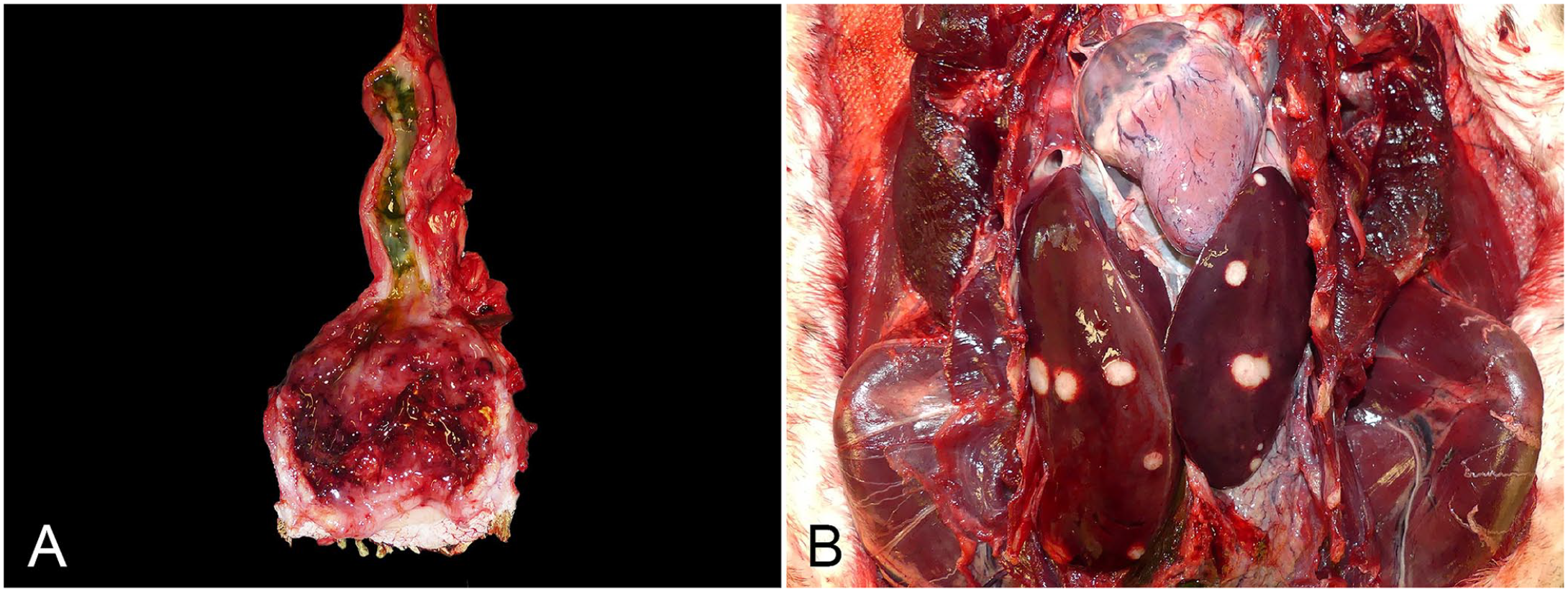
Gross presentation of a cloacal adenocarcinoma with metastases in a Humboldt penguin.
On microscopic examination of H&E-stained sections, the cloacal mass was diagnosed as a transmural adenocarcinoma based on well-differentiated epithelial cells arranged in acini with an abundant extracellular mucinous matrix, frequent lymphovascular invasion, and scirrhous response. Mesenteric carcinomatosis and hepatic and pulmonary metastases were additionally confirmed.
Opportunistically found on histopathology of the right optic nerve, immediately adjacent to the globe, was a well-demarcated, 2.6 × 1.2 × 0.5-mm, unencapsulated, infiltrative, variably cellular mass expanding the subdural space and wrapping and merging with the leptomeninges. Based on the presence of this mass in the initial slide of the right globe, the corresponding tissue paraffin block was melted and the entire optic nerve with the optic disc, adjacent sclera, and retina was trimmed out, sectioned longitudinally, and re-embedded to allow for additional detailed assessment. The mass was composed of spindle-to-polygonal cells arranged in short bundles and sheets with occasional whorls, supported by a collagenous stroma (Fig. 2A). These cells had indistinct borders, abundant fibrillar eosinophilic cytoplasm, oval-to-elongated nuclei with coarsely stippled, open chromatin, and mostly one, prominent, deeply basophilic nucleolus (Fig. 2B). Anisocytosis and anisokaryosis were moderate with a regular presence of multinucleate cells and no mitotic figures in ten 400× fields (2.37 mm2). Regularly interspersed among the neoplastic cells were short bundles of collagen, often arranged in concentric whorls. Infrequently within these whorls were linear-to-irregular, deeply eosinophilic concretions, interpreted as mineralization. In deeper sections, the neoplasm was continuous with the leptomeninges. The histomorphology was consistent with a meningothelial meningioma arising from the optic nerve sheath. Significant changes were not found in the adjacent optic nerve parenchyma to indicate compression or in the brain and optic chiasm to suggest extension from an intracranial tumor. In the right globe, the only other histologic change was increased cellularity of spindle cells and collagen density in the axial anterior corneal stroma without any associated blood vessels or inflammatory cells, interpreted as stromal fibrosis. The left globe and left optic nerve were within normal limits.
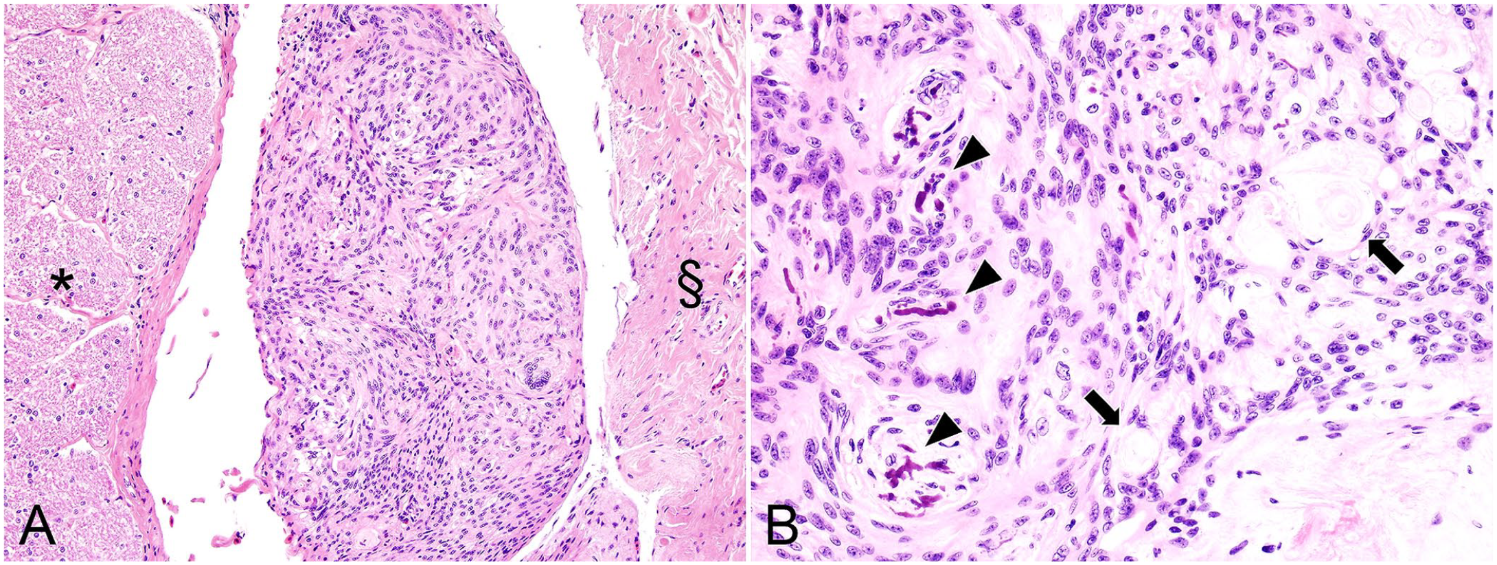
Optic nerve meningioma in a Humboldt penguin.
Immunohistochemistry (IHC) for cytokeratin (CK) AE1/AE3, vimentin, S100, and Ki67 was performed on serial sections of the neoplasm (BOND-MAX fully automated IHC staining system; Leica) at the New York State Animal Health Diagnostic Laboratory, Cornell University (Ithaca, NY, USA). Appropriate Humboldt penguin and canine positive controls were utilized together with negative controls in which the primary antibodies were replaced by non-immune mouse or rabbit serum (Table 1). The neoplastic cells had uniform and robust cytoplasmic immunoreactivity to vimentin (Fig. 3A, 3B); immunolabeling with CK AE1/3 (cytoplasmic; Fig. 3C, 3D) and S100 (cytoplasmic and rarely nuclear; Fig. 3E, 3F) was inconsistent and generally weak. The adjacent, normal leptomeninges had robust and consistent immunolabeling with both vimentin and CK AE1/3 (Fig. 3A, 3C). Cross-reactivity was not found with Ki67, which was also applied to sections of the cloacal adenocarcinoma and non-neoplastic intestinal mucosa. The IHC results supported the original diagnosis of optic nerve meningioma.
Details of immunohistochemical stains performed on sections of meningioma.
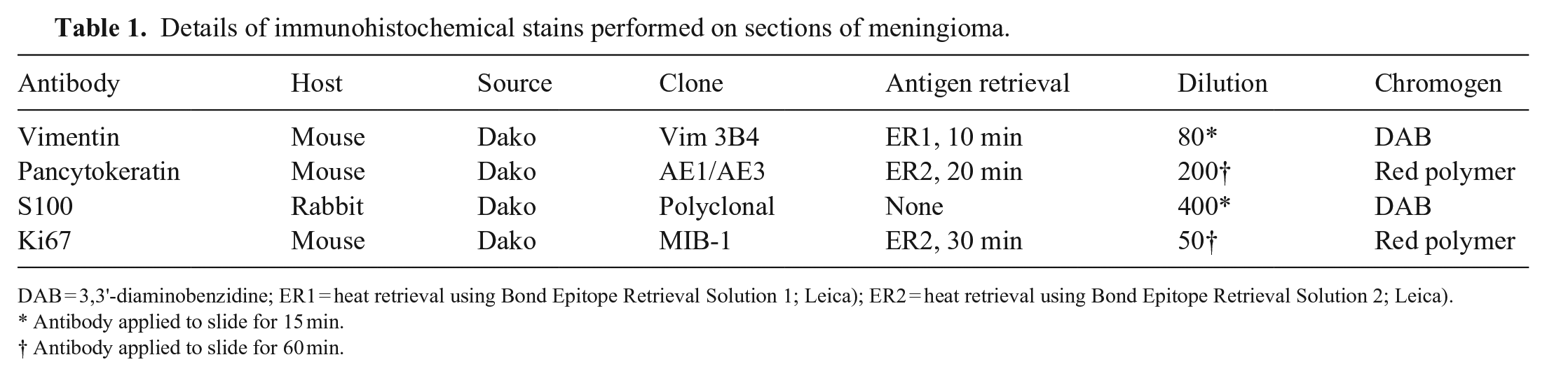
DAB = 3,3'-diaminobenzidine; ER1 = heat retrieval using Bond Epitope Retrieval Solution 1; Leica); ER2 = heat retrieval using Bond Epitope Retrieval Solution 2; Leica).
Antibody applied to slide for 15 min.
Antibody applied to slide for 60 min.
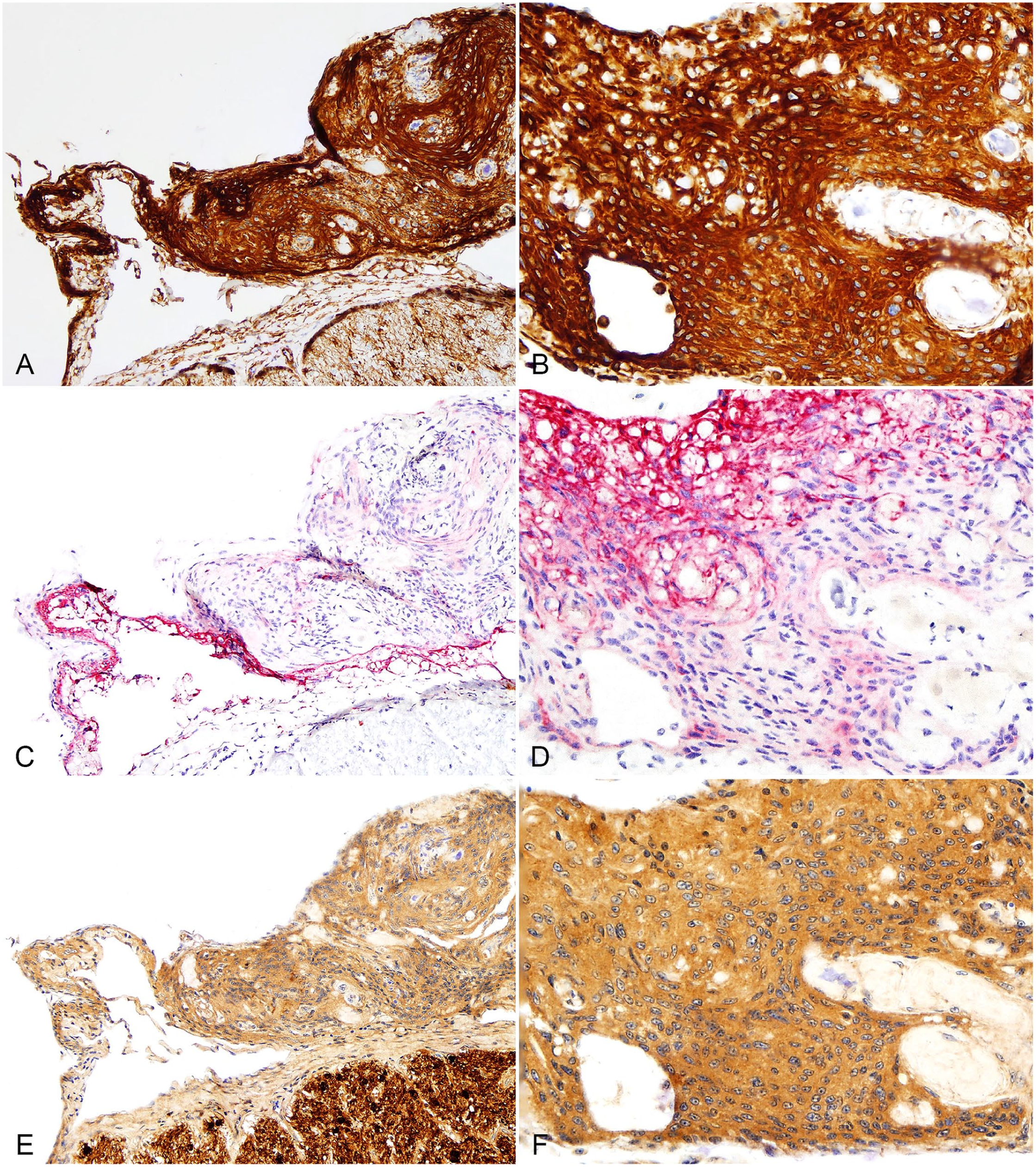
Immunohistochemical profile of an optic nerve meningioma in a Humboldt penguin.
Spontaneous neoplasms in birds are relatively uncommon, with a prevalence of 5.8% and 9.2% at 2 diagnostic pathology services, among 9,574 and 827 avian cases, respectively.7,11 The reported incidence of neoplasia in captive penguins is even lower, 15 and the recorded neoplasms, in descending frequency, include malignant melanoma,5,12 squamous cell carcinoma, 8 adenocarcinoma,3,16,19 lymphoma, 14 cholangiocarcinoma, retrobulbar round-cell tumor, 18 ovarian tumor, 6 leiomyoma, 4 and lens tumor. 1 In the order Sphenisciformes, Humboldt penguins appear to be one of the species overrepresented for tumor development,3,5,12,14,19 which may in part reflect the large number of this species under human care. Our case was unique in that there were 2 concurrent neoplasms, of which 1 was an optic nerve meningioma, a previously unreported type of tumor in birds, to our knowledge. We retrieved no cases of optic nerve meningiomas in birds through a search of Google Scholar and PubMed, using a combination of search terms “optic nerve/retrobulbar/orbital”, “meningioma”, and “avian/birds”, suggesting that this condition has not been reported in the avian species.
Meningiomas in veterinary species are common in the dog and cat and thought to arise from the arachnoid (meningothelial) cells lining the arachnoid membrane and pia mater. These neoplasms are extremely rare in birds with only 2 intracranial cases reported in Rhode Island Red–type and White Leghorn–type pullets in a single publication from 1976. 17 In the retrobulbar or orbital region, meningiomas may arise as a primary tumor from neoplastic transformation of arachnoid cap cells within the optic nerve sheath or as a secondary extension of an intracranial tumor along the optic nerve. 10 Optic nerve meningiomas are uncommon in the veterinary field, with most cases occurring in dogs 10 and rarely in the cat, 9 rat, 20 and cow. 13 Although these cases are generally considered spontaneous, 1 of the 2 cases reported in the rat was chemically induced by crystalline nickel subsulfide. 20
In comparison to common histologic features of canine retrobulbar meningiomas, our case had some similarities and disparities. Most canine cases are classified as a meningothelial subtype based on the predominant pattern of neoplastic proliferation, 10 as seen in our case. However, islands of bony and cartilaginous metaplasia and a myxomatous matrix, frequently reported in dogs, were not found in our case, whereas mineralization without bone, which was present in our case, is not a common feature in dogs. 10 The immunohistochemical profile in our case was consistent with what has been reported in the literature for canine cases, 2 with robust and diffuse immunolabeling for vimentin and weak and patchy immunolabeling for pancytokeratin and S100. Interestingly, the normal leptomeninges in our penguin case had diffuse immunoreactivity for pancytokeratin, in contrast to canine or feline leptomeninges, which normally have patchy immunolabeling.
Potential differential diagnoses in our case based on location and cellular morphology include epithelioid variants of a peripheral nerve sheath tumor and histiocytic sarcoma that could be primary or metastatic. A primary optic nerve meningioma was favored given the continuation of the neoplasm with the leptomeninges, clear demarcation from the optic nerve itself, immunoreactivity to pancytokeratin, and lack of a similar neoplasm in the rest of the body.
Birds with retrobulbar tumors typically have clinical signs such as exophthalmos, epiphora, scleral hyperemia, and unilateral blindness.7,18 The only possibly related ocular sign in our case was intermittent blepharospasm in the right eye, which was considered to be the result of an unknown corneal disease, supported by the histologic presence of anterior stromal fibrosis. There was no evidence of exophthalmos or vision loss, which was in accordance with the small size of the mass being localized to the optic nerve and lack of histologic evidence of optic nerve degeneration. Therefore, the optic nerve meningioma in our case was considered an incidental finding. Based on the presence of a cloacal adenocarcinoma with carcinomatosis and metastases, it is also possible for other, more subtle signs to have been obscured by these debilitating conditions. Diagnostic imaging, such as computed tomography and magnetic resonance imaging, might have been helpful in antemortem diagnosis. Additional cases are needed to capture the trend of clinical signs and histologic features of optic nerve meningiomas in birds.
Footnotes
Acknowledgements
We thank the necropsy and histology laboratory technicians at the New York State Animal Health Diagnostic Center at Cornell University College of Veterinary Medicine for assisting in the necropsy and subsequent histologic processing.
Declaration of conflicting interests
The authors declared no potential conflicts of interest with respect to the research, authorship, and/or publication of this article.
Funding
The authors received no financial support for the research, authorship, and/or publication of this article.