
Abstract
Osteochondromatosis is a condition in which multiple benign, cartilage-capped tumors arise from the surface of bones formed by endochondral ossification. The current report describes the presence of 4 prominent exophytic masses, measuring between 4 and 13 cm in diameter, arising from the surface of the ribs, and located within the thoracic cavity, in a 2-year-old female domestic pig (Sus scrofa domesticus). Histological studies revealed that masses were well-differentiated, cartilage-capped proliferations with an orderly pattern of endochondral mineralization toward deeper areas. The observed gross and microscopic findings are characteristic of osteochondromatosis.
An osteochondroma is a benign, cartilage-capped tumor arising from the surface of bones formed by endochondral ossification.9,12 Osteochondromas may occur in 2 forms: solitary or multiple. Despite their identical histological appearance and biological behavior, the multiple form of the condition is termed osteochondromatosis or multiple cartilaginous exostoses. 12 This condition is usually recognized as an incidental finding during routine controls, radiographic examination, or at necropsy. Occasionally, clinical signs might occur and are due to compression or distortion of adjacent structures. Osteochondromatosis is infrequently reported. To date, it has been described in human beings, 14 horses, 6 dogs, 1 a macaque, 7 and cats, 8 although feline osteochondromatosis has significant differences in regard to etiology, biological behavior, and pathology compared to other species affected.11,12 The present report describes a case of osteochondromatosis in swine.
A 2-year-old female domestic pig (Sus scrofa domesticus), belonging to a batch of 133 pigs (mainly sows), was slaughtered in an officially inspected abattoir in Catalonia (Spain). During the postmortem inspection, 4 prominent exophytic, cauliflower-shaped masses, ranging between 4 and 13 cm in diameter were noticed in the thoracic cavity arising from the surface of the ribs (Figs. 1, 2). The masses had a smooth surface and were attached with a broad sessile base to the costal cartilage or the costal body of the ribs. Nodules were present in the left (3) and right (1) hemithorax. The masses were solid, had a hard consistency, and had bluish-white and reddish areas on the outer surface. During the inspection, no other abnormalities were observed in the carcass or in the internal organs, but the carcass was considered not adequate for consumption due to the observed lesions.
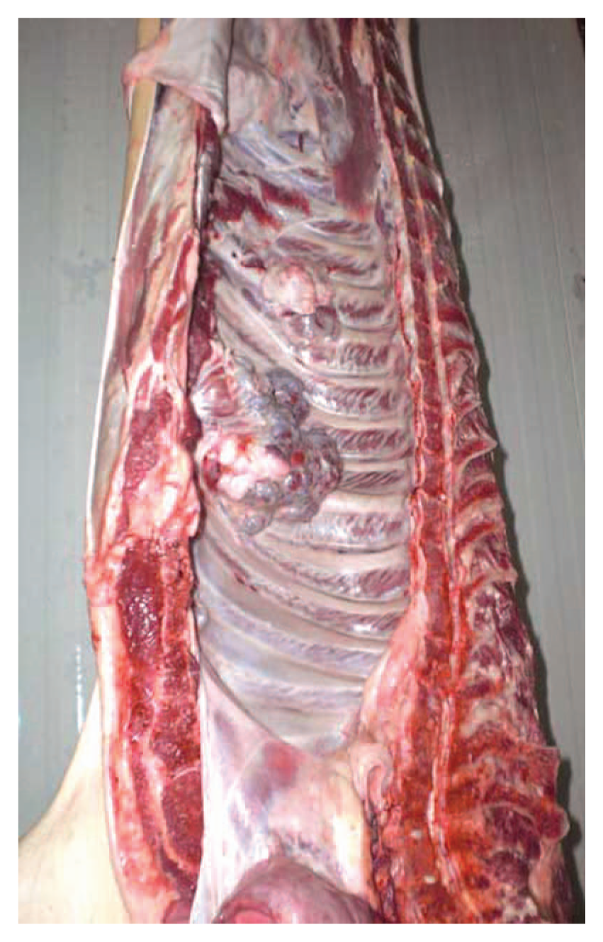
Two-year-old female domestic pig (Sus scrofa domesticus); carcass. Two prominent exophytic, multilobular masses arising from the left ribs are seen within the thoracic cavity.
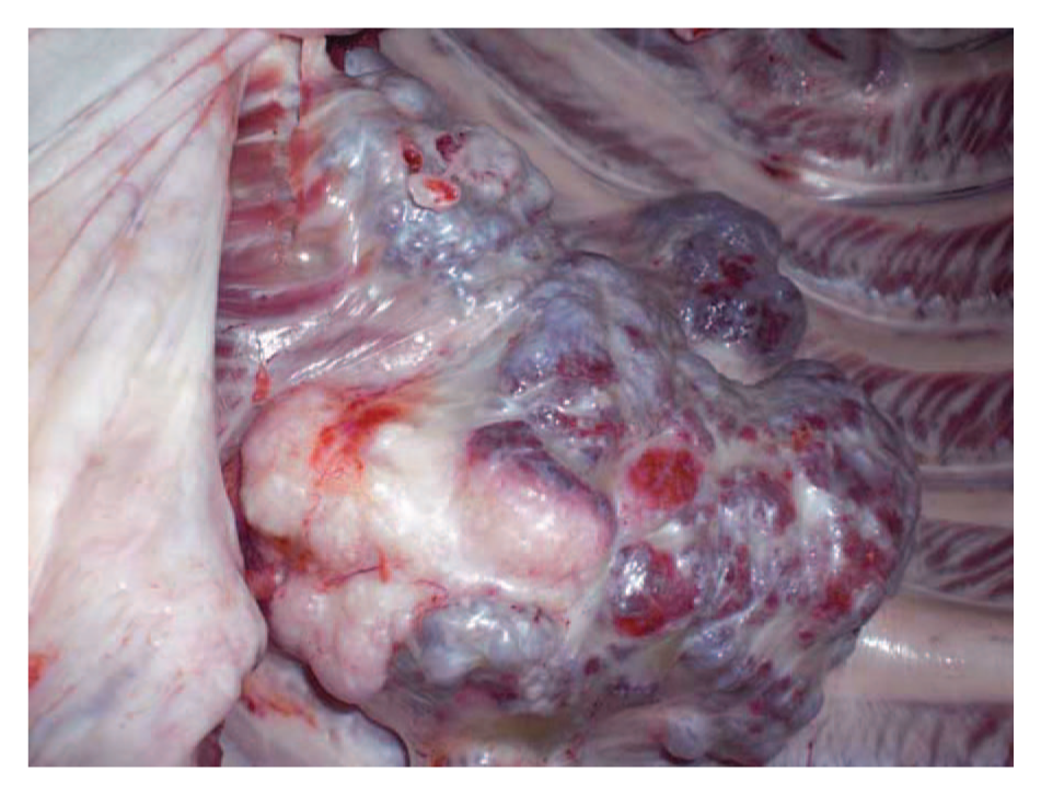
Two-year-old female domestic pig (Sus scrofa domesticus). A closer view of one of the masses showing smooth surface and white-bluish areas, reflecting the presence of the cartilaginous component.
One of the masses was collected, fixed in 10% buffered formalin, and submitted to the Slaughterhouse Support Network (Servei de Suport a Escorxadors, Universitat Autònoma de Barcelona, Barcelona, Spain) for diagnosis. Once in the laboratory, the sample was decalcified in 5% formic acid over 7 days and routinely processed for histology. Four-µm-thick tissue sections were processed for hematoxylin and eosin staining.
Histological examination revealed that the studied tissue was a multilobulated mass with multiple irregular islands of well-differentiated hyaline cartilage and bony trabeculae, the latter originating from endochondral mineralization. Discontinuously, hyaline cartilage covered the surface of the mass and had an orderly pattern of mineralization and transition into mature trabecular bone toward the central and deeper areas of the mass, mimicking the structure of a growth plate (Fig. 3). Above the cartilage, a variably thick perichondrial membrane was also present. Chondrocytes were enmeshed within an abundant hyalinized amphophilic extracellular cartilage matrix. Between the cartilage and bone trabeculae, there were large spaces containing loose tissue with a moderate number of fusiform cells resembling fibroblasts, adipocytes, and thin-walled blood vessels. In the deeper sections, bone marrow was present among the bony and cartilaginous trabeculae. Chondrocytes were well-differentiated and showed hypertrophy in the areas close to endochondral ossification. No mitoses were seen. Multifocally, some chondrocytes within irregular trabeculae had necrotic features. Bony trabeculae often showed active remodeling, with abundant osteoclasts and rows of plump osteoblasts lining their surface (Fig. 4). Based on gross and histological features, a diagnosis of osteochondromatosis was made.
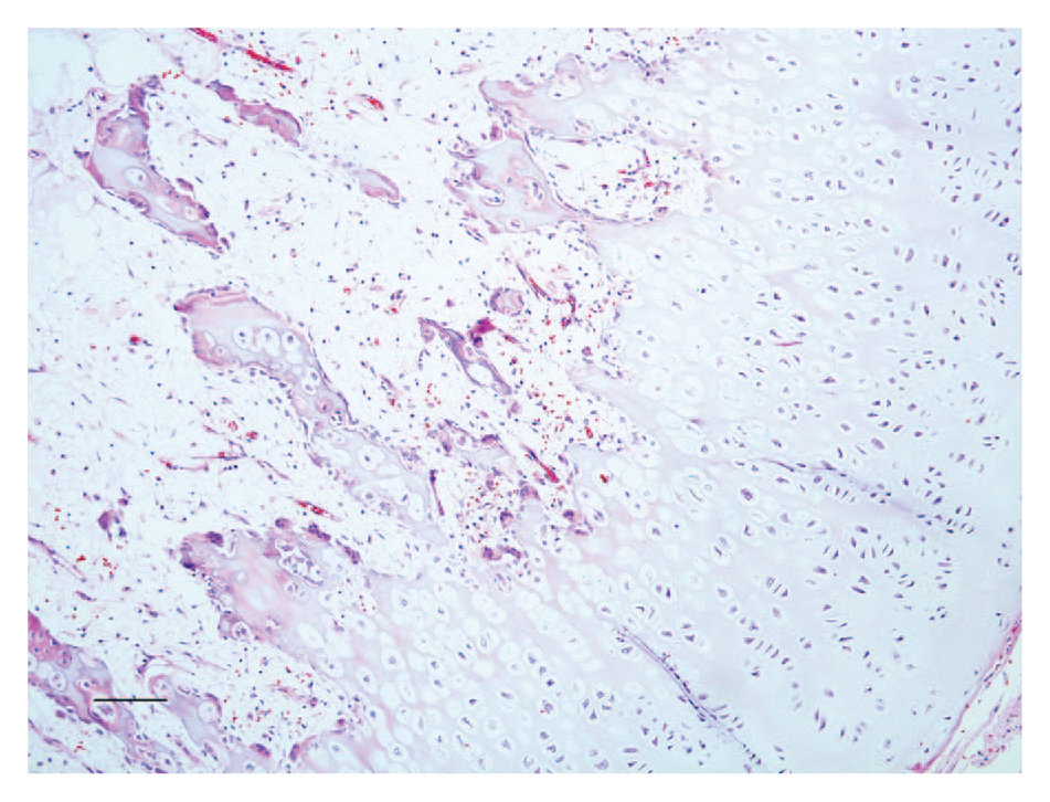
Two-year-old female domestic pig (Sus scrofa domesticus). Cartilage-capped proliferation showing an orderly pattern of endochondral mineralization toward the central areas of the mass. Hematoxylin and eosin. Bar = 100 µm.
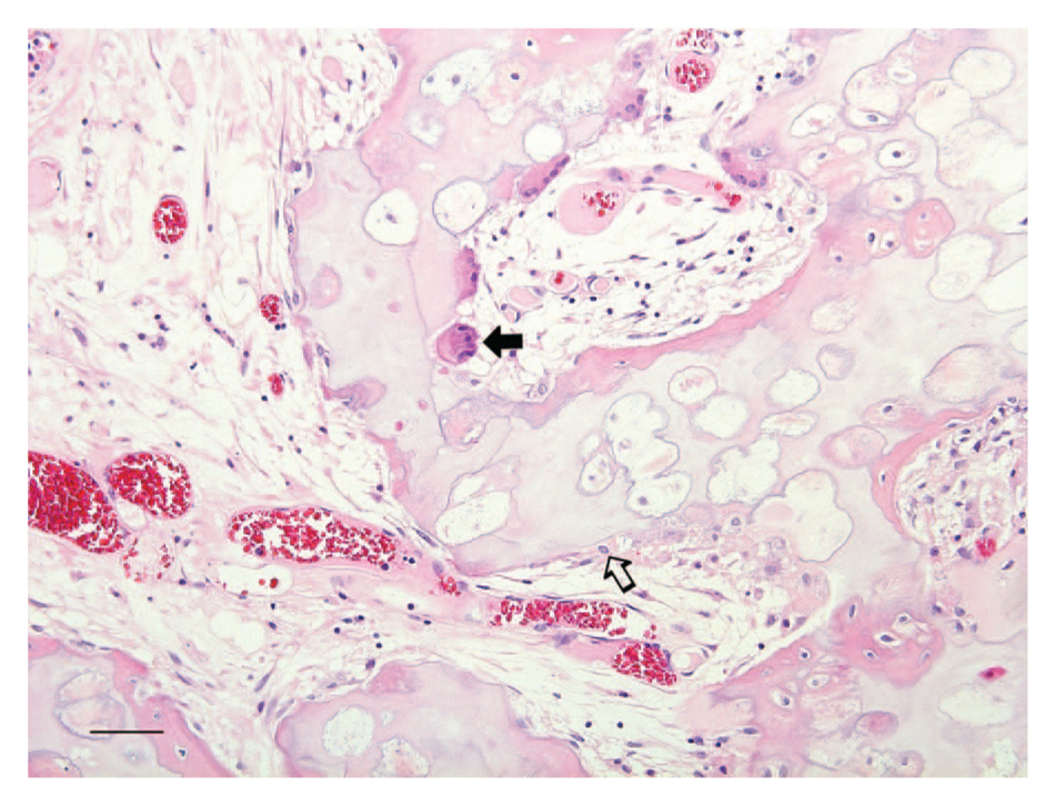
Two-year-old female domestic pig (Sus scrofa domesticus). Irregular cartilage trabeculae undergoing endochondral ossification. Abundant active osteoclasts (black arrow) and plump osteoblasts (open arrow) indicate active bone remodeling. Hematoxylin and eosin. Bar = 50 µm.
In human beings, dogs, and horses, osteochondromatosis typically occurs in young individuals, and is known to be inherited in an autosomal dominant pattern.9,12 In dogs and horses, the lesion most commonly arises from scapula, ribs, vertebrae, and pelvis. 12 In those species, it is not clear if this condition should be considered true neoplasia because its growth stops once bone growth ceases and the cartilage cap is replaced by bone. Similarly, in the present case, the masses were seen on the rib surface from a young adult animal (in pigs, growth plate closure occurs around 3.5 years of age). 13 In contrast, osteochondromatosis in cats has many differences and is not considered to be analogous to the condition observed in horses and dogs. Thus, osteochondromatosis in cats may occur in mature animals and can involve bones derived from intramembranous ossification, which has not been reported in horses and dogs. Moreover, in cats, the lesions are reported to enlarge progressively and without interruption, being therefore more consistent with true neoplasia.11,12 In addition, several authors have associated this lesion with infection by Feline leukemia virus, and it is not considered to be hereditary.3,8,12 Another differential feature of this condition in cats is the fact that the mass is usually not connected with the marrow cavity of the adjacent bone. In the present case, deep sections of the mass showed bone marrow among cartilaginous and bony trabeculae, suggesting a communication between the mass and the bone marrow of the rib. Finally, stromal cells are reported to have higher pleomorphism and atypical features in cats. 12 In the present case, in all the studied sections, stromal cells had a low degree of pleomorphism. Therefore, according to all these findings, the osteochondromatosis presented herein resembles the condition described in horses and dogs rather than the one reported in cats.
The scientific literature contains a single description, written in German, of synovial osteochondromatosis in swine. 15 However, synovial osteochondromatosis is considered to be a different condition than osteochondroma and osteochondromatosis because it arises from synovial membranes instead of bone surfaces, and is considered to be a metaplasia of synovial cells.9,12
In the present case, differential diagnoses included chondroma and chondrosarcoma. Chondroma is a rare benign neoplasm of cartilage that is referred to as enchondroma if it originates within the bone medullary cavity, or ecchondroma if arising from cartilage elsewhere in the skeleton. 12 Few cases of chondromas are reported in several veterinary species,2,3,5 and no descriptions were found in pigs at the time of this writing. Histologically, chondromas consist of irregular lobules of hyaline cartilage, which may also show foci of endochondral ossification and mineralization. 11 However, chondromas miss the growth plate-like organization of the cartilaginous matrix that characterizes osteochondromas and that was observed in the present case.2,9
Malignant transformation of osteochondroma to either chondrosarcoma or osteosarcoma has occasionally been described in older dogs 4 and human beings. 10 However, in the present case, no features of malignancy such as binucleated chondrocytes, tumor cells with plump nuclei and prominent nucleoli, or mitotic figures were observed.9,11 Nevertheless, it must be taken into account that, histologically, osteochondroma can be very difficult to differentiate from low-grade chondrosarcoma, which may show few indications of malignancy and may closely resemble benign tumors of cartilage.11,12 Therefore, in the present case, although malignant transformation to chondrosarcoma was not observed, its occurrence cannot be totally excluded. In conclusion, gross and microscopic findings in the present case support a final diagnosis of osteochondromatosis in swine.
Footnotes
Acknowledgements
The authors thank Paula Agunin and Gemma Garcia (ASPC slaughterhouse veterinary inspectors) for macroscopic pictures and case description. The authors thank also the Slaughterhouse Support Network (Servei de Suport a Escorxadors, SESC-CReSA), which is funded by Agència de Salut Pública de Catalunya (ASPC), Departament de Salut, Generalitat de Catalunya.
Declaration of conflicting interests
The author(s) declared no potential conflicts of interest with respect to the research, authorship, and/or publication of this article.
Funding
The author(s) received no financial support for the research, authorship, and/or publication of this article.