
Abstract
An 8-year-old female elk (Cervus elaphus canadensis) cow, presented for chronic severe weight loss and unthriftiness, was diagnosed with Babesia odocoilei infection based on blood smear evaluation, polymerase chain reaction (PCR), and DNA sequence analysis. Subsequently, velvet antler from a male that died acutely on the same farm was also PCR positive for Babesia spp. Both animals originated from a game ranch of Saskatchewan-bred and -raised animals with no known history of tick exposure, but with a history of numerous sudden deaths of unknown etiology. The presence of B. odocoilei in Canada might be a result of a recent introduction that could have deleterious effects on local wild ungulates or may represent discovery of a previously unrecognized endemic disease in local wildlife.
An 8-year-old elk (or wapiti; Cervus elaphus canadensis) cow was presented to a local veterinarian in July 2012 with a history of marked weight loss and lethargy. The elk resided on a 130-ha wild game ranch north of Prince Albert, Saskatchewan, Canada, in a herd of approximately 30 animals; none of the animals were vaccinated and none had been dewormed recently. Approximately 95% of the animals had been born on the property; the remainder was bred on neighboring elk farms. Ticks had not been noted on the animals, but a large infestation of horse flies (Tabanus spp.), with approximately 100–200 flies per animal, was observed. Within the previous 1–2 weeks, 9 bull elk were found dead in various locations around the property. Unlike the elk cow, the bulls had been in excellent body condition. No necropsies had been performed due to rapid decomposition of the tissues in the summer temperatures. Due to the risk of anthrax (Bacillus anthracis) as a cause of sudden death in Saskatchewan, nasal swabs from 3 dead bulls were submitted to Prairie Diagnostic Services Inc. (PDS), Saskatoon, Saskatchewan, for testing; all were negative.
Based on examination from a distance, the cow was assessed to be in very poor body condition, with a poor coat. The animal was euthanized by gunshot to the neck, and a necropsy was performed in the field. Minimal to no fat was noted in the omentum, pericardium, and perianal tissues, and the blood appeared watery. No other significant lesions were found. Tissues fixed in 10% neutral buffered formalin were submitted to PDS for histologic examination, and fresh liver was submitted for micronutrient analysis. Frozen tissues, including spleen, were held for potential future testing. Blood, collected into an ethylenediamine tetra-acetic acid (EDTA) tube from a vessel severed during necropsy, was submitted for complete blood cell count (CBC). Histologic examination of the tissues revealed numerous large Sarcocystis spp. cysts in the heart and skeletal muscle and mild periacinar vacuolation of hepatocytes. Other findings were mild and nonspecific; no specific diagnosis to explain the clinical signs could be elucidated. The liver was deficient in copper, at 2.54 ppm (normal: 20–120 ppm, deficient <10 ppm), and was considered marginal for manganese (0.95 ppm; normal: 2.0–6.0 ppm, deficient <0.9 ppm).
Results of CBC evaluation, determined using a hematology analyzer, a are reported in Table 1. There was marked macrocytic anemia with moderate regeneration and abundant basophilic stippling. Low to moderate numbers of ghost cells were noted, indicating intravascular hemolysis. A mild left shift and mild monocytosis were also present, consistent with established and ongoing inflammation. On smear evaluation, 1.5% of erythrocytes contained single or paired piriform and ring-shaped organisms, often at the periphery of the erythrocyte (accolé position), and rare Maltese cross arrangements (Fig. 1); morphology was consistent with that previously described for Babesia odocoilei, with the accolé position being typical of this Babesia spp. 8
Complete blood cell count results from an 8-year-old elk (Cervus elaphus canadensis) cow.*
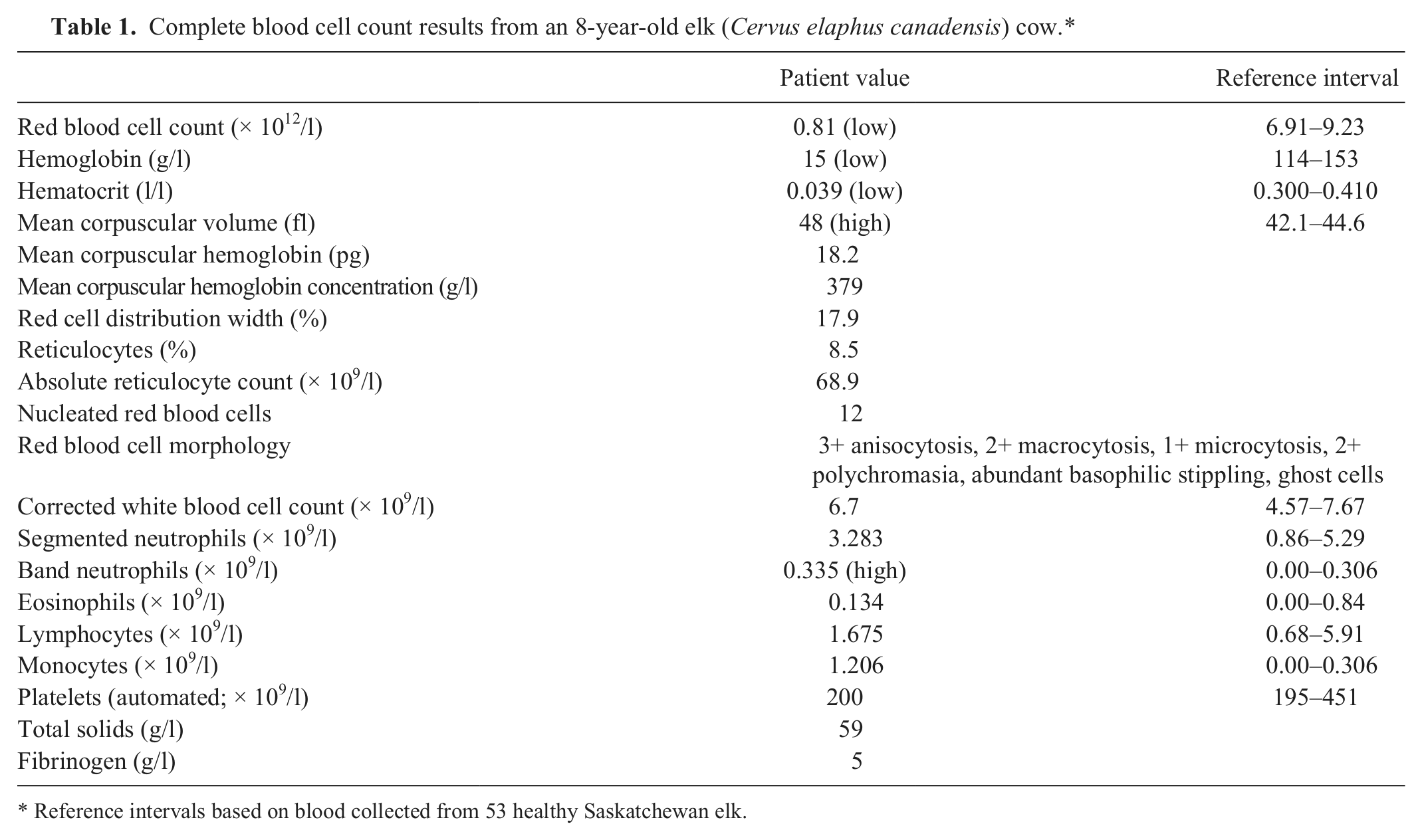
Reference intervals based on blood collected from 53 healthy Saskatchewan elk.
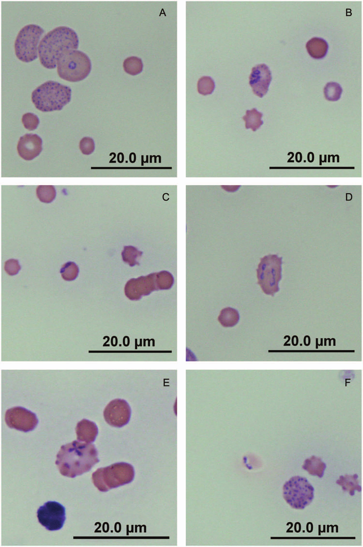
Direct smear of peripheral blood from an 8-year-old elk (Cervus elaphus canadensis) cow. Erythrocytes containing various forms of Babesia odocoilei organisms:
To confirm the identity of the agent, polymerase chain reaction (PCR) and sequencing were performed. DNA was extracted from fresh frozen spleen and EDTA-blood by digestion using a lysis buffer containing 100 mM NaCl, 500 mM Tris, and 10% sodium dodecyl sulfate. Proteinase K (50 mg) was added, the samples were incubated overnight at 56°C, protein was extracted with phenol–chloroform, and DNA was precipitated with ethanol. Pellets were resuspended in 30 µl of Tris–EDTA buffer. Polymerase chain reaction amplification using broad range primers targeting the eukaryotic 18S ribosomal RNA (rRNA) gene (primers Bab A and Bab B; Table 2) b was performed in a 50-µl PCR reaction. 11 The PCR assay was optimized using a final concentration of 3 mM MgCl2 and 1.25 units of Taq polymerase at an annealing temperature of 60°C. A band of the expected size (1,723 bp) was produced in the patient samples (Fig. 2, lanes 3 and 4). 11
Polymerase chain reaction and sequencing primers used in the current study.

Agarose gel electrophoresis of products from polymerase chain reaction (PCR) targeting the 18S ribosomal RNA gene. Lane 1: no template control; lane 2: 100-bp ladder; lane 3: ethylenediamine tetra-acetic acid (EDTA)-blood from an 8-year-old elk (Cervus elaphus canadensis) cow with a 1,723-bp amplification product; lane 4: fresh frozen spleen from the same patient as in lane 3 with a 1,723-bp amplification product; lane 5: velvet antler from a bull of unknown age with a 1,723-bp amplification product; lane 6: EDTA-blood from the second euthanized elk cow with a negative PCR; lane 7: fresh spleen from a euthanized bull with a negative PCR.
Amplified DNA from the Babesia-positive EDTA-blood sample was purified using a commercial kit c and submitted for sequencing at Macrogen Inc. (Seoul, Korea) using the Bab A, Bab528FIN, and Bab B primers b (Table 2) to ensure complete coverage of the 18S rRNA. 11 Bab528FIN was originally designed to target Babesia spp. 11 Raw sequence data were assembled and edited, 22 resulting in 1,607 bp of high-quality sequence. The sequence was 100% identical to previously published sequences for B. odocoilei from elk in Wisconsin (GenBank accession no. AY294206.1) and Texas (AY339760.1, U16369.2); reindeer (Rangifer tarandus tarandus) from Wisconsin (AY237638.1), Pennsylvania (AY661506.1), and New York (AY661505.1, AY661504.1); white-tailed deer (Odocoileus virginianus) from Texas (AY046577.1); caribou (Rangifer tarandus caribou) from Wisconsin (AY339761.1); and muskoxen (Ovibos moschatus) from Minnesota (AY661508.1) over a 1,607-bp region of high-quality sequence. The sequence from the patient sample also shared 99% identity with the California reindeer Babesia isolates RD1 and RD61 (AF158711.1 and AF411337.1, respectively) over a 1,597-bp and 1,591-bp region, respectively, of high-quality sequence. The nucleotide sequence for the partial 18S rRNA gene of B. odocoilei from the current case has been deposited in GenBank (accession no. KC460321).
Shortly after the elk cow had been diagnosed, a bull of unknown age was found dead on the same property. No necropsy had been performed, but the producer had frozen a portion of velvet antler from the animal. Tissue was scraped from the blood-filled center of the antler and PCR was performed, resulting in a product of expected size (1,723 bp) for Babesia spp. (Fig. 2, lane 5). An additional cow and bull elk were also euthanized, by gunshot to the neck, for lameness and poor body condition. Oral ulceration in the former and lung abscessation in the latter, with identification of Fusobacterium spp., suggested necrobacillosis as a likely cause of chronic poor-doing in these elk. Polymerase chain reaction on EDTA-blood from the cow (Fig. 2, lane 6) was negative for Babesia spp., as were splenic tissue from both the cow and the bull (Fig. 2, lane 7). Five months after the initial diagnosis of babesiosis, there have been no further deaths in the herd, and the remaining elk are in good condition.
Babesia odocoilei (phylum Apicomplexa, order Piroplasmida, family Babesiidae) is a tick-borne intracellular protozoal parasite of white-tailed deer, 6 but also causes naturally acquired babesiosis in many other cervid species and occasional bovid species, including North American elk, 7 caribou, 11 reindeer, 9 desert bighorn sheep (Ovis canadensis nelsoni), muskoxen, 20 markhor goats (Capra falconeri), yak (Bos grunniens), and muntjac (Muntiacus reevesi). 4 Naturally acquired infections of B. odocoilei was first reported in Texas 6 and have also been reported in numerous locations in the United States, primarily the north-central and the northeastern regions.1,4,7,9,19,20 To the authors’ knowledge, there have been no prior reports in Canada.
Little is known about competent vectors for B. odocoilei, but the black-legged tick (Ixodes scapularis) has been identified to both carry and transmit the organism. 24 Ixodes scapularis is found endemically in the eastern and southern United States, with the largest populations found in the northeast.5,21 In Canada, I. scapularis was thought to be limited to the northern shores of Lake Ontario and Lake Erie, as well as the southern portion of Nova Scotia3,16; however, new endemic populations have been discovered in southern Manitoba, Quebec, and New Brunswick, as well as identification of new sites within Ontario and Nova Scotia. 17 Although there are no known established populations of Ixodes spp. ticks in Saskatchewan, adult I. scapularis have been collected from northern pocket gophers (Thomomys talpoides) in the province. 2 Given the limited geographic range of terrestrial hosts, initial identification of a non-native tick species is typically the result of “adventitious” ticks originating from migratory birds, a recognized source of I. scapularis larvae and nymphs to Canada.14,17 Various statistical models, created to help predict migration and expansion of tick habitat due to climate change,13,16,18 have demonstrated that I. scapularis is likely to move north and west, with some models predicting a potential for tick populations to become established as far north as the Great Slave Lake region of the Northwest Territories by the 2080s while the current endemic populations may multiply 2- to 4-fold. 18 Increasing temperatures could also lead to prolonged seasonal tick activity and decreased interstadial development rates, increasing risk of tick exposure to both human and animal populations. Adaptations, such as expression of antifreeze glycoproteins thought to aid survival through the winter, may also contribute to expansion of tick populations into formerly uninhabitable environments. 15
The source of infection for the 2 elk in the current report is unknown. The lack of endemic Ixodes spp. tick populations suggests the possibility of an alternate vector. One such possibility is Dermacentor spp., which are commonly found within the province and have been implicated as a vector of equine babesiosis. 23 In 1 report of babesiosis in elk, Dermacentor albipictus were the only ticks found on the animals. 10
Host transmission of B. odocoilei occurs through blood feeding by an infected tick, whereby sporozoite-containing saliva is introduced into the host’s blood stream. 25 Following engulfment by an erythrocyte, the sporozoite undergoes asexual reproduction and begins the cyclical development of trophozoite and merozoite piroplasms. Although a few trophozoites will enlarge and become potential gametocytes, most cause non–immune-mediated hemolysis as they are released and invade other uninfected erythrocytes.4,12 In clinically affected elk, the proportion of infected erythrocytes varies, but has been reported as high as 15–20%. 7
Intra- and extravascular immune-mediated hemolysis, initiated by the host’s immune response, is a major contributor to anemia in the host. 12 In the current case, the elk cow had severe anemia, at a level that seemed inconsistent with the clinical description of the animal. Hematocrit has been reported to range from 28.8% to 33.7% in clinically affected elk,1,7 and as low as 11% in a clinically affected reindeer, 4 values that are significantly higher than the 3.9% hematocrit in the elk cow. Although blood from this animal was not collected by venipuncture, there was no evidence of sample contamination by lymph or other body fluids during collection, and the EDTA tube was filled with an appropriate volume of blood, making dilution by the anticoagulant unlikely. Acute anemia of this degree would be inconsistent with life in many species; therefore, while the anemia was likely severe in the patient of the current report, it is possible that the hematocrit of the sample evaluated may not have been an accurate reflection of the in vivo red blood cell count. Reticulocytosis has not been described in previous reports of babesiosis in elk, even when the hematocrit was moderately decreased.1,7 As a result, the ability of elk to mount reticulocytosis in response to anemia has been questioned. 7 In the current case, reticulocytosis was present, accompanied by abundant basophilic stippling, typical of a ruminant regenerative response.
Clinical babesiosis does not always occur upon first exposure to the parasite; rather, the animal may initially develop a subclinical infection that progresses to clinical disease in the face of concurrent stress.6,11,19 Stressed animals, as well as animals in a naïve population, tend to have a more severe course of disease with a significantly increased risk for fatal babesiosis. 4 Some immunocompetent white-tailed deer have no clinical signs and experience only a mild transient decrease in hematocrit, while others have a chronic illness characterized by pyrexia, emaciation, anemia, and a low level of parasitemia (0.5–1.5% of erythrocytes).9,19 Infection rates as high as 100% have been reported in some elk herds,7,20 with only a small percentage of the animals demonstrating clinical babesiosis. 9 Given the number of acute deaths in the herd of the current report, with most of the animals being in good body condition, an outbreak in a naïve population seems likely. However, the initially diagnosed cow had signs consistent with the chronic form of disease, including poor body condition, anemia, and a low circulating parasitemia of 1.5%. It is possible that there was chronic subclinical babesiosis in the herd with a stressful event inciting acute clinical, and fatal, disease. Horsefly numbers were unusually high this season and were considered a potential source of stress. If the latter scenario is correct, babesiosis may have been present within Saskatchewan for longer than initially suspected. As the herd was bred and raised locally, introduction of infected animals from endemic areas is not explanatory. Therefore, the potential for babesiosis to be a previously unrecognized endemic disease in Canadian cervids should be considered.
To date, North American elk have only been reported to be infected with B. odocoilei; however, PCR is required to confirm the species involved, as well as to differentiate from other morphologically similar organisms such as Theileria spp. Polymerase chain reaction analysis has typically been performed on peripheral blood1,4,7,9,11,20; in the present case, both fresh frozen spleen and velvet antler were also successfully used to amplify Babesia 18S rRNA. The option to use these tissues is advantageous in situations where peripheral blood has not been collected and only banked tissues are available for diagnostic testing. Velvet antler could prove particularly useful, as it is an easy sample for producers to collect. In cases of hemolytic anemia, there are no consistent changes on CBC or biochemistry panel that are suggestive of B. odocoilei infection unless the organism is observed on peripheral blood smears. Hyperbilirubinemia and bilirubinuria may be noted secondary to extravascular hemolysis, and acute renal failure secondary to hemoglobinuria may also occur. 4 Gross and histologic findings are not specific for babesiosis, but splenomegaly, splenic hemosiderosis, hepatic centrilobular degeneration, and hemorrhage within the heart and adrenal glands have been observed in affected elk. 7 The affected elk cow of the present report had hepatic changes consistent with those listed previously. This likely resulted from decreased hepatic perfusion and oxygenation secondary to hemolytic anemia, as has been suggested in previous reports. 7
In summary, when clinical signs are compatible, babesiosis should be included as a potential differential diagnosis in cervids in Saskatchewan and other nonendemic regions of Canada. Where babesiosis is suspected, both blood smear evaluation for parasitemia as well as PCR on EDTA blood, fresh frozen spleen, or velvet antler are recommended for diagnosis. It is uncertain as to when or how B. odocoilei was introduced to Canada and to the herd of the current report, specifically. Introduction of B. odocoilei could have deleterious consequences for a naïve wild ungulate population. However, existence of a previously unidentified endemic B. odocoilei infection in local wildlife cannot be excluded, given that blood is rarely collected for analysis from wild ungulates and the lack of significant findings on necropsy. Research is needed to assess the disease status of Saskatchewan wild ungulates to determine which scenario is underway.
Footnotes
Acknowledgements
The authors wish to thank Bonnie Chaban and Champika Fernando for their technical expertise and input during optimization of the PCR assay, Dr. Janet Hill for her assistance and expertise with molecular diagnostics and DNA sequence analysis, and Dr. Peter Surkan for his assistance with the herd investigation and sample collection.
a.
Cell Dyn 3500R, Abbott Diagnostics, Abbott Park, IL.
b.
Sigma-Aldrich Canada Ltd., Oakville, Ontario, Canada.
c.
EZ-10 Spin Column PCR Purification kit, Bio Basic Inc., Markham, Ontario, Canada.
Declaration of conflicting interests
The author(s) declared no potential conflicts of interest with respect to the research, authorship, and/or publication of this article.
Funding
The author(s) disclosed receipt of the following financial support for the research, authorship, and/or publication of this article: Funding was provided by the Saskatchewan Ministry of Agriculture, Disease Investigation Unit.