
Abstract
Surveillance for Mycobacterium avium subsp. paratuberculosis (Map) infection in small ruminants of Grenada was undertaken using a commercial enzyme-linked immunosorbent assay (ELISA). Among the 479 sheep tested, 11 (2.3%) were ELISA positive while only 1 out of 260 goats (0.3%) was ELISA positive. Five of the 12 ELISA-positive animals were also positive in a commercial agar gel immunodiffusion (AGID) assay, and 4 of these showed acid-fast rods consistent with Map in fecal smears. Two sheep that were test-positive by ELISA, AGID, and fecal smears were euthanized and necropsied. Both had gross and histological lesions of paratuberculosis affecting the ileocecal area of small intestines and adjacent lymph nodes. These tissues were successfully cultured in 2 of 3 variants of Middlebrook 7H10 medium. The identity of acid-fast organisms isolated from the tissues was confirmed as Map by multiplex conventional polymerase chain reaction. Using IS1311 amplification and Hinf I restriction digest analysis, isolates were identified as cattle (C) strains of Map. The current study describes Map infection in Grenada and confirms the presence of C type in sheep on the island of Carriacou. The low seroprevalence in clinically normal animals on the islands of Grenada and Carriacou suggests that control measures implemented in the near future may have a good chance of preventing spread of the infection.
Small ruminants are important production animals in the state of Grenada due to their manageable size and feed requirements plus their ability to utilize sloped terrain and limited pasture areas. The rearing of sheep and goats for both meat and milk is increasing in Grenada (Annual Report of Ministry of Agriculture, Forestry and Fisheries, Grenada, West Indies, 2009). Pasture land suitable for grazing livestock in Grenada totals approximately 11,655 hectares of the Caribbean tri-island nation.
Paratuberculosis is an infectious and sometimes fatal disease affecting all ruminant species, 16 and spread of Mycobacterium avium subsp. paratuberculosis (Map) could significantly hamper the livestock industry of Grenada. The pathogen is known to have multiple types, which preferentially infect specific host species. Type I (S strain) is found most often in sheep and occasionally other hosts, and type II (C strain) is found in cattle as well as many other ruminant species, including sheep, goats, and deer. Studies have investigated the prevalence of Map infection in domestic agriculture and wildlife species in North and South America and Europe,1,7,12 but at present no information is available on herd prevalence of paratuberculosis in sheep and goats in Grenada.
Map culture is a widely used diagnostic test 11 that can be applied to various species and sample types (feces, tissue, water, soil). However, as culture is slow and labor intensive, herd surveillance with enzyme-linked immunosorbent assay (ELISA), which targets the serum antibody produced in response to Map infection, 9 is often used to reduce time and expense in herd prevalence studies. A national serological survey was thus undertaken to determine the prevalence of Map infection in small ruminants in Grenada.
Grenada has an estimated small ruminant population of 3,000 sheep and 2,500 goats. Serum samples from 739 animals (85 rams, 386 ewes, 207 does, and 61 bucks; 13% of the small ruminant population) from the islands of Grenada and Carriacou were obtained between December 2009 and January 2011, from clinically normal animals of at least 6 months of age. Sixty-five production units (sheep and/or goats owned by 1 person or family) were selected from the 6 parishes of Grenada and 9 production units were selected from Carriacou. Of a total of 74 production units, 38 were sheep only, 20 were goat only, and 16 were mixed sheep and goat units. Production units ranged in size from 1 to 150 adult animals. For production units with more than 10 animals, 50% of the animals were sampled. All animals were sampled if the production units consisted of fewer than 10 animals.
A commercially available absorbed ELISA for Map antibodies a reported by the manufacturer to be effective in sheep and goats and evaluated previously 6 was used and interpreted according to manufacturer’s instructions based on a sample-to-positive ratio with a positive cutoff of 70. The ELISA-positive samples were also tested by agar gel immunodiffusion (AGID) using a commercial test kit b as per the manufacturer’s instructions. Fecal smears from ELISA-positive animals were stained and examined microscopically for acid-fast organisms consistent with Map.
Two clinically normal ewes, from different production units on Carriacou, which were each ELISA, AGID, and fecal smear positive, were euthanized and necropsied. From each, the ileocecal area and adjacent ileocecal lymph nodes were collected and divided; 1 portion was fixed in 10% buffered formalin for routine histology and acid-fast staining of sections while the other portion was retained for culture at 80C, each tissue in a separate sterile container. In addition, portions of ileocecal lymph nodes were also retained at 80C for direct polymerase chain reaction (PCR). The culture media employed were modified Middlebrook 7H10 agar slants c (medium A), a commercial radiometric liquid medium d (medium B), and a commercial nonradiometric liquid medium e (medium C) following recommendations and formulae as previously described. 15 For media A and B, the 4 tissue samples from each animal were prepared as follows: approximately 1-cm square piece of tissue was minced with sterile scissors, placed in a sterile bag f with phosphate buffered saline and homogenized in a stomacher g for 2 min on the highest setting. The bag’s contents were then transferred to a 50-ml conical tube containing 30 ml of sterile water and mixed on a rotary shaker at 1 × g for 30 min. Samples were then held stationary for 30 min at room temperature for particulate matter to settle. The top 20 ml was then transferred to another 50-ml conical tube and centrifuged at 2,000 × g for 20 min in a benchtop centrifuge. h The supernatant was removed, and the pellet was resuspended by vortexing in 0.75% hexadecylpyridinium chloride (HPC). i This HPC–tissue mixture was held at room temperature for 3 hr after which it was centrifuged at 2,700 × g for 20 min. The pellet was resuspended by vortexing in 1 ml of an antibiotic cocktail comprised of 10 mg of vancomycin, 5 mg of amphotericin B, and 10 mg of nalidixic acid. After an 18-hr incubation at 37°C, the mixture was used to inoculate media A (100 µl/slant) and B (500 µl/bottle), and then incubated at 35–37°C for up to 16 weeks, with the latter read weekly on a commercial instrument j as previously described. 3 Residual decontaminated tissue preparations for media A and B were retained at 80C. For medium C, tissues were prepared according to the manufacturer’s instructions, e and medium C was inoculated with 100 µl of tissue preparation, and incubated with hourly monitoring over 49 days. Direct PCR on ileocecal lymph nodes was undertaken using 0.5 g of tissue that was homogenized as per the instructions given by the manufacturer of a commercial kit, k and subjected to DNA extraction using the same commercial kit. k The direct PCR was a commercial real-time PCR l targeting the hspX gene 4 and performed as per manufacturer’s instructions.
A 5-target multiplex PCR was used to test DNA extracted from culture-positive samples as previously described. 14 The 5 possible targets were a 16S ribosomal RNA gene target common to all mycobacteria, a chromosomal DNA target (DT1) found in Mycobacterium intracellulare, Mycobacterium avium subsp. sylvaticum, and 2 serotypes of Mycobacterium avium subsp. avium, and 3 mycobacterial insertion sequences (IS1311, IS901, and IS900) that allow identification of Map and discrimination among other members of the M. avium complex. To characterize a specific Map strain, IS1311 amplicons were subjected to HinfI m restriction digestion, and the products of the amplification were visualized and interpreted as previously described 14 after electrophoresis in ethidium bromide–stained gels (Fig. 1).
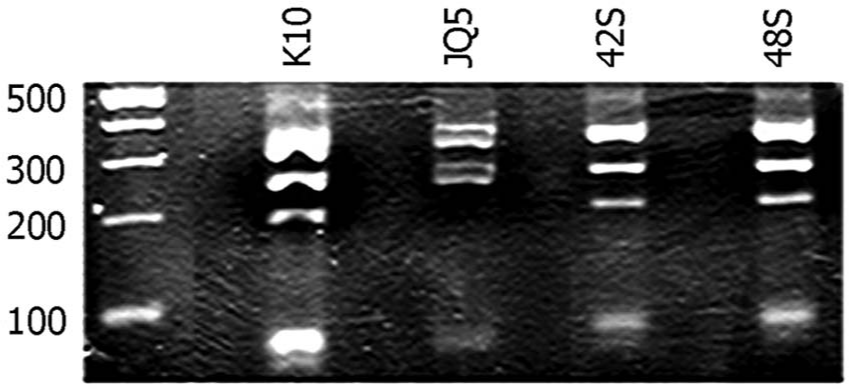
Genotyping of sheep isolates. Ethidium bromide–stained 3% agarose gel of IS1311 polymerase chain reaction–restriction enzyme analysis (PCR-REA) with HinfI (NEB, USA). In each lane, digested products are shown. The PCR-REA analysis of IS1311 shows reference Mycobacterium avium subsp. paratuberculosis (Map) strains from cattle (K10) and sheep (JQ5) in addition to newly identified isolates from sheep in the current study (42S and 48S). A 100-bp molecular size marker is included.
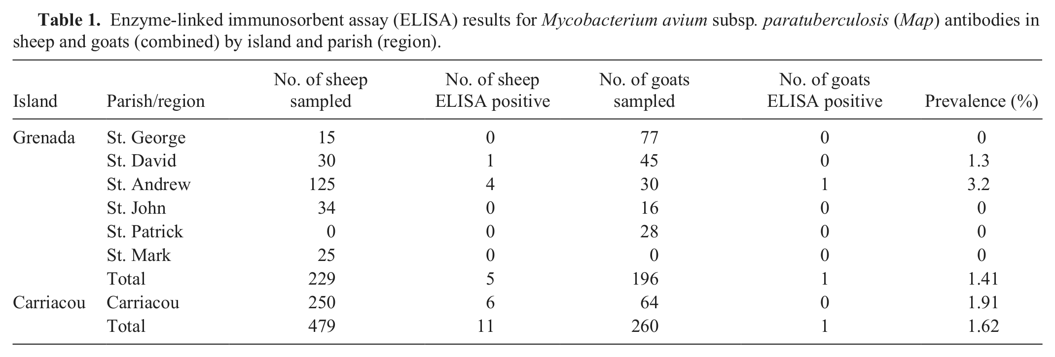
The ELISA-positive animals came from 2 parishes of Grenada (St. Andrew and St. David) and 1 from Carriacou parish (Table 1). Of the 479 sheep tested, 11 were ELISA positive (5 males and 6 females). Only 1 out of 260 goats was ELISA positive (a doe). Six of the 12 ELISA-positive animals also tested positive by AGID. Four of these 6 showed scant numbers of acid-fast organisms on fecal smears; the remaining 8 ELISA positives were smear-negative.
Enzyme-linked immunosorbent assay (ELISA) results for Mycobacterium avium subsp. paratuberculosis (Map) antibodies in sheep and goats (combined) by island and parish (region).
At necropsy of 2 ELISA-, AGID-, and fecal smear–positive sheep from Carriacou, gross and microscopic findings were consistent with paratuberculosis, with portions of the small intestines from both animals revealing mild thickening, corrugation, and edema, especially at the ileocecal junction. Mesenteric ileocecal lymph nodes were enlarged, edematous, and hemorrhagic with a few microgranulomatous lesions. Histological studies of these tissues showed significant lymphocytic infiltration plus multiple diffuse chronic granulomatous inflammatory foci and noncapsulated granulomas with epithelioid macrophages and a few Langhans giant cells. Ileocecal sections and adjacent mesenteric lymph nodes revealed acid-fast organisms located within a few macrophages.
Direct PCR on ileocecal lymph nodes from both sheep were negative. On culture, modified 7H10 agar slants produced colonies resembling Map from all 4 tissues (2 tissues from each case) within 8 weeks of incubation. All 4 cultures in medium B signaled positive after 5 weeks of incubation. None of the cultures in medium C signaled positive by the end of the standard incubation period (49 days). Aliquots from medium B cultures were acid-fast, and 16S rRNA, DT1, and IS900 positive by multiplex PCR. At a later date, when the ability to genotype the isolates using methods outlined previously 5 became available, the frozen tissue preparation was thawed and incubated in medium B as described above until signaling positive (within a month). These were subcultured onto modified 7H10 (medium A) plates to produce sufficient mycobacterial growth for specific Map strain identification. Pure colonies appeared within 3 months. Restriction enzyme analysis of Map from these colonies demonstrated that both sheep had been infected by a C-type strain of Map.
The surveillance study indicated that Map infection is present in Grenada’s small ruminant industry. The current prevalence appears to be low, with a 1.6% animal-level Map seroprevalence in small ruminants from Grenada and Carriacou. The infection also appeared localized in 2 out of 6 Grenada parishes. The true prevalence may be higher because the diagnostic sensitivity of ELISA is low in clinically normal, relatively young ruminants as were tested in the current study (30% of animals were 1.0–1.6 years old). Although only type C strain was isolated from sheep in the present study, in Europe, S strains have been more common in sheep. 13 The prevalence rates reported in small ruminants in some other countries is daunting: 73.7% in sheep in Italy, 1 46.7% in sheep in Portugal, 2 52% in sheep and 50% in goats in Cyprus, 8 48–57% in goats and 42.4% in sheep in Brazil. 10 In infected sheep flocks with S strains, animals can shed very high numbers of organisms, leading to a very high degree of environmental contamination. The low seroprevalence of Map in clinically normal animals on the islands of Grenada and Carriacou suggest that prompt steps taken to prevent spread of infection are likely to be successful. The current study from the Caribbean region may provide a baseline for annual screening of this disease using ELISA.
Footnotes
Acknowledgements
Diagnostic assistance from the staff of the Johne’s Testing Center, School of Veterinary Medicine, University of Wisconsin–Madison is greatly appreciated. Authors are grateful to Drs. Alfred Chikweto, M. I. Bahiyat, R. S. Pawaiya, Bowen Louison, and Derek Thomas for their valuable help rendered during the study.
a.
POURQUIER ELISA Paratuberculosis Antibody Screening Kit, Institut Pourquier, Montpellier, France.
b.
Rapid Johne’s Test, ImmuCell Corp.,Portland, ME.
c.
Middlebrook 7H10 agar slants, BD Diagnostic systems, Franklin Lakes, NJ.
d.
BACTEC 12B medium, BD Diagnostic Systems, Sparks, MD.
e.
BACTEC MGIT ParaTB medium, BD Diagnostic Systems, Sparks, MD.
f.
Whirl-pak, Nasco, Ft. Atkinson, WI.
g.
Seward Stomacher 80 Biomaster, Seward Ltd., Worthing, UK.
h.
Sorvall ST-H750 rotor centrifuge, Thermo Fisher Scientific, Barrington, IL.
i.
Acros Organics/Fisher Scientific, Pittsburgh, PA.
j.
BACTEC 460 instrument, BD Diagnostic Systems, Sparks, MD.
k.
DNeasy Blood and Tissue kit and Handbook, Qiagen Inc., Valencia, CA.
l.
Tetracore real-time PCR kit, Tetracore Inc., Rockville, MD.
m.
New England Biolabs Inc., Ipswich, MA.
Declaration of conflicting interests
The author(s) declared no potential conflicts of interest with respect to the research, authorship, and/or publication of this article.
Funding
The author(s) disclosed receipt of the following financial support for the research, authorship, and/or publication of this article: This research was funded through the Small Research Grant Initiative of St. George’s University, Grenada.