
Abstract
From 2009 to 2011, 163 sheep and 96 goat abortion submissions were received at the Animal Health Laboratory, University of Guelph, Ontario, Canada, for gross and histologic examination, as well as real-time polymerase chain reaction (PCR) testing for Chlamydophila abortus and/or Coxiella burnetii. Additional testing included immunohistochemistry for Toxoplasma gondii and Chlamydophila spp., routine bacterial culture and selective culture for Campylobacter spp., examination of modified acid-fast–stained placenta smears, enzyme-linked immunosorbent assay testing for Chlamydophila spp., and virus isolation. The final diagnosis made for each case by individual pathologists, based on gross and histologic lesions, as well as ancillary testing, was used as a standard to determine the significance of C. abortus and C. burnetii infection. Coxiella burnetii was identified by real-time PCR in 113 of 163 (69.0%) and 72 of 96 (75%) sheep and goat abortion submissions, respectively, but was considered to be significant in causing abortion in only 11 of 113 (10%) sheep and 15 out of 72 (21%) goat submissions that tested positive. Chlamydophila abortus was identified by real-time PCR in 42 of 162 (26%) and 54 of 92 (59%) sheep and goat submissions, respectively, but was considered the cause of the abortion in 16 of 42 (38%) sheep and 34 of 54 (63%) goat submissions that tested positive. Optimal sensitivity and specificity cut points for the real-time PCR copy number for C. abortus and C. burnetii were determined using the final pathology diagnosis as the reference test.
Abortion in sheep and goats has become increasingly important as the potential zoonotic significance of commonly involved pathogens, in particular Coxiella burnetii and Chlamydophila abortus, becomes better understood. Coxiella burnetii is well known as the cause of Q fever in human beings. An outbreak of Q fever in human beings occurred from 2007 to 2009 in the Netherlands, during which C. burnetii abortions were seen on 30 small ruminant farms over a 4-year period. The proximity of these farms was felt to be the main cause of 3,523 reported human cases of Q fever. 30 Chlamydophila abortus is associated with abortion and pelvic inflammatory disease in women. 35 Toxoplasma gondii, Listeria monocytogenes, Campylobacter fetus ssp. fetus, and Campylobacter jejuni are also well-recognized causes of abortion in small ruminants, 21 as well as abortion, fetal infection, and enteritis in humans.
The presence of DNA from both C. burnetii and C. abortus within the genital tracts of healthy (nonaborting) small ruminants has been documented.4,19,23 In the fall of 2008, implementation of polymerase chain reaction (PCR) testing for C. abortus and C. burnetii on routine diagnostic small ruminant abortion cases submitted to the Animal Health Laboratory (AHL) at the University of Guelph, Ontario, Canada, resulted in increased identification of both agents, although more traditional test methods such as histology, culture, and modified acid-fast smears (MAF) either did not confirm the significance of these agents, or concurrently identified other significant pathogens such as T. gondii, Campylobacter spp., or L. monocytogenes. In an attempt to clarify the significance of various test results in the diagnosis of small ruminant abortions, a prospective study was instituted using traditional testing methods as well as quantitative real-time PCR in an attempt to determine cut points for significance of real-time PCR results for both C. abortus and C. burnetii in small ruminant abortions. In addition, the project served as a survey of the causes of small ruminant abortions in Ontario. The final diagnosis in each submission (the cause of the abortion) was assigned to a submission based on individual pathologists’ review of the clinical history, gross examination, histologic examination, and review of ancillary microbiology tests and immunohistochemistry (IHC).
Materials and methods
Submission of samples
Between April 2009 and June 2011, written communications with sheep and goat producer groups and veterinarians were used to encourage submissions of abortion and stillbirth samples to the AHL for no-charge testing. To qualify for subsidized testing, submissions were required to include at least a piece of chorioallantois with a cotyledon. One or 2 fetuses were often also submitted. If fetuses without placenta were submitted from a herd-mate abortion, such fetuses were treated as a separate submission but included in the study. After gross examination, tissues were collected and sent for additional testing as outlined below.
Gross necropsy
Submitted abortions were assigned to individual pathologists based on a duty roster, and the pathologist acted as the case coordinator for each submission. The placenta was examined and abnormalities noted. If a fetus was present, then the crown-rump length, decomposition state, and sex of the fetus were recorded as well as abnormalities. Tissues were collected for histology and the various microbiology tests listed below. In addition to carrying out gross and histologic examinations, pathologists interpreted results of microbiologic tests and provided a summary diagnosis for each case based on these interpretations.
Histology and immunohistochemistry
Tissues collected included placenta, and when available, eyelid, brain, thyroid, thymus, lung, heart, liver, kidney, spleen, adrenal gland, small intestine, and skeletal muscle. Following fixation in 10% buffered formalin for at least 48 hr, tissues were routinely processed and paraffin-embedded. The paraffin blocks were cut at 4 µm and then stained with hematoxylin and eosin for histologic examination. Automated IHC for C. abortus and T. gondii was carried out on placental sections using mouse anti-Chlamydophila spp. monoclonal antibody a (1:75 dilution) and rabbit anti–T. gondii polyclonal antibody b (1:800 dilution) with a goat anti-rabbit alkaline phosphatase–linked polymer detection system. c Known C. abortus– and T. gondii–positive tissues were used as controls for each IHC run, and a negative reagent control was prepared for each test slide by substituting antibody diluent or normal rabbit serum for anti-Chlamydophila spp. or anti–T. gondii primary antibodies, respectively.
Bacteriology
Routine aerobic cultures were performed on placenta, and if present, stomach content and lung. Samples were plated on blood agar and MacConkey (MAC) agar plates. Blood agar plates were incubated at 35°C in the presence of 5% CO2 whereas MAC plates were incubated at the same temperature but in an aerobic atmosphere. For Campylobacter spp., culture of blood agar and charcoal-containing plates were used. Both plate types were incubated under microaerophilic conditions generated by Campy gas packs, d but using different temperatures. Blood agar plates were incubated at 35°C whereas charcoal-containing plates were incubated at 42°C. To detect C. abortus and C. burnetii on smears from placenta, MAF was used. 29
Chlamydophila spp. antigen detection ELISA
When available, lung and spleen samples were homogenized in phosphate buffered saline, and a 0.1-ml aliquot was tested. A commercial test for Chlamydophila spp. e was used as per the manufacturer’s instructions.
Real-time PCR testing
Chorioallantois, and when available, stomach content and lung, were tested for both C. abortus and C. burnetii. DNA was extracted from 25 mg of tissue samples either robotically f or manually using a DNA extraction kit g according to the manufacturers’ instructions. Previously published real-time PCR assays were performed with minor modification for the testing of the ompA gene of C. abortus 23 and insertion sequence IS1111 specific for C. burnetii. 20 The C. abortus real-time PCR was performed with a real-time PCR instrument h in a 25.0-μl reaction volume containing 0.5 µM of forward primer Cab-F (5’-GCGGCATTCAACCTCGTT), 0.5 µM of reverse primer Cab-R (5’-CCTTGAGTGATGCCTACATTGG), 0.1 µM of TaqMan probe Cab-P (FAM-TGTTAAAGGATCC TCCATAGCAGCTGATCAG-TAMRA), 10 µl of LC 480 probe master, 2.0 µl of DNA template, and 5.2 µl of PCR grade water. The PCR reaction started with a DNA denaturing and polymerase activating temperature of 95°C for 10 min, followed by 45 cycles of denaturing at 95°C for 15 sec, and annealing and extension at 60°C for 60 sec. The C. burnetii real-time PCR was performed with a real-time PCR instrument h in a 20.0-μl reaction volume containing 0.5 µM of forward primer Cb-F (5’-GGGTAAAACGGTGGAA CAACA), 0.5 µM of reverse primer Cb-R (5’-ACAACCCCC GAATCTCATTG), 0.1 µM of TaqMan probe Cbls-P (FAM-AACGATCGCGTATCTTTAACAGCGCTTG-TAMRA), 10 µl of LC 480 probe master, 2.0 µl of DNA template, and 5.0 µl of PCR grade water. The PCR reaction started with a DNA denaturing and polymerase activating temperature of 95°C for 10 min, followed by 45 cycles of denaturing at 95°C for 1 sec, and annealing and extension at 62°C for 62 sec. For both real-time PCR assays, temperature transition was 4.8°C/sec for denaturing and 2.5°C/sec for annealing and extension. Fluorescence was acquired at the end of extension (single mode) using a channel setting of 465 nm/510 nm. Copy number of the real-time PCR target was determined by the absolute quantification program of the real-time PCR instrument h using serial dilution of known numbers of Escherichia coli plasmids contained with the target PCR amplicon. For both tests, all reagents were purchased from a real-time PCR reagent manufacturer, i and the primers and probes were manufactured commercially. j
For absolute quantification of both real-time PCR tests, primers up- and downstream of C. burnetii IS1111 PCR target and C. abortus ompA PCR target were used to amplify the targets to include the primer sites. The PCR products were ligated using a commercial system. k The ligation was used to transform DH5α E. coli, which were plated on lysogeny broth agar plates with ampicillin (100 µg/µl) and X-gal (40 µg/µl). DNA were extracted on white colonies from the plates, and the cloning results were confirmed by EcoRI digestion and DNA sequencing. The concentration of plasmid DNA was determined by photospectroscopy and confirmed by quantification on a 1% agarose gel. A dilution series was made with the cloned PCR products, and a standard curve was generated for absolute quantification. The PCR copy numbers are reported in this article as the number of copies/µl.
Virus isolation
When available, lung, spleen, skin, and thymus were submitted for virus isolation in cell culture. Tissues from the same animal were pooled, homogenized in virus transport media, filtered through a 0.4-micron filter, and inoculated onto secondary bovine spleen cells grown on coverslips as previously described, 8 for 2 passages of 1 week each. Cells were examined daily for cytopathic effect. Before being considered negative, at the end of the 2 weeks the coverslips were removed aseptically, and the cells fixed in cold acetone for 10 min. The fixed cells were stained with a pool of 2 monoclonal antibodies l directed at Bovine viral diarrhea virus (BVDV)–like viruses, using a secondary fluorescein isothiocyanate–conjugated goat anti-mouse immunoglobulin G m and evaluated under a fluorescent microscope for intracellular fluorescence characteristic of noncytopathic BVDV-like viruses. BVDV-like virus infection was confirmed by IHC on fetal tissues using anti-BVDV monoclonal antibody.
Final diagnosis
For the current study, the final diagnosis assigned by the pathologist was used as a case summary. After the completion of all tests, the submission pathologist reviewed all of the test results on a submission, and determined the most likely cause of the abortion. Pathologists did not follow an established diagnostic algorithm in defining a diagnosis, but did form a weighted opinion based on history, lesions, and microbiology results including culture, ELISA, and other associated testing. Generally, in cases where typical C. burnetii organisms were seen on histology, and there was placentitis, the organisms were considered significant. This also applied to direct acid-fast smears of placenta, even if typical organisms were not seen in histology sections. Necrotizing placentitis with vasculitis was commonly associated with C. abortus. Isolation of bacteria in pure culture commonly resulted in a diagnosis of abortion associated with those bacteria. The presence of specific pathogens would also often result in a diagnosis, especially if present in large numbers and in stomach or fetal lung. This was especially important in fetuses with severe autolysis or when specific lesions were not seen. Protozoal abortion diagnosis was based on the presence of characteristic histologic lesions, often in brain, or more often the demonstration of organisms using IHC. If BVDV was isolated, it was deemed to be the likely cause of the abortion. If the pathologist mentioned an etiology or multiple etiologies as being significant or possibly significant in the final diagnosis or case comments, these were all considered a diagnosis regardless of the qualifier used (i.e., “suspicious for,” “likely,” or “compatible with”).
Statistical analysis
Performance of IHC, MAF, real-time PCR, and ELISA was analyzed using Stata12 n data analysis and statistical software. For the purposes of analysis, pathology results were dichotomized as positive or negative for C. abortus or C. burnetii infections. Agreement of tests with dichotomous results (IHC, MAF, ELISA, and pathology diagnosis) was determined using Cohen kappa statistic. A nonparametric receiver operating characteristic (ROC) analysis 16 was used to identify optimal sensitivity and specificity cut points for the real-time PCR copy number for C. abortus and C. burnetii, compared to the reference test, pathology diagnosis. The area under the curve (AUC) with 95% confidence intervals (CIs) was determined for the real-time PCR tests. For the purposes of data analysis, the tissue with the highest real-time PCR DNA copy number in a submission was used (usually placenta).
Results
Description of submissions
A total of 259 submissions were received (96 goat and 163 sheep). Of these, 128 of the submissions had comments on the degree of autolysis in the reports: 31 were mildly or minimally autolyzed, 34 were moderately autolyzed, and 41 were described as markedly autolyzed, macerated, or having some degree of mummification. Stage of gestation, estimated using either crown-rump length 33 or by reported due date in history, placed 191 of the submissions between week 15 and week 21, and 41 submissions between week 6 and week 14. Nine submissions had fresh or frozen placenta submitted with formalin-fixed tissues included from the practitioner’s necropsy. For the 229 submissions including placenta(s), 168 also had either at least 1 fetus submitted or fetal tissues submitted from the practitioner’s necropsy. There were 61 submissions with no placenta. These typically were herd-mate abortions, which accompanied a submission with a placenta to qualify for the project. Several were partially scavenged by cats or dogs. Nine submissions had mention of the fetus being frozen before submission; however, this number is likely much higher as fetuses were commonly frozen before shipping or because the abortion occurred during winter months.
Final diagnosis
Summary tables of the pathologists’ final diagnoses after completion of all tests are shown for sheep (Table 1) and goats (Table 2). The pathogen most commonly diagnosed in causing abortions was T. gondii in sheep and C. abortus in goats. Some submissions had more than 1 infectious etiology diagnosed. Of abortion cases in sheep caused by pathogens that were deemed to be significant by the pathologist, 4 of 163 (2.5%) abortions had more than 1 significant pathogen identified (3 with C. abortus and T. gondii, and 1 with C. abortus and C. fetus ssp. fetus). In goats, 8 of 96 (8.3%) abortions had more than 1 pathogen identified as significant by the pathologist (6 had C. abortus with T. gondii, 1 C. abortus with C. burnetii, and 1 had all 3 of these agents).
Diagnosis of cause of sheep abortion by pathologists with associated real-time polymerase chain reaction results.*
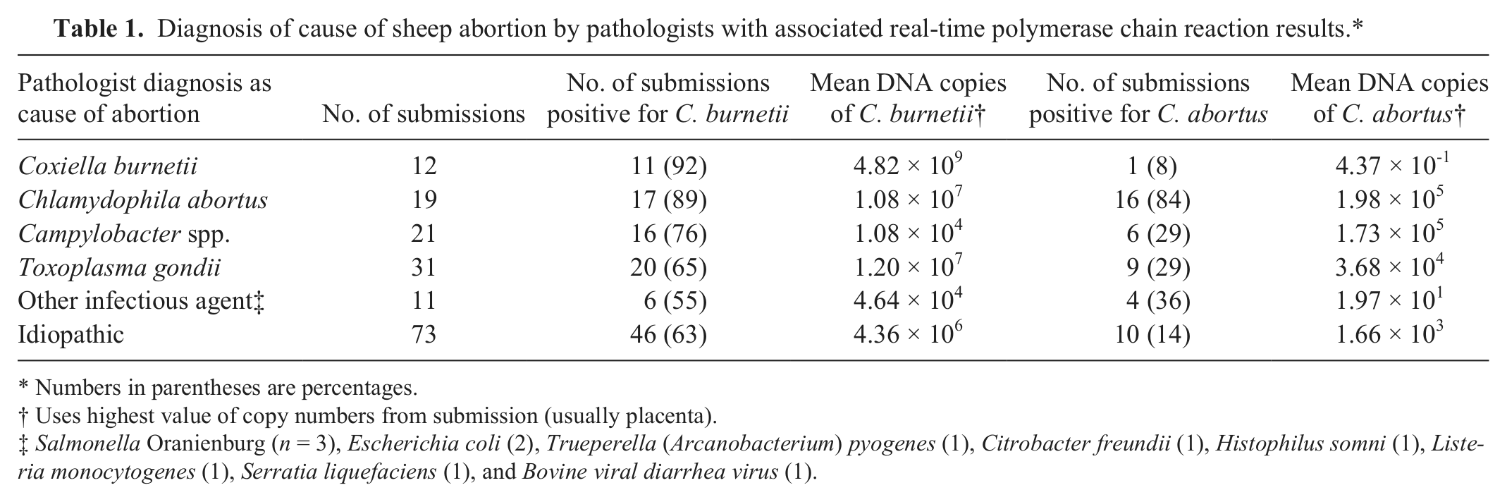
Numbers in parentheses are percentages.
Uses highest value of copy numbers from submission (usually placenta).
Salmonella Oranienburg (n = 3), Escherichia coli (2), Trueperella (Arcanobacterium) pyogenes (1), Citrobacter freundii (1), Histophilus somni (1), Listeria monocytogenes (1), Serratia liquefaciens (1), and Bovine viral diarrhea virus (1).
Diagnosis of cause of goat abortion by pathologists with associated real-time polymerase chain reaction results.*
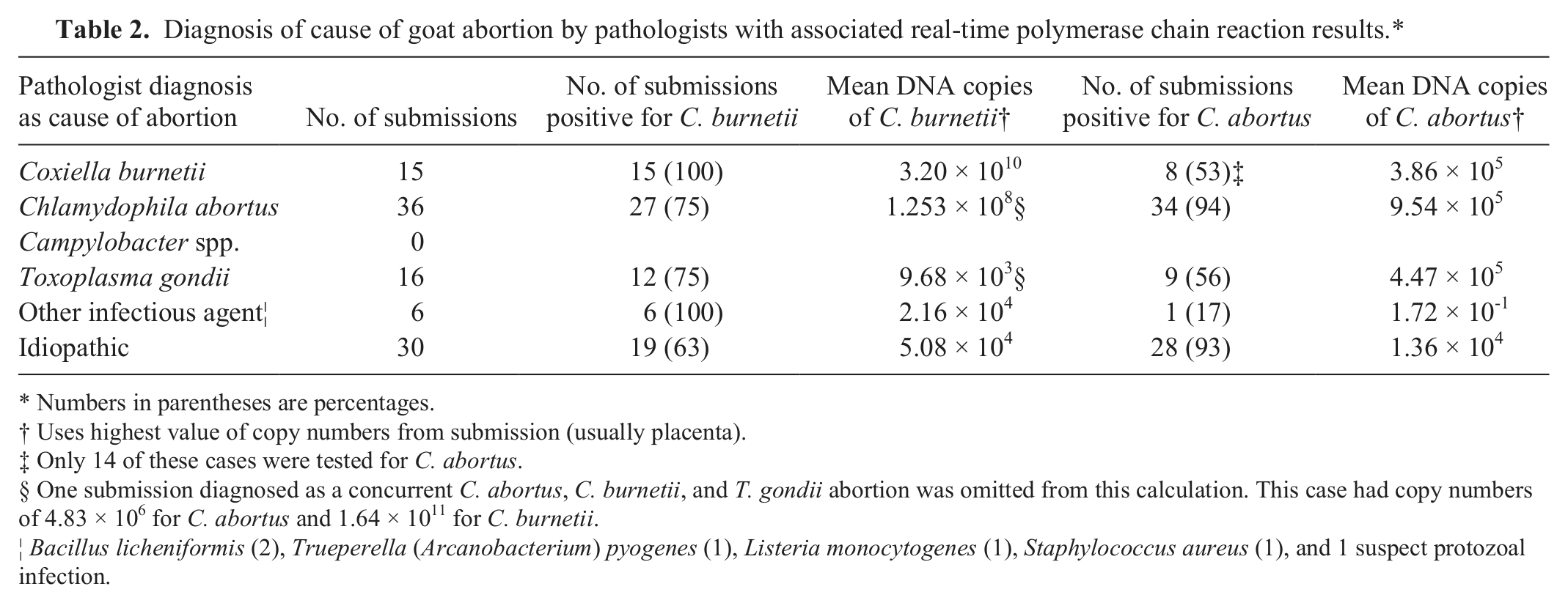
Numbers in parentheses are percentages.
Uses highest value of copy numbers from submission (usually placenta).
Only 14 of these cases were tested for C. abortus.
One submission diagnosed as a concurrent C. abortus, C. burnetii, and T. gondii abortion was omitted from this calculation. This case had copy numbers of 4.83 × 10⁶ for C. abortus and 1.64 × 10¹¹ for C. burnetii.
Bacillus licheniformis (2), Trueperella (Arcanobacterium) pyogenes (1), Listeria monocytogenes (1), Staphylococcus aureus (1), and 1 suspect protozoal infection.
Real-time PCR testing
Coxiella burnetii
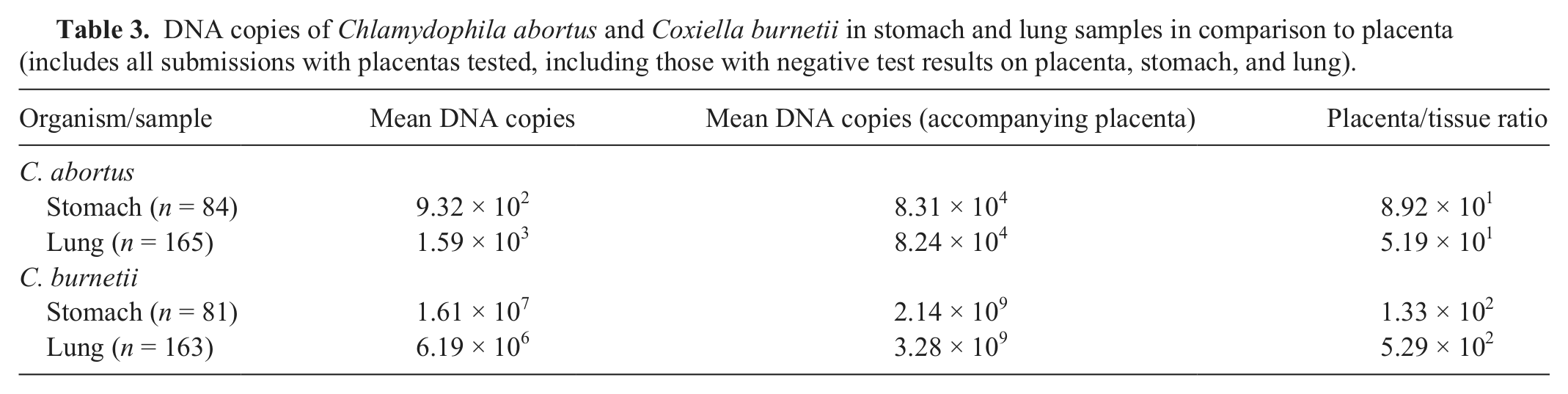
One hundred sixty-three of the sheep abortions and 96 goat abortions were tested. Coxiella burnetii was detected in 113 of 163 (69%) sheep and 72 of 96 (75%) goat submissions. Among the real-time PCR–positive submissions, pathologists considered C. burnetii as the cause of abortion in only 11 out of 113 (10%) sheep and 15 out of 72 (21%) goats. Coxiella burnetii was commonly identified in cases associated with other pathogens in both sheep and goats (Tables 1, 2). When samples were available, the average copy numbers detected in stomach and lung were significantly lower than those in the accompanying chorioallantois (Table 3). When recovered from placenta, matched lung and stomach content samples were also usually positive (Table 4). Summary statistics for C. burnetii are shown (Table 5). A ROC analysis of real-time PCR results using pathology diagnosis as the reference test determined a cut point of 3.78 × 10³ copies that resulted in a sensitivity and specificity of 83.3% (95% CI: 51.6, 97.9) and 88.1% (95% CI: 81.8, 92.8) for sheep, respectively, with an AUC of 0.87 (95% CI: 0.72, 1.00). For goats, the AUC was 0.94 (95% CI: 0.83, 1.00), with a cut point of 6.8 × 10⁴ giving a sensitivity of 93.8% (95% CI: 69.8, 99.8) and a specificity of 91.3% (95%CI: 82.8, 96.4)
DNA copies of Chlamydophila abortus and Coxiella burnetii in stomach and lung samples in comparison to placenta (includes all submissions with placentas tested, including those with negative test results on placenta, stomach, and lung).
DNA copy numbers using real-time polymerase chain reaction from placenta, and associated recovery from stomach content and lung of Coxiella burnetii and Chlamydophila abortus.*
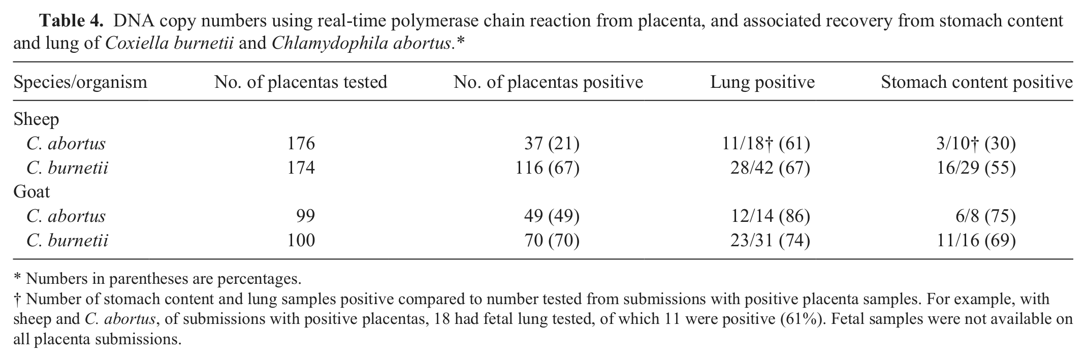
Numbers in parentheses are percentages.
Number of stomach content and lung samples positive compared to number tested from submissions with positive placenta samples. For example, with sheep and C. abortus, of submissions with positive placentas, 18 had fetal lung tested, of which 11 were positive (61%). Fetal samples were not available on all placenta submissions.
Summary statistics for Coxiella burnetii polymerase chain reaction in sheep and goats by pathology diagnosis for abortion caused by this organism.*
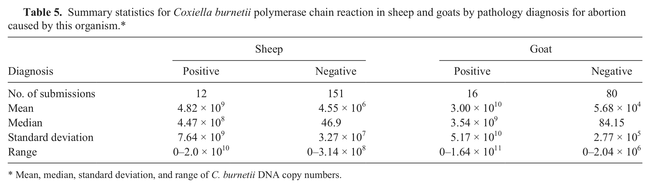
Mean, median, standard deviation, and range of C. burnetii DNA copy numbers.
Chlamydophila abortus
One hundred sixty-two sheep and 92 goat abortions were tested. Of these, 42 of 162 (26%) sheep and 54 of 92 (59%) goat submissions were positive. The percentages of cases where real-time PCR results were positive and were felt by pathologists to indicate the cause of the abortion were 16 of 42 (38%; sheep) and 34 of 54 (63%; goats). Chlamydophila abortus was commonly identified in cases associated with other pathogens in both sheep and goats (Tables 1, 2). When samples were available, the average copy numbers detected in stomach and lung were significantly lower than those detected in the accompanying chorioallantois (Table 3). When recovered from placenta, matched lung and stomach content samples were also usually positive, the exception being with C. abortus in stomach samples from sheep fetuses where the organism was identified in only 3 of 10 samples (Table 4). Summary statistics for C. abortus are shown in Table 6. A ROC curve analysis of real-time PCR results using pathology diagnosis as the reference test determined a cut point of 2.45 DNA copies giving a sensitivity and specificity of 84.2% (95% CI: 60.4, 96.6) and 87.4% (95% CI: 80.8, 92.4) for sheep, respectively, with an AUC of 0.87 (95% CI: 0.79, 0.99). For goats, the AUC was 0.92 (95% CI: 0.86, 0.99) with a cut point of 18.6 giving a sensitivity of 91.7% (95% CI: 77.5, 98.2) and a specificity of 85.7% (95% CI: 73.8, 93.6).
Summary statistics for Chlamydophila abortus polymerase chain reaction in sheep and goats by pathology diagnosis for abortion caused by this organism.*
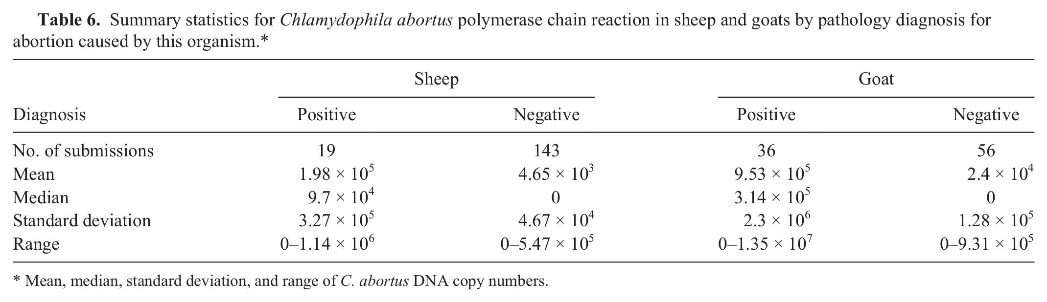
Mean, median, standard deviation, and range of C. abortus DNA copy numbers.
Bacterial culture
Campylobacter spp
In sheep, Campylobacter spp. was isolated in 21 of 163 (13%) of submissions (12 C. fetus ssp. fetus, 8 C. jejuni, and 1 C. coli). No Campylobacter spp. was isolated from goat samples.
Miscellaneous causes of bacterial infectious abortion
Such causes included Salmonella Oranienburg (n = 3), E. coli (2), Trueperella (Arcanobacterium) pyogenes (1), Citrobacter freundii (1), Histophilus somni (1), L. monocytogenes (1), and Serratia liquefaciens in sheep abortions (Table 1), and Bacillus licheniformis (2), T. pyogenes (1), L. monocytogenes (1), Staphylococcus aureus (1) in goat abortions (Table 2).
Virology
A BVDV-like virus was isolated from 1 sheep submission.
Toxoplasma gondii
In sheep, T. gondii was detected in 31 of 163 (19%) submissions, and in 16 of 96 (17%) goat submissions. This pathogen was diagnosed on the basis of lesions and/or IHC and was thought to be significant. It was commonly accompanied by other pathogens. One goat case had diagnosis of “suspect protozoal infection” and was not counted as a T. gondii case.
Multiple pathogen infections
Thirty-seven of 162 (23%) sheep and 43 of 92 (47%) goat submissions that were tested for both C. burnetii and C. abortus had both pathogens identified. Chlamydophila abortus and C. burnetii were commonly detected in the presence of T. gondii as well as other miscellaneous pathogens (Tables 1, 2).
Noninfectious causes
There were 2 cases of congenital goiter and 1 of idiopathic congenital anomalies in submitted sheep abortions.
No diagnosis
Seventy-three of 163 (45%) sheep and 30 of 96 (31%) goat submissions had no cause of abortion determined. This included cases in which low numbers of C. burnetii and C. abortus were identified, but in which the pathologist felt there were no supporting lesions or other supporting tests.
Modified acid-fast smears and IHC
For C. burnetii, MAF-positive cases (n = 29) averaged 1.54 × 10¹⁰ DNA copies with real-time PCR (median = 6.98 × 10⁸) and negative MAF results (n = 199) averaged 5.38 × 10⁸ DNA copies (median = 5.19 × 101). The kappa values and standard error (agreement) comparing agreement for both MAF and IHC with the pathology diagnosis are shown (Table 7). For C. abortus, MAF-positive cases (n = 40) averaged 7.33 × 10⁵ DNA copies (median = 1.98 × 10⁵) with real-time PCR, and MAF-negative cases (n = 181) averaged 5.92 × 10⁴ DNA copies (median = 0). The IHC-positive cases (n = 41) averaged 4.22 × 10⁵ DNA copies with a median value of 1.32 × 10⁵ while negative cases (n = 141) had an average of 2.39 × 10⁴ DNA copies and a median value of 0. The kappa values and standard error (agreement) comparing agreement for MAF, IHC, and ELISA with the pathology diagnosis are shown (Table 7).
Kappa values compared to pathology final diagnosis.*
Numbers in parentheses are standard errors. ELISA = enzyme-linked immunosorbent assay; IHC = immunohistochemistry; MAF = modified acid-fast smears.
Discussion
In sheep abortions in Ontario, at least 1 infectious agent deemed causative was found in 90 of 163 (55%) of submissions in the current study. Toxoplasma gondii was determined to be the most common cause of abortion, representing 33% of infectious abortion diagnoses (31/94 diagnosed infectious causes), followed by Campylobacter spp. (22%), C. abortus (20%), and C. burnetii (12%). Previously published surveys of the cause of sheep abortion in the United Kingdom list Chlamydophila spp. (51%), followed by T. gondii (28%), Campylobacter spp. (12%), Salmonella spp. (3%), and Listeria spp. (3%). 7 A study in Switzerland recorded C. abortus (39%) followed by T. gondii (19%) as most important, followed by C. burnetii (1%) in sheep. 9 In New Zealand, C. abortus and C. burnetii have not been reported, and the main causes of abortion are reported as Campylobacter spp., T. gondii, and Salmonella Brandenburg. 36 In the Netherlands, the most common infectious causes of abortion in sheep were C. abortus, Campylobacter spp., T. gondii, Listeria spp., and Yersinia spp. 34 The cause of abortion in sheep in Ontario is similar to that described in the United Kingdom and Switzerland, with a large percentage of abortions due to T. gondii, C. abortus, and Campylobacter spp. The larger proportion of T. gondii may be associated with technologic advances in diagnostic procedures such as IHC, or better means of control of C. abortus via use of vaccines and antibiotics. The use of selective media in the current study may also have resulted in a higher proportion of Campylobacter spp. cases. It is of interest that C. burnetii was not a major cause of abortion in these other regions compared to Ontario. The reason for this is unknown, but strain differences or husbandry practices may be related. Coxiella burnetii has been recognized as a cause of abortion in Ontario sheep since 1983. 28
In goat abortions in Ontario, at least 1 infectious cause was found in 67 of 96 (70%) of submissions in the present study. Chlamydophila abortus was the most common significant pathogen representing 49% of the infectious abortion diagnoses (36/73 diagnosed infectious causes), followed by T. gondii (22%) and C. burnetii (21%). A comparable study in California also found C. abortus as the most common cause of abortion (38% of diagnosed infectious abortions or 14% of submissions), followed by C. burnetii at 24% of infectious causes. Toxoplasma gondii was diagnosed in 9%, E. coli in 6%, and Campylobacter spp. in 2.5% of diagnosed infectious abortions. 24 The previously mentioned study in Switzerland recorded C. abortus (23%) followed by T. gondii (15%) as most important, followed by C. burnetii (10%) in goats. 9 Results of the current study for the major causes of abortion in goats are similar to both of these studies insofar as the major etiologies of abortion and their ranking.
Establishing a “gold standard” for diagnosis of a disease caused by endemic organisms such as C. burnetii and C. abortus that may be normal resident microflora is difficult, especially when using material submitted through a diagnostic stream. Lesions associated with C. burnetii abortion in sheep and goats have been described usually as very severe placentitis, often confined to the intercotyledonary space, but sometimes with only mild placentitis. 28 Often placental trophoblasts are filled with basophilic intracytoplasmic organisms. Mixed inflammatory cell infiltrates into the intercotyledonary placenta are seen on maternal and fetal sides of the chorioallantois, and vasculitis is present. Foci of hepatic inflammation may be present, and there may be fetal pneumonia. 25 In the present study, pathologists did not follow an established diagnostic algorithm in defining a diagnosis, but did form a weighted opinion based on lesions, history, and microbiology results including culture, ELISA, and other differential diagnostic testing. What ultimately was reported to the farmers and veterinary clinicians was used as the “gold standard.” Cases were frequently reviewed with other pathologists. Admittedly, using the real-time PCR results in part to arrive at a diagnosis and then using that diagnosis as a gold standard to arrive at real-time PCR cut points, is not a recommended practice for diagnostic test evaluation. The present study took this approach because time-sensitive diagnostics were involved, as opposed to research cases, that required an appropriate workup, including real-time PCR. The current study, however, has provided the pathologists in AHL suggested cut points and the related CI for determining the likelihood of abortion as being caused by the pathogen. This approach is not without flaws, and when assessing suggested cut points for diagnosis of C. burnetii or C. abortus, this should be born in mind. Individual laboratories should consider determining appropriate test cut points for their particular population of animals.
The results of the current study emphasize that a simple positive PCR result used as a stand-alone test can be over interpreted, and may lead to misdiagnosis in cases of ovine and caprine abortion, as the presence of the agent (or its nucleic acids) does not necessarily mean presence of the disease. The present study demonstrates that a positive PCR result for C. burnetii or C. abortus needs to be considered in light of other tests, including gross and histologic examination, bacterial and viral culture, and special stains including IHC and acid-fast stains. Determination of DNA copy numbers gives additional weight to real-time PCR results. With both of these organisms, as expected, increased DNA copy numbers were seen in cases where lesions and other testing provided further agreement with their incrimination as the inciting cause of the abortion.
The larger numbers of C. burnetii and C. abortus in placenta samples as compared to stomach content and lung tissue is not surprising as both of these organisms are commonly seen in large numbers in trophoblasts, and C. abortus has been shown to colonize trophoblasts. 27 Stomach content or lung samples can be used as an alternative if placenta is not present, provided the 1.5–2.5 log difference in DNA copy numbers is factored in when interpreting the results.
Testing for C. burnetii in the current study used IS1111 as the target gene, which is a unique transposase that was originally shown to have 21 gene copies present in the C. burnetii genome. 32 It was subsequently shown to have marked variability in gene copies between strains of C. burnetii, varying from 7 to 110 copies. 22 Other genes, such as icd for isocitrate dehydrogenase that are present in only 1 copy, would have been less sensitive, but would probably have led to less variability. 18 If real-time PCR results are to be compared, and cut points established, especially used between laboratories, it would be very important to keep in mind the variability of the numbers of the IS1111 genes between C. burnetii strains, and the difference in the number of gene copies that would be present if using icd. In retrospect, icd would likely have been a better choice for establishment of cut points, especially if one is unaware of the strain of the organism being tested. As different strains may affect sheep and goats, 2 this may result in variation seen in the cut points between the 2 species depending on the strains that are circulating in Ontario herds.
Shedding of C. burnetii has been seen in ewes at parturition, with 15 (48%) ewes infected with the organism on the day of lambing in a study that showed that infection can be found in sheep that is often not associated with abortion. 4 Another study has shown shedding of C. burnetii associated with both reproductive failure in goats as well as normal birthing, and that subsequent shedding of the bacteria after birthing is frequently seen. 3 Results of these and the present study show that diagnosis of C. burnetii abortion should not be based solely on positive PCR results, but that real-time PCR results as well as lesions and other ancillary tests must be used together to formulate a diagnosis. If one accepts use of the final diagnosis as a gold standard, the current results indicated a cut point of 3.78 × 10³ copies for sheep and 6.8 × 10⁴ for goats (95% CI). The AUC measures the accuracy of the ROC analysis. A test with 100% sensitivity and specificity has an AUC of 1.0, whereas a test that does not discriminate between true positives and false positives has an AUC of 0.50. For most diagnostic tests, there is an overlap of values for true-positive and false-positive results.
A MAF smear for C. burnetii performed by an experienced technician in conjunction with the typical histology was found to have substantial agreement with the final diagnosis, with kappa values of 0.69 (sheep) and 0.68 (goats; Table 7). In general, a value of 0.9–1.0 is considered excellent, 0.8–0.9 is considered good, and 0.60–0.80 equals substantial agreement. 31 Other tests such as fluorescent in situ hybridization (FISH) have been used to demonstrate C. burnetii in placentas. 17 One of the advantages of FISH is the direct visualization of the organisms, and it may be possible to roughly assess the numbers of organism associated with the lesions. One disadvantage of FISH is that many diagnostic laboratories are not equipped to do this testing. PCR testing is widely done and increasingly automated. In a diagnostic laboratory situation, although producers are strongly encouraged to submit placenta, it often has been lost to scavengers. Other tissues such as lung and stomach content can be used as an alternative, with high copy numbers giving strong presumptive evidence of fetal infection with the organism.
Diagnosis of C. abortus abortion based only on positive PCR results is also error prone. Shedding of C. abortus in association with both abortion, as well as birth of live lambs has been demonstrated. 13 Similar to C. burnetii, C. abortus abortion diagnosis should also use real-time PCR only as a part of the overall diagnosis, and in conjunction with other tests and herd history. Again, if the final diagnosis is used as a gold standard, at 95% CI, the current study determined cut points of 2.45 DNA copies for sheep and 18.6 DNA copies for goats. These cut points are very low, and suggest that the presence of smaller amounts of C. abortus DNA should be regarded with greater significance than with C. burnetii. Histology along with MAF smears is also convincing, and MAF smear results tend to correlate with numbers found in real-time PCR tests, although with a lower kappa value than seen with C. burnetii. The results of ELISA could also be misleading as a stand-alone test as the presence of the organism does not necessarily equate with disease; associated kappa values were low. This may be in part due to erroneous pathology interpretations. There were 3 sheep submissions in the current study in which there was a pathology diagnosis of C. abortus but that had negative real-time PCR and MAF results that were negative or unreadable due to background staining. In these 3 cases, the pathologist made the diagnosis based on 1) mild placentitis and a positive PCR test to Chlamydophila spp. (although the real-time PCR to C. abortus was negative), 2) “coccoid organisms in trophoblasts” and a positive antigen ELISA, or 3) lesions, including necrosuppurative placentitis with vasculitis, multifocal hepatic and splenic necrosis, and fetal pneumonia with no other pathogens identified except for extremely low C. burnetii real-time PCR results (57.9 DNA copies). Although C. abortus is associated with the production of vasculitis in the placenta of sheep, 6 a review of selected cases in the present study showed that it was not unique to this organism. In the reviewed lesions seen in placentas from bacterial abortions, vasculitis in the chorioallantois was also seen in cases of abortion due to C. jejuni, C. fetus ssp. fetus, and B. licheniformis. This may have contributed to some “false-positive” diagnoses by pathologists, and may explain the low cut point for the C. abortus real-time PCR testing. Coxiella burnetii can often be diagnosed on histology alone if large numbers of the typical organisms are present. In contrast, C. abortus can easily be confused with other bacteria in trophoblasts, and vasculitis, although sometimes mentioned as a typical lesion in C. abortus–infected placentas, is not specific. The presence of coccoid forms in trophoblasts is also not specific for C. abortus. Trophoblasts are phagocytic, and bacteria can commonly be seen in these cells. Some bacteria likely proliferate in trophoblasts including Brucella abortus. 1 A recent study used a similar method to form a diagnosis, using histological examination of placenta to estimate cut points for real-time PCR on C. abortus using a duplex real-time PCR, which included T. gondii. 12 By inoculating ewes and correlating lesions with DNA copy numbers, an estimated cut point of value of at least 106 DNA copies was established for diagnosis of C. abortus using placental samples. The study had similar findings to the present study in that C. abortus and T. gondii were commonly identified with other pathogens in the same abortion case. 12
Campylobacter spp. was only identified in sheep, and several submissions were from the same farm. Most of these were also positive for C. burnetii, and frequently also had C. abortus recovered. Many of the miscellaneous pathogens such as Campylobacter spp., L. monocytogenes, H. somni, Salmonella Oranienburg, and BVDV were all isolated in submissions that had positive real-time PCR results for C. burnetii and/or C. abortus. Identification of these other pathogens is strong evidence that there may have been bystander shedding of the organism frequently as a result of the birthing, and that this would have occurred whether as part of normal parturition, or as part of the abortion process initiated by other pathogens.
Toxoplasma gondii is well recognized as a cause of abortion in sheep14,15 and goats,10,26 and has been estimated to cause approximately 1–2% of neonatal lamb losses in the United Kingdom. 5 Evidence of T. gondii was present in 47 of 259 submissions, often in the presence of other pathogens. In sheep, there is evidence of vertical transmission of the organism,11,37 and a similar situation may be present in goats. The presence of vertical transmission as well as identification of other pathogens in cases in which T. gondii was identified also suggests that identification of the organism in an abortion case does not necessarily mean it was the cause of the abortion.
Determining the cause of a small ruminant abortion still remains a somewhat subjective decision. Use of a highly sensitive test such as PCR for diagnosis of disease caused by infectious agents such as C. burnetii or C. abortus does not distinguish between infection and disease causation. Using DNA copy numbers in conjunction with lesions, acid-fast stains, and bacterial and viral culture gives a better picture of the likely cause of the abortion. More information such as strain differences of C. burnetii and C. abortus and the associated pathogenicity to both small ruminants and human beings may help to determine why disease in contact humans is not common, but can occur as outbreaks in both animal and human health.
Footnotes
Acknowledgements
The authors thank the many producers and veterinarians who submitted case material for this project. The work could not have been done without the able assistance of many people in the AHL receiving room, necropsy room, histology, bacteriology, and virology laboratories. Ana Rita Rebelo and Patricia Bell-Rogers are thanked for their assistance with additional PCR work.
a.
ViroStat Inc., Portland, ME.
b.
BioGenex, Fremont, CA.
c.
Ventana Medical Systems Inc., Tucson, AZ.
d.
Campy Gen Oxoid Canada, Nepean, Ontario, Canada.
e.
Clearview Chlamydia, Inverness Medical Canada Inc., Ottawa, Ontario, Canada.
f.
MagNA Pure LC and MagNA Pure LC DNA Isolation Kit I, Roche Diagnostics, Montreal, Quebec, Canada.
g.
Qiagen DNeasy Tissue kit, Qiagen Inc., Mississauga, Ontario, Canada.
h.
LightCycler 480, Roche Diagnostics, Montreal, Quebec, Canada.
i.
Roche Diagnostics, Montreal, Quebec, Canada.
j.
TIB Molbiol, Adelphia, NJ.
k.
pGEM-T Easy Vector System, Promega Corp., Madison, WI.
l.
15C5 and 20.10.6, Dr. E. Dubovi, New York State College of Veterinary Medicine, Cornell University, Ithaca, NY.
m.
H&L, Invitrogen Corp., Camarill, CA.
n.
StataCorp LP, College Station, TX.
Declaration of conflicting interests
The author(s) declared no potential conflicts of interest with respect to the research, authorship, and/or publication of this article.
Funding
The author(s) disclose receipt of the following financial support for the research, authorship, and/or publication of this article: This work was funded by the Ontario Ministry of Agriculture, Food and Rural Affairs, Animal Health Strategic Investment (project no. 09-09).