
Abstract
The current report describes an outbreak of vesicular disease affecting dairy cows in midwestern Brazil in which a coinfection with 2 poxviruses—Vaccinia virus (VACV) and a parapoxvirus—was demonstrated. Milking cows presented vesicles, painful reddish or whitish papules, and scabby proliferative lesions in the teats and udder, in a clinical course of approximately 10–21 days. Histologically, multifocal areas of moderate to severe acanthosis, spongiosis, hypergranulosis, and parakeratotic or orthokeratotic hyperkeratosis with adjacent focally extensive ulcers were observed in the epidermis. Rounded eosinophilic inclusion bodies were observed in the cytoplasm of epithelial cells of areas with acanthosis or necrosis. Moderate inflammatory infiltrate of lymphocytes, plasma cells, neutrophils, and macrophages were observed in some dermal areas. Two people milking the affected cows developed lesions on the hands, painful papules which progressed to ulcerative and scabby lesions in 4–7 days. Electron microscopy of scabs from 1 cow revealed the concomitant presence of orthopoxvirus and parapoxvirus particles. Scabs from 2 cows were positive by polymerase chain reaction for the parapoxvirus B2L gene; 1 of the scabs was also positive for the VACV vgf gene. Nucleotide sequencing of the B2L amplicon revealed a similarity of 96–99% with Orf virus (ORFV) and lower identity with Pseudocowpox virus (92–95%) and Bovine papular stomatitis virus (85–86%). Nucleotide sequencing of a region of parapoxvirus DNA polymerase gene revealed a high similarity (98–100%) with ORFV sequences. Thus, an unusual coinfection with VACV and a parapoxvirus, likely ORFV, was demonstrated in the outbreak.
Orthopoxvirus and Parapoxvirus are the 2 main genera of the subfamily Chordopoxvirinae, family Poxviridae. The Orthopoxvirus genus includes important viruses that cause proliferative skin disease in animals and/or human beings, namely Vaccinia virus (VACV), Cowpox virus, Variola virus (smallpox, the human kind of which was eradicated in 1980), Rabbitpox virus, and Monkeypox virus. The genus Parapoxvirus contains 4 virus species: the prototype Orf virus (ORFV), Bovine papular stomatitis virus (BPSV), Pseudocowpox virus (PCPV), and Parapoxvirus of red deer in New Zealand (http://ictvonline.org/virusTaxonomy.asp?version=2009). Orthopoxvirus and Parapoxvirus genera viruses are widely distributed and frequently associated with important impacts on public and animal health.9,21
Since 2000, a number of cases and outbreaks of VACV infection affecting dairy cows and/or human beings have been described in southeastern Brazil,8,13,21,24 and also in the midwestern 15 and northern 14 parts of the country. Likewise, cases of parapoxvirus-associated vesicular disease affecting cattle and, occasionally human beings, have also been described in some Brazilian states.3,7,19 In at least one of the outbreaks involving human beings and cows, a mixed VACV and parapoxvirus infection was demonstrated in human lesions. 3 In addition, ORFV infection is endemic in most sheep- and goat-raising Brazilian states, particularly the south and northeast, and occasional human cases have also been described associated with animal handling.16,18
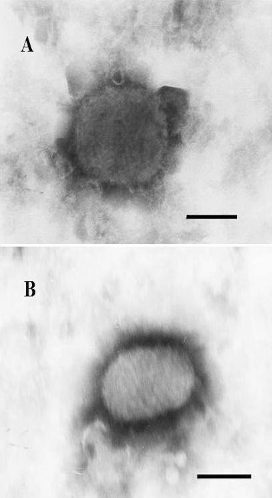
The present report describes an outbreak of vesicular disease in dairy cows in midwestern Brazil in which coinfection by VACV and a parapoxvirus was demonstrated in a cow. The disease affected a farm of milk production (hand milking) located in Jataí county, state of Goiás, Brazil, in September 2011. At the time of the outbreak, the farm held a total of 50 calves, 49 heifers, and 43 milking cows. One calf, 5 heifers, and 40 milking cows presented the clinical signs (Table 1). The main signs included vesicles (4–8 mm in diameter), painful reddish or whitish papules (3–9 mm in diameter; Fig. 1A), and scabby proliferative lesions localized on the teats (heifers and cows), and ulcers restricted to the hard palate and gum (calf). The clinical course ranged from approximately 10–21 days. In addition, some cows presented similar lesions in the udder (Fig. 1A). In general, the teat lesions were multifocal and affected 2 or all teats of each cow, regressing approximately 12–20 days after onset. A few days after the appearance of papules, coalescent scabby erosions and ulcers were noticed. In some cases, a mild serous secretion was observed in the ulcerative lesions that occasionally dried up, creating crusts. Affected cows presented severe local pain that did not allow the completion of milking, leading to interruption of lactation and, occasionally, to secondary mastitis. Some cows showed a mild clinical improvement upon local treatment with 3% iodine solution for 7 consecutive days.
Epidemiological data of an outbreak of Vaccinia virus and parapoxvirus coinfection in dairy cows in midwestern Brazil.
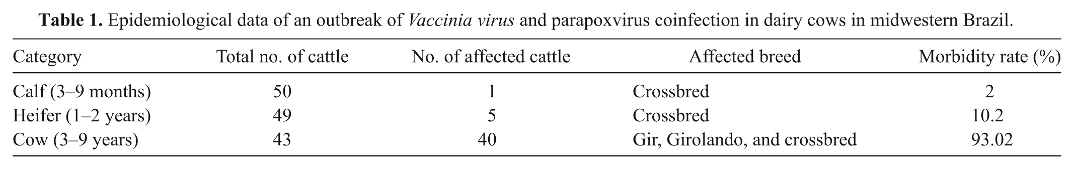
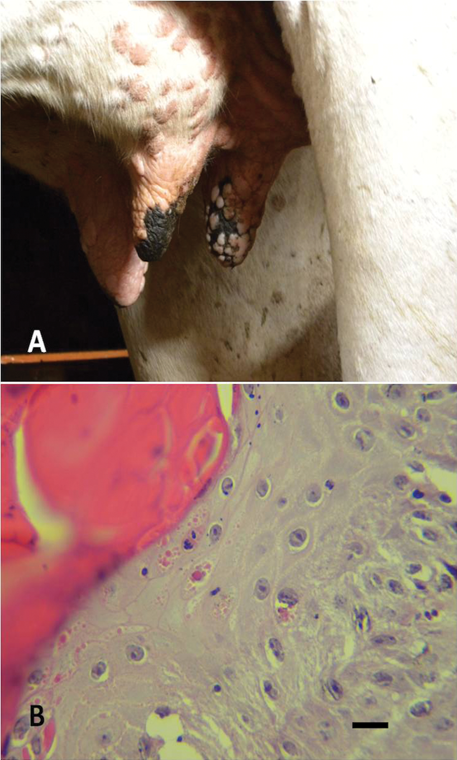
Fragments of affected skin obtained from 3 cows were fixed in 10% buffered formalin and processed for routine histopathological examination upon hematoxylin and eosin staining. Histologically, multifocal areas of moderate to severe acanthosis, spongiosis, hypergranulosis, and parakeratotic or orthokeratotic hyperkeratosis with adjacent focally extensive ulcers were observed in the epidermis. Few 2–8 μm, circular, eosinophilic inclusion bodies were noted in the cytoplasm of epithelial cells of areas with acanthosis or necrosis (Fig. 1B). Detachment of the dermal layer and necrosis with moderate inflammatory infiltrate of lymphocytes, plasma cells, neutrophils, and macrophages were observed in some areas. Adjacent dermis showed the same type of perivascular infiltrate. Eosinophilic extensive areas of serous and cell crusts above the affected epidermis were prominent. The gross and histopathological lesions observed in the present study are similar to other descriptions of parapoxvirus and VACV infections.5,7,16,19
Specimens for virus isolation (vesicular fluid, scabs) were collected in a sterile manner from papules and scabby lesions on teats of 4 affected cows. For virus isolation, scabs were homogenized, resuspended to 10% (weight/volume) in culture medium, and inoculated onto monolayers of Madin–Darby bovine kidney and African green monkey kidney epithelial (Vero) cells. After 3 passages of 5 days each, all samples were negative for infectious virus as ascertained by absence of cytopathic effect on inoculated cultures. Convalescent blood was collected from some affected cows and submitted to virus-neutralizing assay for VACV antibodies. No neutralizing activity against VACV strain P1V 2 was detected in the samples.
Scab samples of 2 animals were submitted to electron microscopy (EM) under negative staining. Strikingly, EM examination confirmed the presence of 2 morphologically distinct poxvirus particles in scabs from 1 animal (SV547/11-Preta). Typical 250–300 nm orthopoxvirus particles with brick-shaped morphology and irregular tubules at surface were observed concomitantly with classical 260 nm × 160 nm ovoid, helical structured parapoxvirus particles (Fig. 4). Thus, the results of EM first demonstrated a mixed OPV and parapoxvirus infection in lesions of 1 affected cow.
Concomitantly, total DNA was extracted from scabs using DNAzol reagent a and submitted to polymerase chain reaction (PCR) for VACV, using primers for vgf gene described previously. 2 Total DNA extracted from Vero cells infected with VACV-P1V was used as positive control. A band of approximately 381 bp, corresponding to the expected amplicon for VACV, was detected in DNA extracted from a scab of 1 animal (SV547/11-Preta).
A PCR for parapoxviruses, agents recently implicated in vesicular disease in cattle from several Brazilian regions,7,19 was performed. By using a set of primers that amplifies a target sequence within the parapoxvirus B2L gene, 11 a band of approximately 590 bp, corresponding to the expected size of the amplicon for parapoxviruses, was detected in scabs from 2 animals (SV547/11-Preta and -Malhada). Because this set of primers is expected to amplify all parapoxviruses, the amplicons were submitted to nucleotide sequencing for identification of the virus species. The PCR products were purified using a commercial kit b according to the manufacturer’s instructions and submitted to nucleotide sequencing. c The obtained sequences were analyzed 22 and aligned 10 using commercial software packages, and compared with other parapoxvirus sequences deposited in GenBank. These sequences were deposited in GenBank under the names SV547/11-Preta (JX268519) and SV547/11-Malhada (JX268520). Nucleotide sequencing of the amplicon revealed a nucleotide similarity of 96–99% with ORFV and lower identity with other parapoxviruses, namely PCPV (92–95%) and BPSV (85–86%). The alignments of parapoxvirus sequences deposited in GenBank and the samples of the current study were used to construct a phylogenetic tree based on the B2L sequence, using the neighbor-joining method with 1,000 bootstrap replicates implemented by MEGA5.0, 23 based on Tamura 3 parameters model, and showed that SV547/11-Preta and -Malhada, clustered with several worldwide ORFV isolates (Fig. 2A). Thus, the results of PCR and sequencing demonstrated the presence of a parapoxvirus closely related to ORFV in lesions of 2 animals, 1 of which was also positive for VACV, demonstrating a coinfection with these agents. The sequences of SV547/11-Preta and -Malhada were highly similar (99%) to a Brazilian ORFV sequence (MT-05) identified in Mato Grosso state, also located in midwestern Brazil. 1 To further confirm the identity of this co-infecting parapoxvirus, a PCR for a highly conserved gene was performed. 4 A 103-bp sequence of DNA polymerase (DNApol) gene was amplified out of 1 sample (SV547/11-Preta). Nucleotide sequencing of the DNApol amplicon revealed a nucleotide similarity of 98–100% with ORFV sequences, 93% with PCPV, and 92% with BPSV. A phylogenetic tree based on the DNApol partial sequence was constructed (Fig. 2B). The SV547/11-Preta sequence clustered in the same branch of several ORFV sequences, confirming its close relationship with ORFV. A partial alignment of B2L and DNApol sequences was performed with several ORFV sequences deposited in GenBank (Fig. 3A, 3B). A few nucleotide substitutions were observed in SV547/11 B2L sequence compared with reference ORFV strain OV-IA82.
Consensus bootstrap phylogenetic tree based on the nucleotide sequences of parapoxvirus B2L (
Partial alignment of nucleotide sequences of B2L (
Poxvirus particles in scab of SV547/11-Preta sample processed for negative-staining electron microscopy.
A 40-year-old female employee of the affected farm presented body pain, general malaise, 40°C fever, and severe lesions (pustules, vesicles, and ulcers) on the hands after milking affected cows. Lesions started as painful papules and pustules, which progressed to ulcerative and scabby lesions by days 3–7. After the onset of the outbreak, a 25-year-old milker from the same farm presented the same signs. Unfortunately, human samples were not obtained, precluding any attempt of agent identification. Treatment with antipyretic and analgesic drugs was performed in human cases. According to the owners, similar signs have never been observed before either in animals or in human beings in the region. In addition to several reports of VACV-associated disease in cattle and human beings in the last decade in Brazil,12,26 a few events of mixed VACV infections have also been reported in cattle 26 and horses.5,6 A mixed VACV and parapoxvirus infection, identified as PCPV, has also been reported in skin lesions of a cow and its milker. 3
Human and bovine poxvirus infections, noticeably VACV, are fairly common in some Brazilian rural areas.12,20,21,24,25 Parapoxviruses (PCPV and BPSV) have also been detected in cases and outbreaks of cattle and human vesicular disease.3,7,19,20 Likewise, ORFV infection is endemic in most sheep- and goat-raising regions, and human occupational disease has been occasionally reported. 16 The association of a parapoxvirus close to ORFV, which is primarily a sheep and goat pathogen, with cattle disease is a rather rare event, with few descriptions in the literature. 17 Hence, the current study reports an unusual event of infection with a parapoxvirus closely related to ORFV in cattle and, most importantly, a rare case of coinfection by 2 poxviruses belonging to different genera. A report of infection by an ORFV-like parapoxvirus in Finish reindeer has been described in the literature, suggesting that the lesions were caused by ORFV or by a parapoxvirus closely related to ORFV. 23
In the present outbreak, although many categories of cattle (calves, heifers, and cows) had been infected, the morbidity rate was higher in the cows (93.2%; Table 1). Most likely, the daily and intensive handling of these animals favors the faster transmission of the disease. The coinfection diagnosed in the present report occurred in the dry season of the year. Other parapoxvirus and VACV Brazilian outbreaks in cattle also have been observed in the same period.13,19
The etiology of the human disease in this outbreak was not determined, yet both VACV and parapoxviruses are zoonotic viruses and have been implicated in occupational human disease in Brazil. In general, VACV and parapoxvirus human disease is associated with hand milking of affected cows, whereas human cases of ORFV infection are usually linked to handling and management of sheep and goats affected by contagious ecthyma. In this sense, the affected farm presented the main epidemiological conditions associated with most VACV outbreaks reported in Brazil (e.g., poor sanitary conditions, hand milking, and routine and consistent animal movement in and out of farm areas).
The origin and primary agent of the outbreak reported herein could not be determined. A sheep or goat origin of the identified parapoxvirus is possible, but other possible origins, including a bovine virus, should not be discarded. Regardless of speculation regarding origin, the available sequence data indicates that the parapoxvirus SV547/11 is an ORFV.
Footnotes
a.
Invitrogen Corp., Carlsbad, CA.
b.
Ilustra GFX PCR DNA kit, GE Healthcare Bio-Sciences AB, Uppsala, Sweden.
c.
MEGABACE sequencer, Amersham Biosciences, Piscataway, NJ.
Declaration of Conflicting Interests
The author(s) declared no potential conflicts of interest with respect to the research, authorship, and/or publication of this article.
Funding
The author(s) declared that they received no financial support for their research and/or authorship of this article.