
Abstract
Bovine papular stomatitis virus (BPSV) is a parapoxvirus associated with papular and erosive lesions on the muzzle, lips, and oral mucosa of cattle. Teats of milking cows are occasionally affected, and the infection is frequently transmitted to human beings. The present report describes an outbreak of BPSV infection affecting cows in midwestern Brazil, with human involvement. The disease was observed in neighboring small hand-milking farms, affecting 20 milking cows. The signs included painful reddish papules, ulcers, and scabby proliferative lesions on the teats, with a clinical course of 7–12 days. Affected cows presented severe local pain, not allowing the completion of milking. Histologically, acanthosis, spongiosis, and parakeratotic hyperkeratosis with adjacent focally extensive ulcers and multifocal inflammatory infiltrate were observed in the epidermis. Eosinophilic inclusion bodies were noted in the cytoplasm of epithelial cells. Personnel milking the affected cows developed lesions on the hands, painful papules that progressed to ulcerative and scabby lesions in 4–7 days. A polymerase chain reaction using a set of pan-parapoxvirus primers for the B2L gene performed on DNA extracted from scabs amplified a 590-bp product, which when sequenced, revealed similarities of 99%, 85%, and 84% with BPSV, Pseudocowpox virus, and Orf virus, respectively. A phylogenetic tree based on the B2L sequence was constructed, showing that the virus clustered with BPSV isolates. Although clinical cases compatible with BSPV infection have been frequently described in Brazil, the present report identifies the agent associated with cattle and human disease in the country.
Parapoxviruses (PPVs) belong to the family Poxviridae, subfamily Chordopoxvirinae, and include 4 virus species that cause nonsystemic, proliferative skin disease in domestic and wild ruminants: the prototype Orf virus (ORFV), Bovine papular stomatitis virus (BPSV), Pseudocowpox virus (PCPV), and Parapoxvirus of red deer in New Zealand. The diseases caused by PPVs are widespread around the world and can occasionally be transmitted to human beings.5,6 Bovine papular stomatitis virus infection is more common in calves, and lesions include papules, often mildly erosive mainly on the muzzle, lips, hard palate, and oral mucosa of calves and, occasionally, on the tongue, esophagus, and forestomach of calves and in the udders of cows. 4 In human beings, BPSV infection is associated with nodules and pustules on the hands and sometimes on the face. 3
Cases clinically compatible with BPSV infection have been frequently reported by veterinarians, yet the agent has not been unequivocally diagnosed in Brazil. Laboratory confirmed cases have been reported in Uruguay, and suspected cases have been described in southern Brazil. 9 In contrast, orthopoxvirus-associated disease in dairy cows and human beings associated with Vaccinia virus (VACV) has become very common in some rural areas of southeast Brazil in the last decades. 15
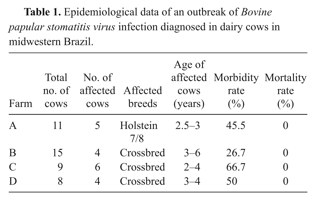
The present report describes an outbreak of BPS disease in dairy cows in midwestern Brazil. The disease affected 4 neighboring small farms (rural settlements) of milk production (hand milking) located in Jataí county, state of Goiás, Brazil, in August 2010. The 4 farms held a total of 43 milking cows, and 20 cows presented the clinical signs (Table 1). The disease was observed almost concomitantly in the 4 farms and was restricted to milking cows. The main signs were painful reddish papules (3–10 mm in diameter) and scabby proliferative lesions localized on the teats, with a clinical course of 7–12 days. In general, the lesions were multifocal and affected 3 or all teats of each cow, regressing approximately 12–16 days after the onset. A few days after the appearance of papules, coalescent scabby erosions and ulcers were noticed (Fig. 1A). In 3 cases, a mild serous secretion was observed in the ulcerative lesions that occasionally dried up, creating crusts. Affected cows presented severe local pain and did not allow the completion of milking, leading to interruption of lactation and, occasionally, to secondary mastitis. Some cows showed a mild clinical improvement upon local treatment with 3% iodine solution.
Epidemiological data of an outbreak of Bovine papular stomatitis virus infection diagnosed in dairy cows in midwestern Brazil.
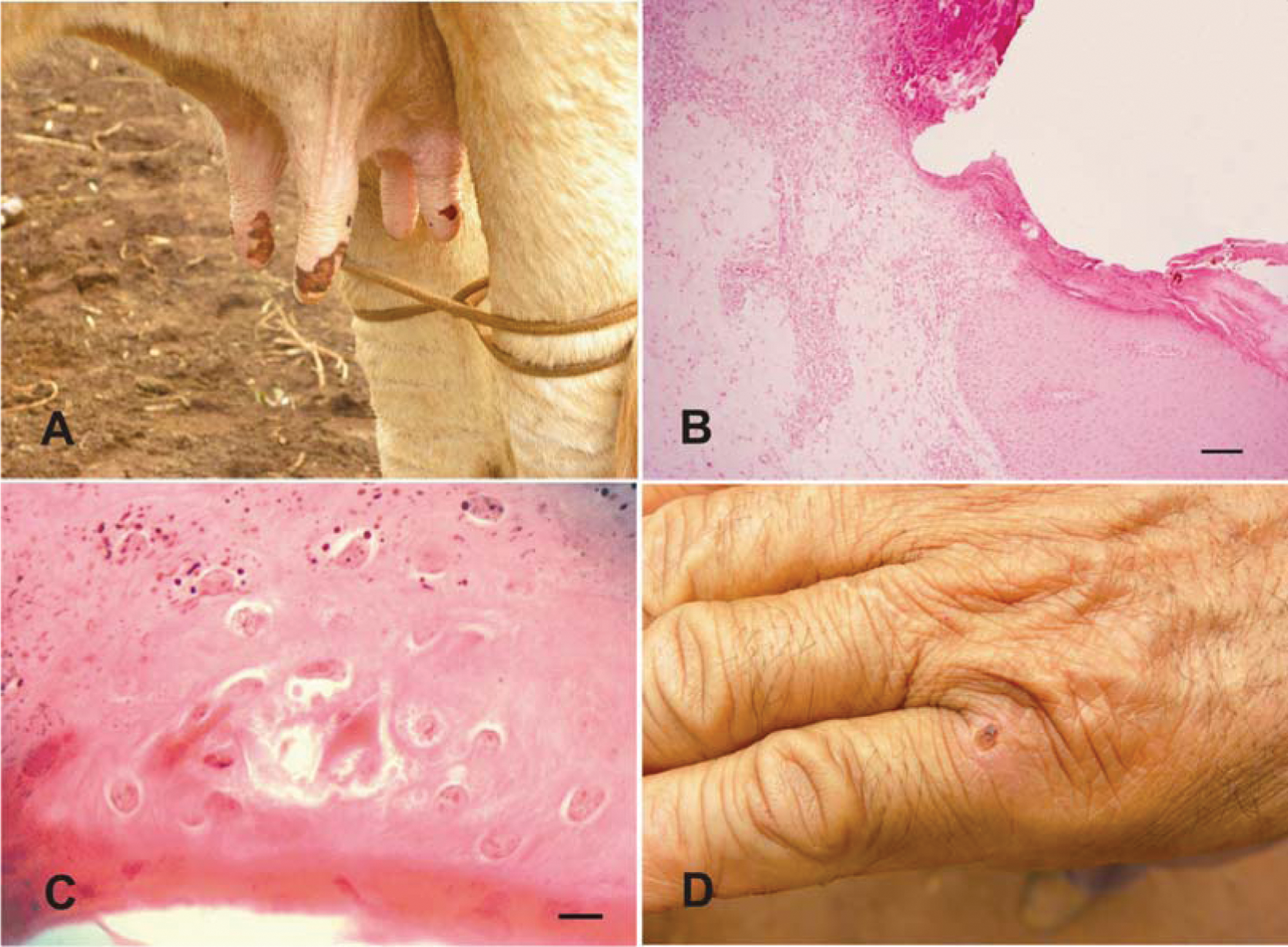
Fragments of affected skin obtained from 3 cows were fixed in 10% buffered formalin and processed for routine histopathological examination upon hematoxylin and eosin staining. Histologically, multifocal areas of moderate acanthosis, spongiosis, and parakeratotic hyperkeratosis with adjacent focally extensive ulcers were observed in the epidermis (Fig. 1B). Numerous 2–6 μm, circular, eosinophilic inclusion bodies were noted in the cytoplasm of epithelial cells of areas with acanthosis or necrosis (Fig. 1C). Detachment of the dermal layer and necrosis with moderate inflammatory infiltrate of lymphocytes, neutrophils, and macrophages were observed in some areas. Adjacent dermis showed the same type of perivascular infiltrate. Reddish extensive areas of serous and cell crusts above the affected epidermis were prominent. The gross and histopathological lesions observed in the present study are similar to other descriptions of BPSV infections.4,8,10
Milkers on 3 farms presented severe lesions on the hands after milking affected cows, starting with painful papules which progressed to ulcerative and scabby lesions by days 4–7 (Fig. 1D). No treatment was performed in human cases. According to the owners, similar signs have never been observed before in the animals or farm workers in the region.
Specimens for virus isolation were collected from papules and scabby lesions on teats of 3 affected cows. Homogenized scab suspensions were inoculated onto monolayers of Madin–Darby bovine kidney and Vero (African green monkey kidney epithelial) cells and subjected to 3 passages of 5 days each. All samples were negative for infectious virus as ascertained by absence of cytopathic effect on inoculated monolayers.
Total DNA extracted from scabs with DNAzol reagent a was tested by PCR for Bovine herpesvirus 2 (BoHV-2), the agent of bovine mammillitis, 14 and for VACV, 1 with negative results for both viruses. Thereafter, DNA extracted from scabs was tested using a PCR designed to detect the B2L gene of all PPVs, using the primers PPP1 forward (5′-3′GTCGTCCACGATGAGCAG) and PPP4 reverse (5′-3′TACGTGGGAAGCGCCTCGCT). 7 Total DNA extracted from BHK (baby hamster kidney)-21 cells infected with the ORFV strain IA-82 was used as positive control. A band of approximately 590 bp, corresponding to the expected size of the amplicon for PPVs, was amplified from DNA extracted from scabs of 2 animals from different herds (data not shown). To determine the specific PPV involved, sequencing of the PCR products was conducted. The PCR products were purified using the a commercial kit a according to the manufacturer’s instructions and then sequenced. b The sequences were analyzed by the Staden package 13 and aligned using commercial software c and compared with other PPV sequences deposited in GenBank. This sequence, designated SV819/10, was deposited in GenBank (accession no. JN629089). The amplicon sequence showed similarities of 99%, 85%, and 84% with BPSV, PCPV, and ORFV, respectively. A phylogenetic tree based on the B2L sequence was constructed using the maximum parsimony method with 1000 bootstrap replicates implemented by MEGA3.1, and showed that SV819/10 clustered with several BPSV isolates (Fig. 2).
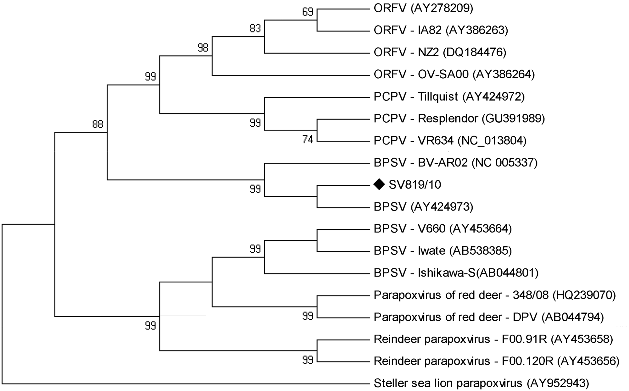
Consensus bootstrap phylogenetic tree based on the nucleotide sequences of parapoxvirus B2L gene. The tree was constructed using the maximum parsimony method with 1,000 bootstrap replicates implemented by MEGA5.03. Values greater than 50% are shown. Bovine papular stomatitis virus SV819/10 (GenBank accession no. JN629089) sample was identified with black marker.
Diagnosis of BPSV infection was presumptive, based on the clinical and histopathological findings. Differential diagnosis included foot-and-mouth disease (currently eradicated from Brazilian territory) infection by PCPV, Bovine viral diarrhea virus (endemic in the country), BoHV-2 infections, and orthopoxvirus diseases (endemic in some areas of Brazil).4,12 In the present case, the amplification of PPV sequences from scabs and the subsequent sequencing of the amplicon provided the definitive identification of BPSV. This is somewhat unusual since PPV infections of the bovine teats and udder have been most commonly associated with PCVP. The failure to isolate virus may be related to inappropriate handling of clinical samples resulting in virus inactivation, lack of susceptibility of inoculated cultures to the agent, or both.
Although distributed worldwide, BPSV infection is considered of little sanitary importance by some authors. 4 However, in the present cases, the disease was economically important for the affected herds, leading to a decrease in milk production and interruption of lactation in many cows.
Virological examination was not performed on human specimens. Nevertheless, the characteristics of the lesions, and the spatial and temporal association between animal and human cases, reinforce the diagnosis of the human lesions. Human BPSV infections usually occur as a result of direct contact with lesions on dairy cows and have been well documented. 3 Thus, milkers and other personnel working with livestock, especially dairy cows, should be aware of the possibility of PPV infection. 11
According to technical and informal reports by veterinarians and farmers, cases of skin lesions on the udder and teats of milking cows, comparable to those described in the present report, are reasonably common in Brazilian dairies, mainly in those managed under poor sanitary conditions. The clinical syndrome is sometimes referred as bovine variola (cowpox), although the agent of true cowpox (Cowpox virus) is considered exotic in the country. Definitive agent identification has been accomplished only in a limited number of cases, most commonly associated with VACV infection 15 and, in 1 case, with PCPV. 2 Thus, the vast majority of the clinical cases observed in the field remain unconfirmed. The present report provides definitive identification of BPSV associated with cattle and human disease and may contribute to an improvement in the diagnosis, leading to a better understanding of the epidemiology and sanitary impact of these vesicular diseases in cattle.
Footnotes
a.
PureLink PCR kit, Invitrogen Corp., Carlsbad, CA.
b.
MEGABACE sequencer, GE HealthCare Bio-Sciences AB, Uppsala, Sweden.
c.
Clone Manager Professional Suite, Align plus 5, version 5.10, Sci Ed Central, Cary, NC.
The author(s) declared no potential conflicts of interest with respect to the research, authorship, and/or publication of this article.
The author(s) received no financial support for the research, authorship, and/or publication of this article.