
Abstract
An adult Madagascar tree boa (Sanzinia madagascariensis) underwent coeliotomy for investigation of a coelomic mass. At surgery, a large mass originating from the peri-pancreatic adipose tissue and involving the gall bladder was removed. The snake did not recover from general anesthesia. A complete postmortem was performed, and samples were submitted to the University of Glasgow for histopathology. On histological examination, the mass was composed of adipose tissue infiltrated with a poorly demarcated spindle cell neoplasm. The neoplastic cells were highly pleomorphic with abundant cytoplasm and frequent clear cytoplasmic vacuoles, suggestive of adipocyte origin. Immunohistochemical characterization of the mass was inconclusive. Metastatic neoplastic cells were present within vessels in the liver, lungs, and brain. As an incidental finding, the gonads contained both maturing ovarian follicles and seminiferous tubules with intact germinal epithelium and evidence of spermatogenesis, along with other features of male and female gonad anatomy. The current report describes a rare neoplasm in snakes within an intersex Madagascar tree boa.
An approximately 14-year-old Madagascar tree boa (Sanzinia madagascariensis) from Bristol Zoo Gardens (Clifton, Bristol, England) presented with a 3-week history of weight loss and an extended over-winter fasting. Prior to this, there had been no reports of significant clinical illness: the snake had been housed with a male and had twice been seen mating in the preceding 2 years. During physical examination, an intracoelomic mass was detected approximately two-thirds down the length of the body from the head. Radiographically, the mass appeared as heterogeneous soft tissue opacity, with multifocal, smaller mineralized opacities. Differential diagnoses for this lesion considered were intestinal impaction, granulomatous inflammation, or neoplasia. Other significant findings included mild erythema of the ventral scales and a moderate loss of condition since the last routine examination. Routine hematology revealed a moderately elevated total white blood cell count with a mild relative heterophilia and moderate azurophilia; a proportion of azurophils appeared reactive.
The snake was placed on a course of antibiotics and given warm baths once daily. Ten days after beginning therapy, the snake underwent dysecdysis. Assisted feeding was attempted and was successful on 1 occasion 2 weeks after the initial presentation. Three weeks following initial examination, no further voluntary feeding was noted, and the animal was tube fed with a proprietary liquid carnivore convalescence diet. a No significant improvement was noted with nursing and medical management, and the snake was taken to surgery for an exploratory coeliotomy to investigate the intracoelomic mass.
At surgery, the mass was situated within peri-pancreatic adipose tissue, adjacent to and closely associated with the body of the pancreas and spreading to surround the gall bladder. The mass was approximately 8 cm × 5 cm, covered by a loosely adherent fibrous membrane, soft to firm, multinodular, and tan, with a fatty homogenous cut surface. Following surgery, the snake failed to start breathing spontaneously and, after a short period of cardiac arrhythmia, suffered cardiac arrest and died.
A complete postmortem examination was performed immediately following death. Significant findings, along with the surgically removed peri-pancreatic mass, included steatitis throughout the coelomic cavity, a thickened ventricular wall in the heart, a swollen liver, and uveitis. Grossly, the animal was phenotypically female, with inflamed ovaries with several maturing follicles and female tubular genitalia.
Samples of the intracoelomic mass and representative samples of internal organs were collected and preserved by immersion in 10% formalin. The tissue samples were routinely sectioned, paraffin embedded, and 4–5 μm sections were prepared and stained with hematoxylin and eosin. Sections were also routinely stained with periodic acid–Schiff (PAS), Grocott methenamine silver (GMS), and Fite acid fast. Immunohistochemistry for vimentin, major histocompatibility complex (MHC) class II, and lysozyme using a horseradish peroxidase–streptavidin method was performed briefly as follows: vimentin b (1:50 dilution) with no antigen retrieval, MHC II b (1:20 dilution) following heat-induced antigen retrieval in sodium citrate buffer at pH 6.0, and lysozyme b (1:1,000 dilution) following enzymatic antigen retrieval with proteinase K for 10 min. Nuclei were counterstained with Gill hematoxylin. An oil red O stain was attempted but could not be completed because the tissue could not be sectioned on cryostat without disintegrating.
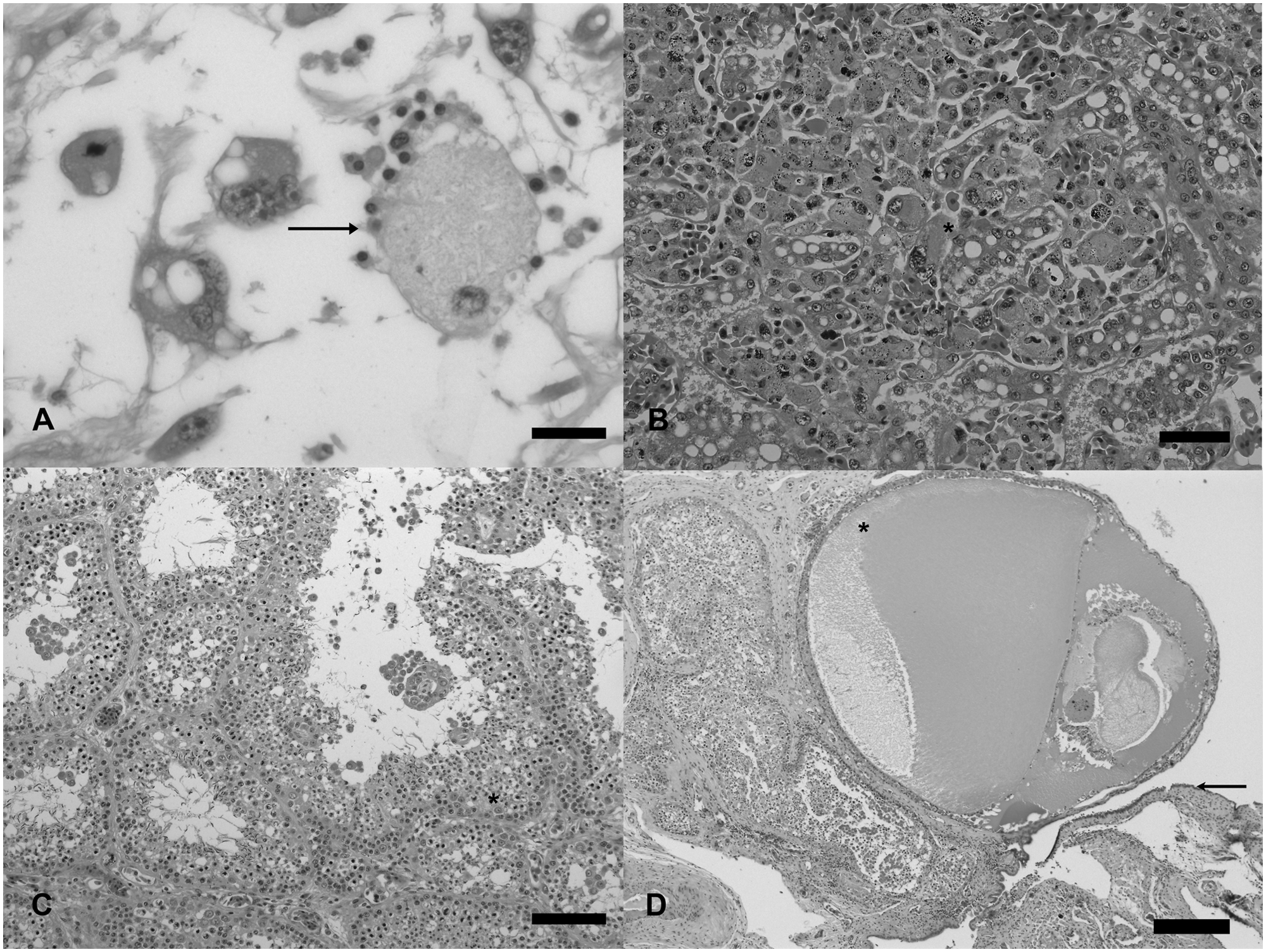
Histologically, the mass was composed entirely of adipose tissue that seamlessly transitioned to anastomosing bands of streaming neoplastic cells. The neoplastic cells were highly pleomorphic and ranged from round, to spindle shaped, to kite-shaped with indistinct cell borders. The neoplastic cells had abundant cytoplasm that often contained abundant, distinct, clear cytoplasmic vacuoles. Nuclei were round to oval or, in some cases, folded and irregular, large and occasionally very large (up to 50 μm in diameter), with up to 4, variably sized, deeply eosinophilic nucleoli. Multinucleated giant cells and karyomegalic cells were often present (Fig. 1A). Within multinucleated giant cells there is often considerable intracellular anisokaryosis with nuclei varying in diameter up to 4 times. Three mitotic figures were seen in 10 high power fields. Occasionally mitotic figures were irregular or bizarre. Within the neoplasm, there was scattered multifocal hemorrhage and necrosis, and moderate, multifocal extramedullary hematopoiesis.
Madagascar tree boa (Sanzinia madagascariensis).
Neoplastic cells were also seen in dilated hepatic sinusoids, scattered within alveolar capillaries and within cerebral vessels. Circulating neoplastic cells had a very similar cytological appearance to cells within the primary neoplasm, displaying marked pleomorphism and multinucleation (Fig. 1B). The cytoplasmic vacuoles with the neoplastic cells did not stain with PAS. No areas of mineralization or osseous metaplasia were recognized in the sections examined, which may explain the mineralized opacities on radiographs.
In addition to the neoplasm, there were disseminated granulomas involving the kidney, liver, spleen, lung, brain, and anterior chamber of the eye characterized by large numbers of epithelioid macrophages, hemosiderophages, and lymphocytes surrounding a central core of necrotic cellular debris. Fungal and acid-fast organisms were not detected in these areas with GMS or acid-fast staining. Swelling of the liver recognized grossly is attributed to a combination of inflammatory and neoplastic infiltrations within the sinusoids and parenchyma, and mild, diffuse hepatocellular vacuolation (Fig. 1B). Each cell contained up to 3 vacuoles, which were large and well defined and interpreted to be lipid. No histological changes were noted in the ventricular myocardium that could be associated with ventricular enlargement seen grossly.
The gonad was composed of mature seminiferous tubules lined by Sertoli cells and between 2 and 3 layers of germinal cells. Within seminiferous tubules there were moderate numbers of spermatids (Fig. 1C). Adjacent to this and in other areas of the gonad there were several ovarian follicles at varying stages of maturation (Fig. 1D). The surface of the gonad was bordered by a single layer of cuboidal epithelium. The presence of male and female gonadal tissue is indicative of an ovotestis.
Neoplasia in captive snakes was once thought to be uncommon (2.3–3.8%), 8 with documentation confined to single case reports that reported tumors of epithelial, mesenchymal, and hematopoietic origin in a wide range of locations. More recently, several case series have suggested that this is an underestimate and that the true incidence is between 12% and 23%.4,13,24,29 The incidence of neoplasia reported is highly variable over time and between institutions: varying between 0% and 14% in one report. 24 Similarly neoplasms of mesenchymal origin are reported to occur at widely different rates between institutions (between 34% and 62% of neoplasms diagnosed).4,24 It has been suggested that this may be the result of inter-institution variation in species and husbandry techniques, the presence of an environmental carcinogen, or an outbreak of an as yet unidentified oncogenic infectious agent (e.g., type C retroviruses). Type C retroviruses have been identified within snake neoplasms of epithelial and mesenchymal origin, although a causal link to neoplastic transformation or with the observed differences in incidence has not been proven.4,5 Sample size or the effect of an as yet unidentified environmental carcinogen may also be significant. Despite this wide variation in incidence, there is a consistently high rate of malignancy of neoplasms irrespective of type with between 70% and 80% of neoplasms detected having histopathological or behavioral criteria of malignancy.4,13,29 This feature is even more striking when mesenchymal neoplasms are viewed alone, with up to 90% of neoplasms showing criteria of malignancy. 24
A subset of highly malignant anaplastic spindle cell sarcomas with neoplastic giant cells, which often metastasize widely, have been identified in a number of snake species. 24 Such neoplasms, however, arise within skin, somatic musculature, or precardial connective tissue and as such appear distinct from the present case.
The neoplasm in the current case shares many similarities to a reported case also in a Madagascar tree boa. 2 In both cases, the neoplasm arose within the peri-pancreatic adipose tissue, was composed of highly pleomorphic mesenchymal cells, and displayed a solid sarcomatous primary phase with an intravascular metastatic phase. However, in the previous case, the neoplasm was cytologically biphasic, a feature not recognized in the present case, with spindloid cells within the primary neoplasm and round to polygonal, discrete cells within vessels. A transitional cellular phenotype was also detected in some areas, which supported the diagnosis of a biphasic tumor. Striking similarities between the 2 cases, however, suggest that this type of neoplasm may be a distinct entity within the Madagascar tree boa and may represent a genetic susceptibility in this species.
Accurate characterization of anaplastic spindle cell tumors in snakes 24 has been impeded by the highly anaplastic nature of the tumor cells and lack of validated immunohistochemical markers in reptiles. In the current case, further immunohistochemical characterization of the neoplasm was attempted but the results were inconclusive. Primary antibodies raised in mammals against mammalian antigen are routinely used in research settings to detect a wide range of reptilian target antigens,1,3,18 but are rarely conclusive in reported diagnostic cases.2,16,22 The inconclusive results in the present case were attributed to either the degree of anaplasia of the neoplastic cells altering antigen expression or a lack of protocol optimization for use in reptile tissue. Because the neoplasm appeared to have a high lipid content grossly and due to the presence of PAS-negative intracytoplasmic vacuoles, a tentative diagnosis of liposarcoma was made.
In addition to the neoplasm in the current case, several granulomas were noted within multiple organ systems. Systemic heterophilic or histiocytic granulomatous inflammation has been reported in snakes and other reptiles as the result of a diverse range of causes including bacterial, mycobacterial, chlamydial, protozoal, fungal, algal, and parasitic infections 28 and as the result of persisting foreign bodies. 11 In the present case, no bacteria, mycobacterial, or fungal elements were detected with special stains. No fresh tissue was available for culture or molecular microbiology. One case of concurrent mesenchymal neoplasia with a confirmed systemic fungal infection has been reported previously in a captive-bred mangrove snake (Boiga dendrophila). In this previous report, fungal elements could be seen prominently within disseminated granulomatous lesions stained with hematoxylin and eosin; however, no causal link was noted between the inflammatory and neoplastic lesions. 15 In the absence of detectable infectious organisms, the systemic inflammatory process may represent a paraneoplastic process. Granulomatous inflammation has been reported in mammals and human beings as the result of paraneoplastic production of granulocyte–monocyte colony-stimulating factor.7,23
Hermaphroditism is the developmental abnormality that results in part or all of the gonads of both sexes being present within an individual. 27 The condition has been routinely reported in several mammalian species21,27 and intermittently reported in amphibians 6 and several species of reptile: tortoises,9,20 2 lizards,17,30 turtles, 26 and a snake. 12 It is also frequently cited as an effect of chronic environmental exposure to certain classes of pesticides and herbicides also known as environmental steroid mimetics, fish and amphibians being particularly susceptible. 14 In a brief report, 12 a phenotypically female boa constrictor with ovotestes comprising immature follicles and inactive, hypoplastic seminiferous tubules is described. However, tissues were not available for a complete external examination. In the present case, the boa had female tubular genitalia. In contrast to the previous case report, the gonad in the present case appeared to be well developed, with maturing ovarian follicles and mature sperm within seminiferous tubules. A wide range of morphologies and functionality has been described in intersex animals. It is difficult to know if this snake was truly infertile. However, the snake was housed with a male and never subsequently laid eggs.
Sexual differentiation is a complex polygenic process that is incompletely understood in mammals and reptiles. Gonadal sex in mammals is determined by the presence or absence of a testis-determining factor (Sry) located on the Y-chromosome. This master gene initiates testicular differentiation from the primordial genital ridge in the embryo. In the absence of Sry, gonads will develop as ovaries. However, abnormalities in many other genes have been associated with intersex conditions, and many intersex animals do not have an abnormality in known sex-determining genes. The development of tubular and external genitalia occurs as the result of genetic and hormonal signaling pathways initiated by the gonad. Intersex phenotypes have been reported in both XX and XY individuals. 27
The mechanisms of sexual determination in reptiles are less well understood and in many instances involve environmental as well as genetic influences. However, all snakes, including boids, exhibit genetic sexual differentiation with little or no influence from environmental factors such as temperature that may affect gonadal sex in other species. 25 Unlike colubridae and viperidae, male and female boids are karyotypically identical. 19
Snakes have Z and W sex chromosomes and, in contrast to mammals, the male is the homozygote, with ZZ being the male genotype. In the current case, the testis-determining pathway is the default with an “ovarian determining factor” present on the W chromosome. Homologues of mammalian sex-determining genes have been identified in reptiles but their function or significance is yet to be determined. 25
Early reports suggested that all hermaphrodite reptiles were genotypically male. 10 However, as the mechanisms for sex determination in both mammals and boids share significant similarities, it is likely that hermaphrodites can be produced from both ZZ and ZW genotypes. Without further genotyping, it is only possible to speculate on the underlying cause of the intersex phenotype seen in the present case.
Footnotes
a.
Carnivore Critical Care, Oxbow Animal Health, Murdoch, NE.
b.
Clone V9, anti-human HLA-DR antigen alpha chain clone TAL-IB5, clone EC 3.2.1.17; Dako UK Ltd., Ely, Cambridgeshire, UK.
Declaration of conflicting interests
The author(s) declared no potential conflicts of interest with respect to the research, authorship, and/or publication of this article.
Funding
The author(s) received no financial support for the research, authorship, and/or publication of this article.