
Abstract
The current report summarizes the incidence and variety of neoplasms in pot-bellied pigs as documented by the pathology group at the University of Tennessee, College of Veterinary Medicine (UTCVM) between 2004 and 2011. Sixty-three pot-bellied pig cases (53 necropsies and 10 surgical biopsies) were identified from the UTCVM case database. Of these, 22 cases from 21 patients (34.9%) were given a diagnosis of neoplasia, including 10 females, 3 spayed females, 2 males, and 7 neutered males. The mean age of affected animals was 11.3 years. The incidence of neoplasia among the necropsy cases was 28% (15/53), and in the surgical biopsy material, the incidence was 70% (7/10). Reproductive and gastrointestinal tissues were most commonly affected, and malignancies were frequently identified, with hepatic and intestinal carcinomas predominating. Multiple neoplasms were often identified in patients submitted for necropsy, and in 4 out of 11 of the necropsy cases, at least 1 of the neoplasms was a malignancy. Six necropsy cases revealed metastatic spread.
In the past 10 years (2001–2011), the incidence of reported neoplasia in pet pot-bellied pigs has increased, mostly as a consequence of increased life spans resulting from improved veterinary medical care. The majority of the literature regarding neoplasia in this species refers to their propensity to develop genital tract tumors, most of which are of smooth muscle origin and involve either the uterus or the ovary.1,2,7,13 While some smooth muscle tumors are malignant, the overall behavior of these tumors typically involves locally invasive growth rather than metastases. In addition, uterine adenomas and adenocarcinomas have been reported in older, intact females,1,5,7 as well as an interstitial cell tumor in an older, intact male. 17
Other pot-bellied pig organ systems with consistent reporting of neoplasia include the gastrointestinal tract and liver where gastric carcinoma, small intestinal carcinoma, hepatocellular carcinomas, biliary carcinomas, and hepatomas have been found.10,12 In addition, oral cavity tumors, specifically squamous cell carcinoma, have also been reported.9,19 The objective of the current study was to determine the incidence of neoplastic processes within a population of pet pot-bellied pigs seen at the University of Tennessee, College of Veterinary Medicine (UTCVM; Knoxville, Tennessee) over the past 10 years.
The pathology database of UTCVM was searched for cases with a diagnosis of neoplasia, from either surgical biopsy or necropsy, from the past 10 years (2001–2011). Slides from all identified cases were reviewed by a board-certified pathologist to verify the diagnosis. A specific porcine tumor classification system was not available. Basic clinical and historical information for each case was also recorded.
Immunohistochemistry was performed on a single vertebral round cell tumor case (case 7). Cluster of differentiation (CD)3, a CD79, b and multiple myeloma oncogene-1 (MUM-1) c antibodies were used, with porcine lymphocytes and plasma cells serving as positive controls. These same cells served as negative controls by not receiving the primary antibodies in each case. All test cases were cut at 5 µm and placed on charged slides, air dried, and then heated at 60°C for 15 min. All slides were deparaffinized with xylene and then rehydrated through graded ethanols to deionized water. Heat-induced epitope retrieval was performed using ethylenediamine tetra-acetic acid d buffer at pH 9 in a steamer for 25 min at 95°C. All slides were rinsed in deionized water and soaked in Tris buffered saline–Tween for 10 min before being placed into the autostainer. e Three percent hydrogen peroxide block was applied for 5 min to the CD3 and CD79 slides, and all 3 antibody cases received a 5-min serum-free protein block. f CD3 was applied for 30 min at a 1:100 dilution, CD79 was applied for 30 min at a 1:1000 dilution, and MUM-1 was applied for 30 min at a 1:75 dilution. The labeled polymer anti-mouse commercial horseradish–peroxidase system g was applied for 30 min for CD3 and CD79, and a second commercial horseradish–peroxidase system h was applied for 30 min for MUM-1. DAB (3,3’diaminobenzidine tetrahydrochloride) i was applied as the chromogen for 10 min. All slides were then subsequently rinsed with deionized water, stained for 5 sec in hematoxylin, blued in ammonia water, dehydrated through ethanols, cleared with xylene, and coverslipped.
The authors identified 63 pot-bellied pig cases (53 necropsies and 10 surgical biopsies) from the UTCVM database. Of these 63 cases, 22 cases (incidence 34.9%, 15 necropsies and 7 surgical biopsies) from 21 patients had a diagnosis of neoplasia. The incidence of neoplasia among the necropsy cases was 15 out of 53 (28%) and from the surgical biopsy material the incidence was 7 out of 10 (70%). One animal (case 8) with a surgical biopsy result was subsequently necropsied.
The signalment, types, and associated characteristics of the neoplasms are listed in Table 1. The average age of the necropsy animals was 12.2 years, while that for the surgical biopsy patients was 9.6 years. The mean age of all affected animals was 11.3 years.
Neoplasms of pot-bellied pigs: a retrospective case series (2004–2011).*
Bold text in rows indicates the case has multiple neoplasms listed in the table. hpf = high power field; N = necropsy; B = biopsy; F = female; M = male; NR = not reported; FS = female spayed; MN = male neutered.
Necropsy animals consisted of 5 intact females, 3 spayed females, 1 intact male, and 6 neutered males. Surgical biopsy cases were composed of 5 females (1 of which was spayed prior to becoming a necropsy specimen) and 1 each of an intact male and a neutered male. Total cases consisted of 10 females, 3 spayed females, 2 males, and 7 neutered males.
Of the 7 surgical biopsy cases, 4 cases, including 1 animal with 2 tumors, were benign and 3 were malignant. Leiomyomas were the most common neoplasm, which were found in extragenital sites including the mesentery and perineal locations. One case had an accompanying mammary adenoma (Fig. 6), suggesting a possible increase in sex hormone levels. An interstitial testicular tumor was considered benign based on histomorphologic features. The malignant tumors were uterine leiomyosarcoma and dermal and nasal squamous cell carcinomas. The pig presented for necropsy 3 years following an initial diagnosis of a mesenteric leiomyoma for a perineal leiomyoma and concurrent hepatocellular carcinoma.
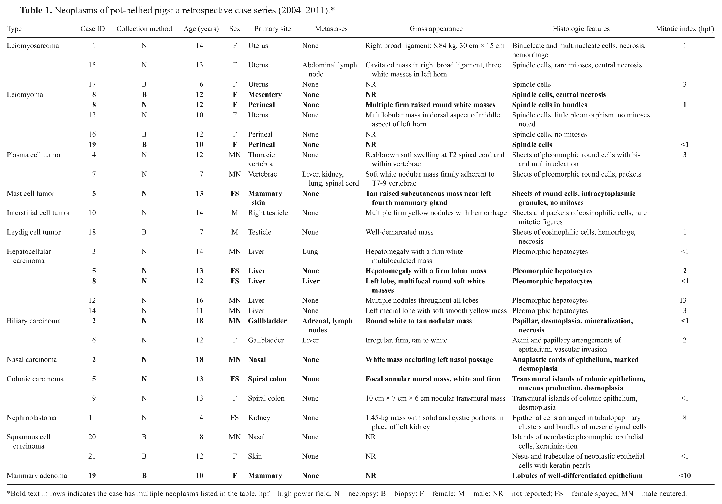
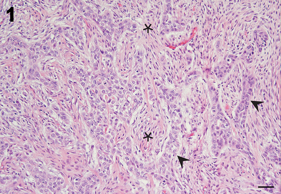
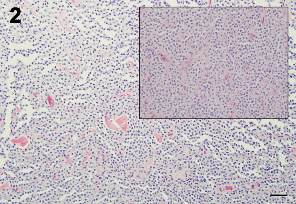
Eleven necropsy cases had a single neoplasm, and 4 animals had 2 separate neoplasms for a total of 19 tumors. Four (24%) were benign, and 15 (76%) were malignant. Of the benign tumors, 2 were leiomyomas, 1 a cutaneous mast cell tumor, and 1 a testicular interstitial cell tumor. Malignancies consisted of 2 leiomyosarcomas, 5 hepatocellular carcinomas (Fig. 5), 2 cholangiocarcinomas, 1 renal nephroblastoma, 2 plasma cell tumors (Figs. 3, 4), 2 colonic adenocarcinomas, and 1 nasal adenocarcinoma (Fig. 1). Seven malignancies had evidence of metastases: both cholangiocarcinomas, 2 of the hepatocellular carcinomas, and 1 each of the colonic adenocarcinomas, plasma cell tumors, and a leiomyosarcoma. While 1 hepatocellular carcinoma and 1 cholangiocarcinoma had metastasized locally throughout the liver, the other 5 cases involved more distant metastases: adrenal and lymph node for a cholangiocarcinoma; lung for a hepatocellular carcinoma; liver, kidney, lung, and spinal cord for a plasma cell tumor; lymph node for a colonic adenocarcinoma, and abdominal mesentery for a leiomyosarcoma. The site of origin of the neoplasms was as expected for the tumor type, except that 1 leiomyoma was perineal, the plasma cell tumors involved the vertebrae, and the mast cell tumor was found on the ventral abdominal skin (Fig. 2). In 2 of the leiomyosarcomas and 1 leiomyoma there was evidence of concurrent uterine cystic endometrial hyperplasia.
Pot-bellied pig; case 2; nasal adenocarcinoma. Nasal turbinate architecture is effaced by a proliferation of cords and trabeculae of epithelial cells (arrowheads) with features of pleomorphism and abundant desmoplasia (asterisks). Bar = 25 µm.
Pot-bellied pig; case 5; cutaneous mast cell tumor. Encapsulated nodule composed of sheets of well-differentiated mast cells. Bar = 50 µm. Inset: cellular features of mild anisocytosis and anisokaryosis and limited granularity are features of these mast cells.
Pot-bellied pig; case 7; plasma cell tumor. Large neoplastic cellular mass (arrowhead) ventral to the thoracic vertebra. Similar neoplastic cells fill vertebral marrow spaces. Bar = 200 µm.
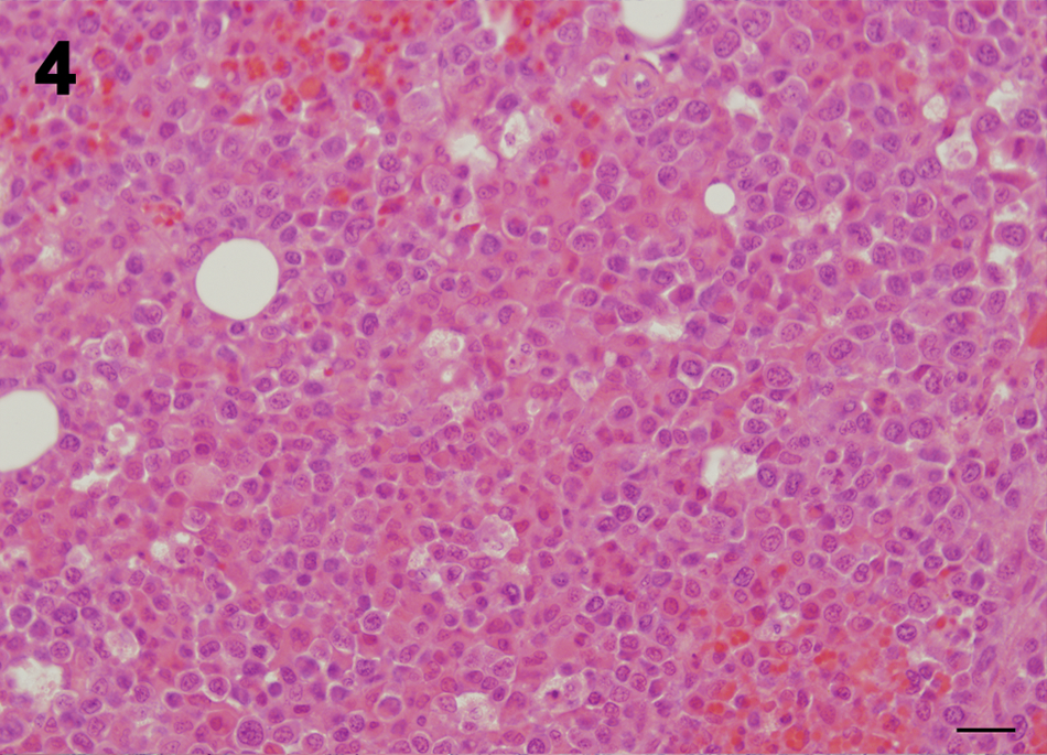
Pot-bellied pig; case 7; plasma cell tumor. Neoplastic cells arranged in sheets. Cells are round with distinct cell borders, eccentrically placed nuclei, and prominent eosinophilic cytoplasmic globules. Bar = 20 µm.
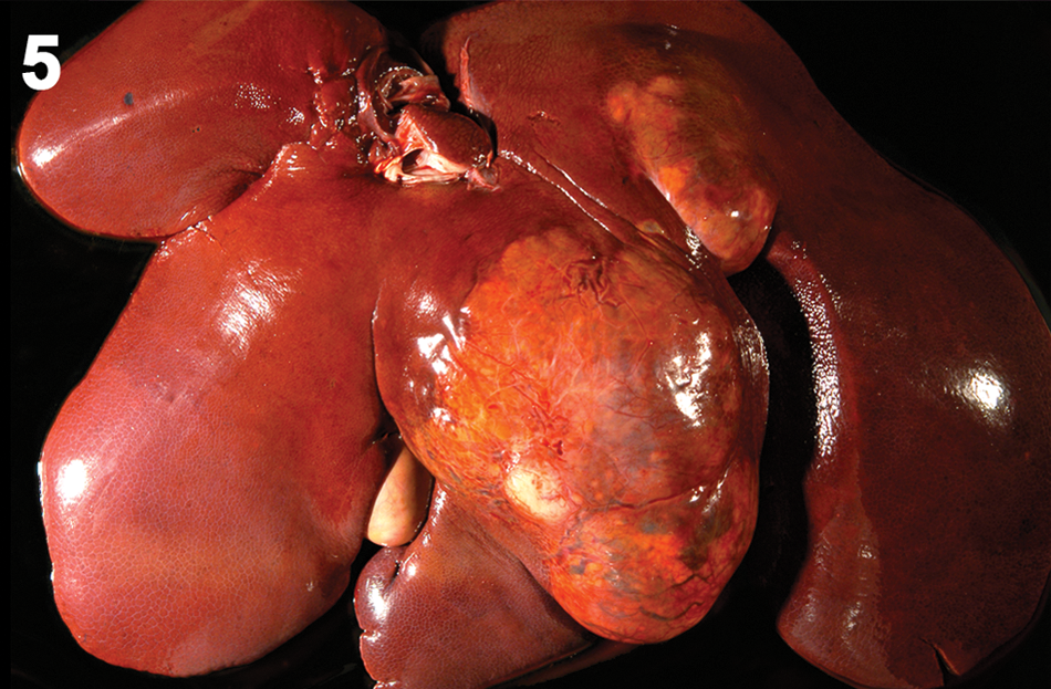
Pot-bellied pig; case 14. Hepatocellular carcinoma occupying the left medial liver lobe.
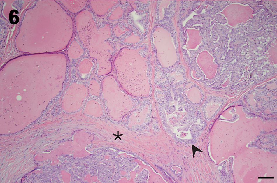
Pot-bellied pig; case 19; mammary adenoma. The mammary gland nodule is composed of tubulopapillary arrangements of cuboidal epithelium and abundant proteinaceous fluid (arrowhead). Fibrous tissue separates lobules (asterisk). Bar = 100 µm.
A large percentage (70%) of surgical biopsies from pot-bellied pigs at UTCVM yielded a diagnosis of neoplasia. There was a lesser but still impressive incidence of neoplasia in the pot-bellied pig at necropsy (28%), and the overall percentage of affected animals was 34.9%. The mean age of pot-bellied pigs with neoplasia in the present study was 11.3 years, confirming that it is often a disease of geriatric animals. Improved husbandry and an active rescue program within Tennessee and neighboring states help ensure that these pet pigs achieve a longer life span.
A number of the neoplasms were found in the reproductive tract of intact male and female pigs. Much has been published previously to suggest that uterine and ovarian smooth muscle tumors may be a result of constant hormonal stimulation achieved in older intact individuals.2,7 Indeed, the incidence of neoplasia cases involving the reproductive tract in the current study reflects the older intact subset of animals. The involvement of tissues besides the uterus, including the abdomen, mesentery, and perineal locations, has been previously published; these neoplasms can arise from smooth muscle from any origin including a blood vessel. When uterine smooth muscle neoplasms were present, cystic endometrial change was noted concurrently in 3 of the cases. 7 Similarly, interstitial cell tumors have been reported in geriatric intact males. 17 While little is known about the behavior of interstitial cell tumors, the histologic features in the single case reported herein are suggestive of a benign course.
Of the 11 necropsy cases, 4 had multiple unrelated tumors, 1 of which was malignant in each case. This suggests a relatively high tumor burden and the possibility of environmental, age, dietary, or genetic predispositions to tumor development in these pigs. The gastrointestinal tract was the source of 47% (9/19) of the tumors. Hepatic tumors, both biliary and hepatocellular in origin, have been previously reported in pigs and composed 37% (7/19) of these cases.10,12 Similarly, colonic adenocarcinomas have been previously reported, and in this series, both originated in the spiral colon. 4 Gastric and small intestinal carcinomas have been reported, but none were identified in the present study. 10 In 2 previously reported cases, colonic adenocarcinomas were accompanied by intestinal T-cell lymphosarcoma as determined by positive CD3 expression. 4 No lymphosarcomas were confirmed in the cases reported herein despite performing immunohistochemistry on 1 of the vertebral round cell tumors. The reason for this absence is not known, but few cases have been reported in pot-bellied pigs, and in domestic pigs the lymphosarcoma is often a tumor of young, rather than geriatric, animals. 8
Nephroblastomas are the most common primary renal tumors in pigs. 6 In 1 large study of 74 cases, 66 (89%) were unilateral and 8 (11%) were bilateral, with metastasis occurring in 2 cases. Although thought to be insidious in nature, nephroblastomas are often found at slaughter as incidental changes. There was a single unilateral nephroblastoma in the current series, but renal parenchyma was entirely replaced, and hence it was considered a malignant variant.
Multiple myeloma is rare but reported in swine. One case involved the spinal cord, sternum, and pelvis, and another was previously diagnosed in visceral and peripheral lymph nodes in a 6-month-old, crossbred hog, primarily because of highly developed rough endoplasmic reticulum. 16 Packeting of round cells and binucleate and trinucleate morphology are also characteristic of multiple myeloma. Both multiple myeloma cases in the current study involved the vertebral bodies. In 1 of the 2 cases described herein, immunohistochemical expression for lymphocyte markers (CD3 and CD79) was negative, and equivocal expression for MUM-1, a plasma cell marker verified in dogs, 15 was noted.
Mast cell neoplasia, either systemic3,18 or cutaneous, 11 has been previously reported in miniature pigs and domestic pigs, respectively. The case reported in this series was a benign cutaneous mast cell tumor in the ventral abdominal skin.
Ethmoid mucosal tumors of pigs have also been previously reported. 14 Nasal adenocarcinoma has been reported and is considered an endemic form among pigs from Brazil, China, and Ghana. 20 There was a single nasal adenocarcinoma in the current necropsy series and a nasal squamous cell carcinoma in the biopsy series. Although squamous cell carcinoma has been most commonly reported in the oral cavity of pot-bellied pigs,9,19 the 2 cases in the present study were nasal and dermal in origin, respectively.
Mammary neoplasms are infrequent in pigs and pot-bellied pigs. The features of the mammary adenoma described herein closely resembled those seen in other species. Overproduction of sex hormones was not determined in the current case, but the animal had a concurrent leiomyoma.
At the time of death or euthanasia of the animal, 7 out of 16 (44%) malignancies had metastasized either locally (2) or to more distant sites (5).
In conclusion, the current study represents one of the largest retrospective studies of neoplasia in pot-bellied pigs. Reproductive system neoplasms in both sexes are seen quite frequently. The gastrointestinal tract, including the liver and intestine, is also a preferred location for cancerous development in these animals, and most of these are malignancies. Often at the time of necropsy, metastases are widespread. Immunohistochemistry can be adapted for tumor identification in this species.
Footnotes
Acknowledgements
Misty Bailey is acknowledged for excellent editorial support and Anik Vasington for graphics assistance.
a.
Monoclonal mouse CD3 antibody F72.38, Dako North America Inc., Carpinteria, CA.
b.
Monoclonal mouse CD79 antibody HM57, Dako North America Inc., Carpinteria, CA.
c.
Monoclonal mouse MUM-1 antibody MUM-1p, Dako North America Inc., Carpinteria, CA.
d.
Fisher Scientific, Waltham, MA.
e.
Autostainer S3400, Dako North America Inc., Carpinteria, CA.
f.
Dako North America Inc., Carpinteria, CA.
g.
Envision+System HRP Anti-mouse, Dako North America Inc., Carpinteria, CA.
h.
Anti-mouse, Biocare Medical, Concord, CA.
i.
DAB, Dako North America Inc., Carpinteria, CA.
Declaration of conflicting interests
The author(s) declared no potential conflicts of interest with respect to the research, authorship, and/or publication of this article.
Funding
The author(s) received no financial support for the research, authorship, and/or publication of this article.