
Abstract
Various neoplasms have been reported in Vietnamese pot-bellied pigs (Sus scrofa) with few reports of hepatocellular tumors. Twenty-two pot-bellied pigs diagnosed with hepatocellular carcinoma at necropsy over a 3-year period at one institution are described, representing 29% of the total pot-bellied pigs necropsied. The average age of affected pigs was 16.6 years with 15 males and 7 females. The most common clinical signs were decreased appetite (16/22) and weight loss (7/22). Grossly, the majority were massive tumors (13/22) with fewer nodular tumors (8/22) and 1 diffuse tumor. Massive tumors were typically multilobulated, very large, and encompassing 1 or more adjacent liver lobes, and were soft to firm and tan-yellow to orange-brown. Nodular tumors had multiple, 1–15 cm in diameter, discrete nodules in multiple liver lobes. Gross evidence of abscesses, necrosis, hemorrhage, or cysts associated with the tumor was occasionally described. Half of the cases had possible intrahepatic metastasis, and extrahepatic metastasis was identified in 3 cases, including to the hepatic lymph node (1/3), lung (2/3), spleen (1/3), and kidney (1/3). Histologically, all tumors had a trabecular or solid pattern, or a combination. An adenoid pattern was only identified in small regions of a few tumors. The neoplastic cells were relatively well-differentiated with moderate pleomorphism and a low mitotic index. Other histologic features within the tumors included intracellular glycogen or lipid accumulation, extramedullary hematopoiesis, foci of coagulative necrosis, and bile stasis. Aged pot-bellied pigs can be predisposed to hepatocellular carcinomas, which are locally aggressive and can metastasize within the liver and to other organs.
There are few reports of neoplasia in Vietnamese pot-bellied pigs (Sus scrofa), including uterine leiomyoma/leiomyosarcoma,9,10 oral squamous cell carcinoma,5,12 gastric carcinoma, 7 intestinal adenocarcinoma, 7 cholangiocellular carcinoma, 7 endometrial adenocarcinoma, 4 chronic lymphocytic leukemia, 3 and possible splenic hemangiosarcoma. 8 There are rare reports of hepatocellular adenoma and carcinoma in Vietnamese pot-bellied pigs,5-7 and metastasis of hepatocellular carcinoma to the lungs has been documented. 7
Vietnamese pot-bellied pigs, also often referred to as miniature pet pigs, are a popular household pet. 11 Unlike domestic pigs raised for slaughter, pot-bellied pigs have an average lifespan of 20–25 years 11 ; therefore, they provide an opportunity to study the pathology of aging pigs. The purpose of the current retrospective study is to identify and describe a population of aging Vietnamese pot-bellied pigs with hepatocellular carcinoma.
The large animal pathology database at New Bolton Center at the University of Pennsylvania, School of Veterinary Medicine (Philadelphia, Pennsylvania) was searched for Vietnamese pot-bellied pigs submitted for necropsy from February 2008 through January 2011; 76 pot-bellied pigs were identified. The necropsy records were reviewed from all pigs with primary liver neoplasia in the final diagnosis (26 cases). Cases were excluded if the diagnosis was primary biliary neoplasia (2 cases) or undifferentiated disseminated carcinoma (1 case). The cases with reported primary hepatocellular tumors were re-evaluated. The tissue sections taken at the time of necropsy had been handled routinely with 4–5-µm sections prepared from formalin-fixed, paraffin-embedded tissue samples and stained routinely with hematoxylin and eosin stain. Histopathology of the livers and of available tissues with metastatic disease was reviewed by a board-certified anatomic pathologist (JL Haddad) to confirm or amend the original diagnosis. An additional case was excluded because the histopathology was not diagnostic for a hepatocellular neoplasm on re-evaluation. For 1 case with reported hepatic lymph node metastasis, the histologic evidence of metastatic disease could not be confirmed on re-evaluation; however, the original diagnosis of metastasis was included in the results of the study. Criteria used for diagnosis of hepatocellular carcinoma, as opposed to benign hepatocellular neoplasia, were based on guidance from the literature and included thickness of hepatocellular trabeculae within the tumor (>3 cells thick), variability of trabecular thickness, nuclear atypia and variability, multinucleation, regions of a solid growth pattern, invasion of adjacent parenchyma, or extrahepatic metastasis.1,2 Based on these criteria, the remaining 22 pot-bellied pigs were identified as having hepatocellular carcinoma and were designated for inclusion in the study.
Information was taken from the necropsy records including signalment (age and sex), documented clinical signs, gross appearance of the liver, locations of metastatic disease (if present), gross and histologic descriptions, diagnoses of concurrent diseases, and whether death was natural or the animal was euthanized. The gross pattern of the hepatocellular carcinoma (massive, nodular, or diffuse) was categorized based on the description provided in the necropsy report. Massive tumors were defined as those encompassing 1 or multiple adjacent lobes, nodular tumors were defined as those with multiple nodules in multiple liver lobes, and diffuse tumors were defined as indistinct infiltration throughout the liver.1,2 Evidence of possible intrahepatic metastasis was defined by the presence of discontinuous involvement of multiple liver lobes, regardless of overall tumor pattern.
The liver histopathology for each case was reviewed by a board-certified anatomic pathologist (JL Haddad), and features of the neoplasm were documented. A trabecular pattern was defined as plates of neoplastic hepatocytes separated by sinusoidal spaces, an adenoid pattern was defined as acinar formation by neoplastic hepatocytes, and a solid pattern was defined as sheets of neoplastic hepatocytes without separation by sinusoids.1,2 The mitotic index was defined as the number of mitotic figures in neoplastic hepatocytes in 10 consecutive high power fields. Additional histologic findings within the neoplasm were identified and recorded. Hepatocellular glycogen was identified as swelling and clearing of the hepatocyte cytoplasm, and lipid was identified as discrete clear cytoplasmic vacuoles.
Based on the inclusion criteria, 22 pot-bellied pigs with hepatocellular carcinoma were identified, which was 29% of the pot-bellied pigs necropsied during this timeframe. The average age of affected pigs was 16.6 years with a range of 10–20 years; 2 pigs were only identified as aged pigs without a specific age listed on the necropsy submission form. The pigs included castrated males (13/22, 59%), intact females (5/22, 23%), intact males (2/22, 9%), and spayed females (2/22, 9%). Of all of the pot-bellied pigs necropsied during this timeframe (76 pigs), 39 were female (51%) and 27 were male (49%). The most common clinical signs reported were decreased appetite (16/22, 73%) and weight loss (7/22, 32%). The duration of these clinical signs, when reported, ranged from days to months. Three pigs had no reported clinical signs possibly related to their liver tumor. Additional rarely reported signs included bleeding from the colon and vomiting. Most of the pigs were euthanized (15/22, 68%), several died naturally (6/22, 27%), and, in 1 case, the manner of death was not indicated in the necropsy report (1/22, 5%).
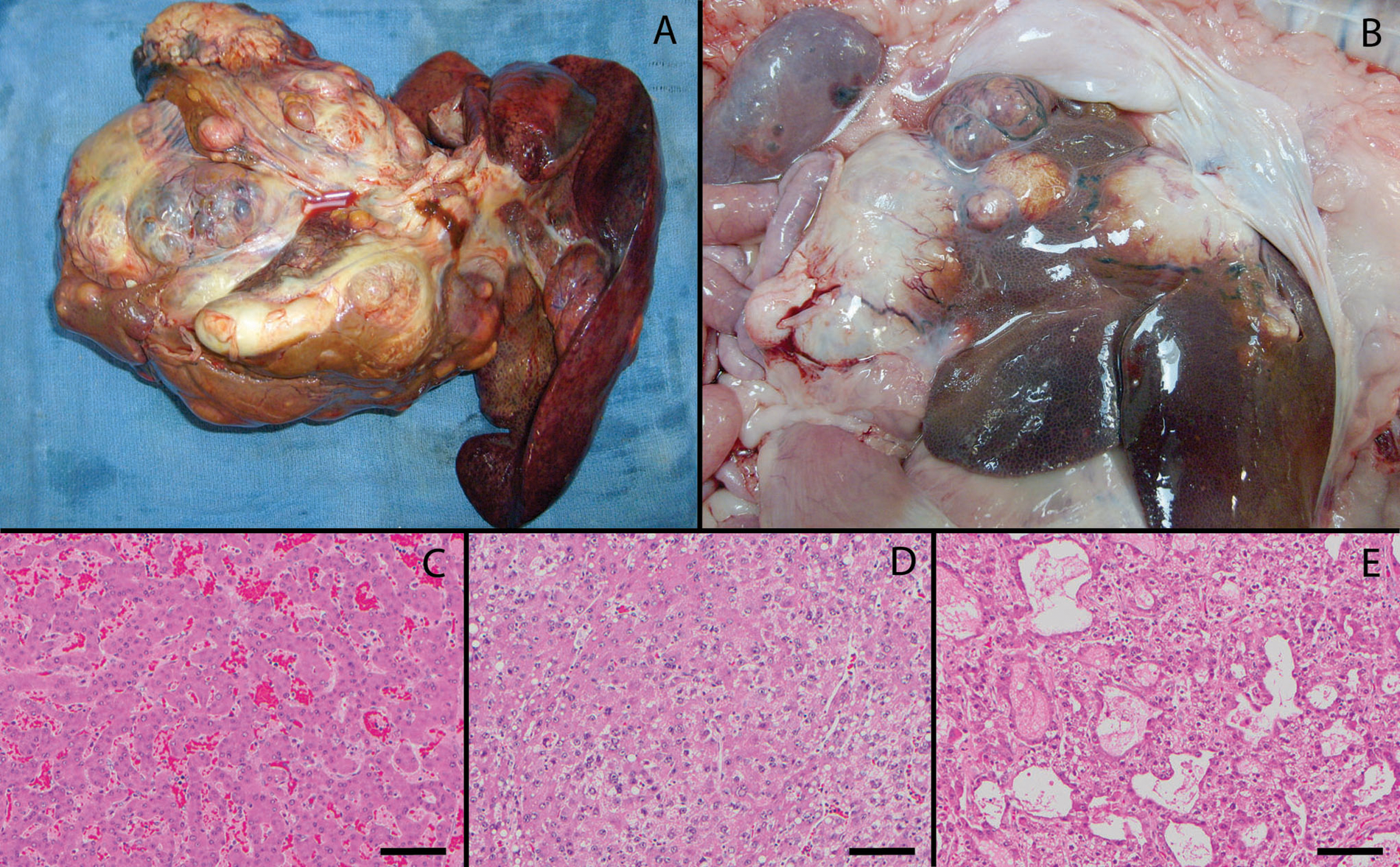
On gross examination, the majority of cases had massive tumors (13/22, 59%) with fewer nodular tumors (8/22, 36%) and 1 diffuse tumor (1/22, 5%). Massive tumors were typically multilobulated, very large, and encompassing 1 or more adjacent liver lobes, and were soft to firm and tan-yellow to orange-brown (Fig. 1A). Nodular tumors had multiple, 1–15 cm in diameter, discrete nodules in multiple liver lobes; nodules were soft to firm and varied in color including orange-tan, brown-tan, white-tan, and tan-green (Fig. 1B). The single case with the diffuse tumor had massive enlargement of the liver with an irregular contour, and the liver was mottled orange-brown to tan and friable. Gross evidence of abscesses (6/22, 27%), necrosis (4/22, 18%), hemorrhage (3/22, 14%), or cysts (2/22, 9%) associated with the tumor was occasionally described. Other concurrent gross findings in the livers included choleliths (4/22, 18%), abscesses unassociated with the tumor (3/22, 14%), pus within the gallbladder or bile ducts (2/22, 9%), liver lobe torsion (1/22, 5%), a biliary cyst (1/22, 5%), and necrotic material in the gallbladder (1/22, 5%). Possible intrahepatic metastasis, which included cases with a nodular pattern or with a massive tumor and additional nodules in other lobes, was identified in half of the cases (11/22, 50%), and extrahepatic metastasis was identified in few cases (3/22, 14%), including to the hepatic lymph node (1/3), lung (2/3), spleen (1/3), and kidney (1/3). Cases with extrahepatic metastasis included 1 diffuse, 1 nodular, and 1 massive tumor. In the case with lymph node metastasis, the hepatic lymph node was enlarged to approximately 3 times the normal size with multiple white-tan foci disrupting the nodal architecture. In the case with pulmonary and renal metastasis, the lung had multiple, firm, irregular, red-brown masses throughout all lobes with foci of yellow oozing material on cut surface, and the kidney had a 3-mm tan depressed region in the cortex. In the case with pulmonary and splenic metastasis, no gross description was available for the metastases.
Common concurrent conditions identified at necropsy in these pot-bellied pigs included degenerative joint disease (11/22, 50%), renal cysts (9/22, 41%), glomerular disease (7/22, 32%), dental disease including tusk abscesses and periodontitis (5/22, 23%), and amyloidosis (3/22, 14%). Of the intact females, 2 pigs had uterine leiomyomas (2/5, 40%).
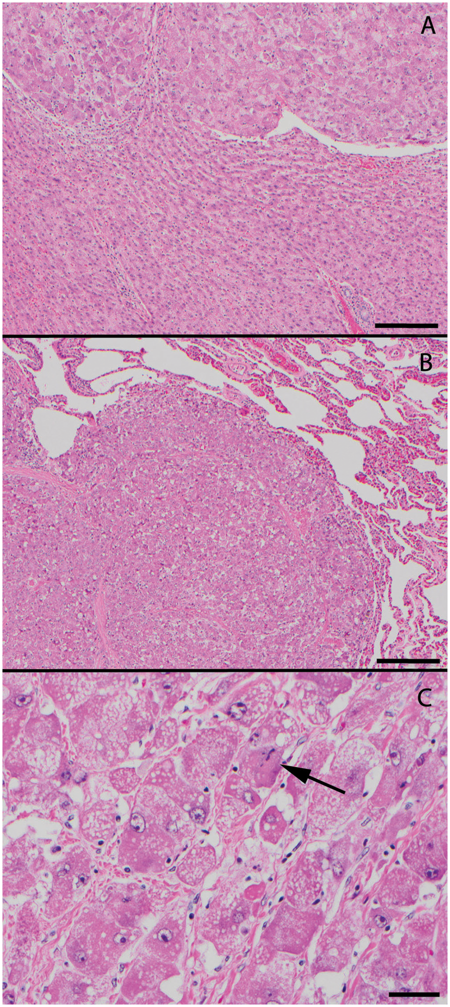
Histologically, most tumors had a combination of trabecular and solid areas (20/22, 91%); of these, slightly more had trabecular as the predominant type (11/20, 55%) and slightly fewer had solid as the predominant pattern (9/20, 45%). Two tumors had only a trabecular pattern (2/22, 9%). Within several of the tumors that had a combination of solid and trabecular areas, an adenoid pattern was identified in small regions of the tumor (6/20, 30%; Fig. 1C, 1D, 1E). In cases with adjacent liver included in the histologic section, there was often compression or infiltration of the adjacent liver parenchyma (Fig. 2A). Neoplastic cells were identified histologically at sites of metastatic disease (Fig. 2B). Cases with extrahepatic metastasis included 2 tumors with a predominance of a trabecular pattern and 1 tumor with a predominance of a solid pattern. Overall, the neoplastic hepatocytes were relatively well-differentiated with moderate pleomorphism, variable degrees of binucleation and multinucleation, and a low mitotic index. The mitotic index ranged from 0–3 with zero mitotic figures in many cases (12/22, 55%; Fig. 2C). Other frequently identified histologic features within the tumors included intracellular glycogen accumulation (14/22, 64%), intracellular lipid accumulation (14/22, 64%), extramedullary hematopoiesis (12/22, 55%), foci of coagulative necrosis (10/22, 45%), and bile stasis (8/22, 36%).
The overall incidence of hepatocellular carcinoma in this population of Vietnamese pot-bellied pigs was 29%, which appears to be higher than many reports in various species, 2 suggesting a predilection in pot-bellied pigs. The average age and range of ages affected by this tumor vary with species, and domestic pigs have been reported to have hepatocellular carcinoma at less than 6 months old 2 ; however, the study population described in the present report was an aged group of pot-bellied pigs with a range in age of 10–20 years, with an average of 16.6 years. In dogs, hepatocellular carcinoma is more common in males than females, but no sex predilection has been reported for the other domestic species. 2 The pigs in the current study were predominantly male (15/22, 68%), which is increased compared to the proportion of males in the pot-bellied pig necropsy population at this institution during this timeframe (37/76, 49%).
The massive form of hepatocellular carcinoma was more common in this group of pigs, as it is in dogs. 2 It is difficult to definitively distinguish between intrahepatic metastasis of the hepatocellular carcinoma and multiple de novo sites of tumor formation within the liver. For this study, possible intrahepatic metastasis was defined as multiple nodules in multiple liver lobes. Therefore, the proportion of cases with intrahepatic metastasis (50% of cases) may have been falsely increased due to liberal criteria for possible intrahepatic metastasis. The rate of extrahepatic metastasis of hepatocellular carcinoma is variable in the literature 2 and was 14% in the current study, which is lower than that reported for other species (>25%). 2 The presence of extrahepatic metastasis was not associated with gross tumor pattern in this group of pigs. Metastasis of hepatocellular carcinoma is most commonly to the hepatic lymph node and lung,1,2 which were sites of metastatic disease in this population, along with the spleen and kidney. Because the hepatic lymph nodes were not evaluated histologically in many of the cases in this population, pigs with metastatic disease may have been missed, falsely lowering the rate of extrahepatic metastasis.
Possible etiologies for hepatocellular neoplasia in domestic or laboratory animal species include infectious etiology and toxin or chemical exposure. Infectious agents associated with hepatocellular tumors include Helicobacter infection in mice, hepadnavirus infection in woodchucks, squirrels, certain avian species, and human beings, 13 and Hepatitis B virus and Hepatitis C virus in human beings. 2 There was no evidence of infectious etiology in the histologic sections in the group of pigs described herein; however, additional testing for viral etiology was not performed. Numerous chemical carcinogens have been associated with hepatocellular neoplasia; however, these chemicals are often used in industrial or research settings 2 and therefore it is unlikely these pigs were exposed to such agents. Naturally occurring toxins, such as aflatoxin, pyrrolizidines, and nitrosamines, have been associated with hepatocellular neoplasia.2,6 There was no known exposure of this group of pigs to these toxins; however, complete analysis of the pigs’ feed and environment was not performed.
Limitations to the current study include the retrospective nature, including a few gross necropsy reports with only very brief gross descriptions of the neoplasm, variably thorough investigation of metastatic disease documented at necropsy, and availability of only limited sections of liver and other organs for histologic evaluation. The gross pattern of the neoplasm (massive, nodular, or diffuse) had to be deduced from the gross necropsy description. Because only the available histologic sections taken at necropsy could be reviewed, it was difficult to thoroughly evaluate the sections for invasion of the adjacent parenchyma in these pigs. In addition, determination of the proportions of the tumors that had a solid, trabecular, or adenoid pattern was subjective and based only on the histologic sections available for review. On occasion, the artifactual separation of cells due to autolysis made it difficult to definitively identify trabecular versus solid regions of the tumor. Other limitations include the lack of complete history regarding the environment, travel history, genetic background, or familial relationships between the pigs included in the study and the fact that the pigs were all necropsied at a single institution. A majority of the pigs were submitted by 1 local pot-bellied pig rescue organization; therefore, the population can be biased towards pigs that enter this facility.
Although neoplasia has been uncommonly reported in Vietnamese pot-bellied pigs, the findings of the current study suggest that aged pot-bellied pigs can be predisposed to hepatocellular carcinomas, with a possible male predilection. These tumors can be locally aggressive and can metastasize within the liver and to distant sites.
Footnotes
Acknowledgements
The authors would like to thank Susan Armstrong for her efforts in the rescue and care of many of the pigs in this study and for her submission of these animals for necropsy.
Declaration of conflicting interests
The author(s) declared no potential conflicts of interest with respect to the research, authorship, and/or publication of this article.
Funding
The author(s) declared that they received no financial support for their research and/or authorship of this article.