
Abstract
A 2-year-old female llama (Lama glama), from a private zoological park, with anorexia, ataxia, dyspnea, ascites, and emaciation, was necropsied. Gross inspection, and cytological, histological, and immunohistochemical analyses were performed. A firm, white, multinodular mass, 25 cm in diameter was found in the liver parenchyma. Similar nodules up to 3 cm were found in lymph nodes, lung, diaphragm, and peritoneum. Histologically, the affected organs were replaced by an infiltrative growth of undifferentiated neoplastic cells. Immunohistochemically, neoplastic cells were positive for pancytokeratin (panCK), CK20, and CK19, and negative for thyroid transcription factor 1, CK7, and carcinoembryonic antigen. A diagnosis of poorly differentiated metastatic cholangiocarcinoma was made.
A 2-year-old female llama (Lama glama) from a private zoological park was submitted to the Pathology Department of the Faculty of Veterinary Medicine from Cluj-Napoca (Romania) for necropsy. The animal had a history of depression, anorexia, progressive weakening, ataxia, decubitus ulcers, fever, dyspnea, and abdominal distension. Death occurred 6 weeks after the onset of clinical signs. Necropsy, and cytological, histological, and immunohistochemical examinations were performed. Grossly, the animal was emaciated. Subcutaneous edema of the posterior limbs, abdomen, and thorax, hydrothorax, hydro-pericardium, and severe ascites (approximately 15 liters of fluid) with fibrin clots adherent to the visceral peritoneum, and multiple, acute, gastric ulcers were observed.
A firm, white-gray, multinodular mass, 25 cm in diameter was found in the liver parenchyma. Moreover, multiple small nodules, unencapsulated, with poorly defined margins, and ranging from 0.5 to 4 cm in diameter scattered throughout the liver parenchyma were present. The cut surface of the tumors varied from gray-white to red-brown (Fig. 1). Multiple areas of necrosis, a few microabscesses, and venous thrombosis were also found within the liver parenchyma. Similar nodular lesions affecting the mesenteric, tracheobronchial, and submandibular lymph nodes, lungs (Fig. 2), parietal pleura, pericardium, diaphragm, the serosal surface of the gut, and peritoneum were present. Lesions were umbelicated, firm, and gray, and ranged from 0.5 to 3 cm in diameter. The lymph nodes were markedly enlarged and multifocally replaced by metastatic lesions. No tumors were detected in other organs, including kidney, stomach, pancreas, and mammary glands.
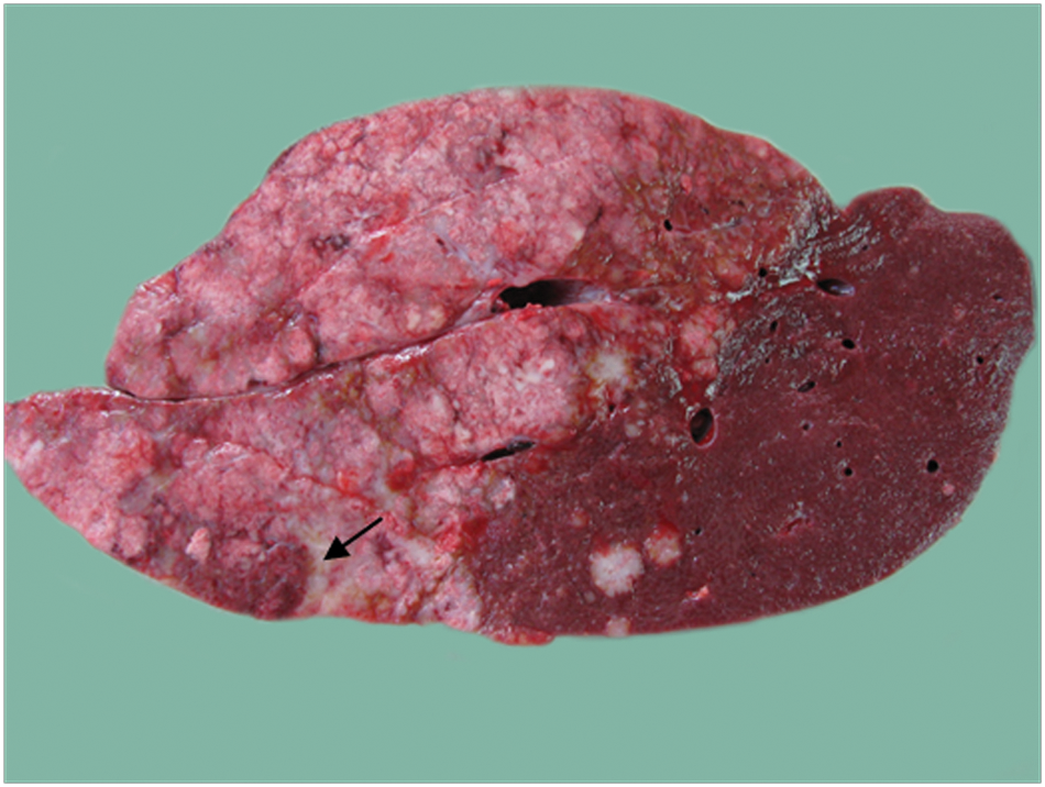
Liver, cut surface aspect; llama (Lama glama); cholangiocarcinoma. The tumor varies from white to gray to yellow, with multiple areas of necrosis and extensive fibrosis (arrow).

Lung; llama (Lama glama); gross aspects. The visceral pleura and pulmonary parenchyma are affected by numerous nodules (arrow).
Samples from liver, lung, lymph nodes, diaphragm, and pericardium were evaluated by cytological, histological, and immunohistochemical examinations. For cytology, multiple smears from the hepatic masses were stained by Dia-Quick Panoptic. a For histology, the samples were fixed in 10% phosphate buffered formalin for 24 hr, embedded in paraffin wax, cut into 3–5 µm sections, and stained with hematoxylin and eosin and periodic acid–Schiff (PAS). Histological interpretation of lesions was done according to the World Health Organization Classification of Tumors of the Alimentary System of Domestic Animals. 6 For immunohistochemistry, sections from liver mass, lung nodules, and normal liver and lung tissue (inner control) were selected. After dewaxing, heat-induced epitope retrieval pretreatment, and endogenous peroxidase inactivation (H2O2 1% in phosphate buffered saline b [PBS]), slides were incubated with goat serum 10% in PBS for reducing background staining. Sections were incubated with the following mouse monoclonal primary antibodies: broad spectrum cytokeratins (panCK), c CK20, c CK19, c CK7, c thyroid transcription factor 1 (TTF1), c and carcinoembryonic antigen (CEA),b for 24 hr at 4°C. After washes, the secondary antibody (labeled streptavidin biotin) was applied, followed by incubation with diaminobenzidine. b Negative controls for each sample were prepared by replacing the primary antibody with mouse immunoglobulin G1 negative control. b Semiquantitative assessment of immunohistochemical intensity was performed and scored as negative (−), weak (+), moderate (++), and intense (+++).
Cytologically, neoplastic cells were cuboidal to columnar, arranged in cohesive groups and tubules, and were characterized by eccentric large regular nuclei, prominent nucleoli, and bluish cytoplasm. More than 70% of cells were anaplastic, with severe anisocytosis and anisokaryosis, multiple and prominent nucleoli, and clear cytoplasm. Multinucleated tumor giant cells, numerous mitotic figures, and many neutrophils were also present.
Histological examination of the hepatic lesions revealed an infiltrative, unencapsulated, and poorly circumscribed neoplasia, composed of poorly differentiated epithelial cells arranged in acini and tubules, separated by abundant amount of fibrovascular stroma (scirrhous response), multifocally invading the sinusoids (Fig. 3), substituting the surrounding parenchyma, and compressing the hepatic cords, causing moderate to severe parenchymal atrophy. The neoplastic cells were pleomorphic, characterized by a columnar or round and polyhedral shape, with fairly distinct cell borders, finely granular cytoplasm, large vesicular nuclei (average 12.70 µm/100 nuclei, range: 10.50–15.90 µm), and several prominent, variably sized nucleoli. Mitotic figures were numerous and bizarre, ranging from 7 to 15 per high power field. Multiple neoplastic emboli within the central and portal veins, thrombosis, hemorrhages, and multiple necrotic foci were scattered throughout the hepatic parenchyma. Periodic acid–Schiff- positive mucous secretion was identified in the neoplastic cells and within the lumen of the neoplastic acini. Pulmonary (Fig. 4), diaphragmatic, lymph nodal, and pleural metastases were characterized by a less prominent scirrhous reaction.
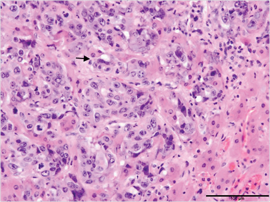
Liver; llama (Lama glama); cholangiocarcinoma. Neoplastic cells are present within the sinusoids (arrow). Numerous mitotic figures, often bizarre, are present. Hematoxylin and eosin. Bar = 100 µm.
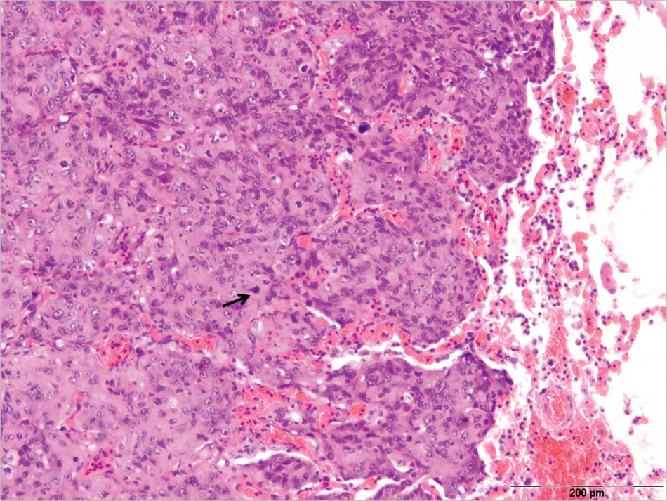
Lung; llama (Lama glama); metastatic cholangio- carcinoma. Alveolar spaces are infiltrated by neoplastic cells arranged in a solid pattern. Multiple mitotic figures are evident (arrow). Hematoxylin and eosin. Bar = 200 µm.
The primary neoplasia was diffusely and intensely positive (+++) for broad spectrum CK and CK20 (Fig. 5), weakly positive (+) for CK19, and negative (−) for CK7, CEA, and TTF1. The pulmonary metastases was intensely positive (+++) for broad spectrum CK and CK20. An intensely positive (+++) for broad spectrum CK was revealed in both alveolar and bronchiolar epithelial cells. In normal liver, an intensely and diffusely expression for panCK and CK20, and mild (+) expression for CK19 were seen only in bile duct cells. The hepatocytes were negative for all markers. The immunohistochemistry results from both normal tissue compared with primary neoplasia are illustrated in Table 1.
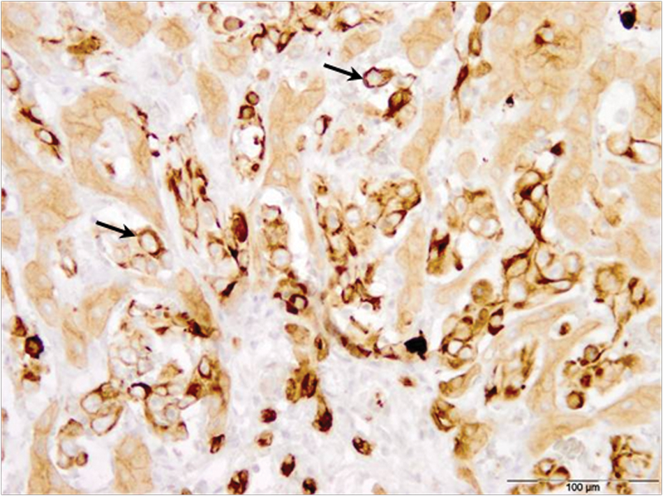
Liver; llama (Lama glama); cholangiocarcinoma. Neoplastic cells are positive for cytokeratin 20 (arrows). Immunoperoxidase–diaminobenzidine stain, Mayer hematoxylin counterstain. Bar = 100 µm.
Immunohistochemistry results of primary and metastases tumor samples and normal liver from a llama (Lama glama).*
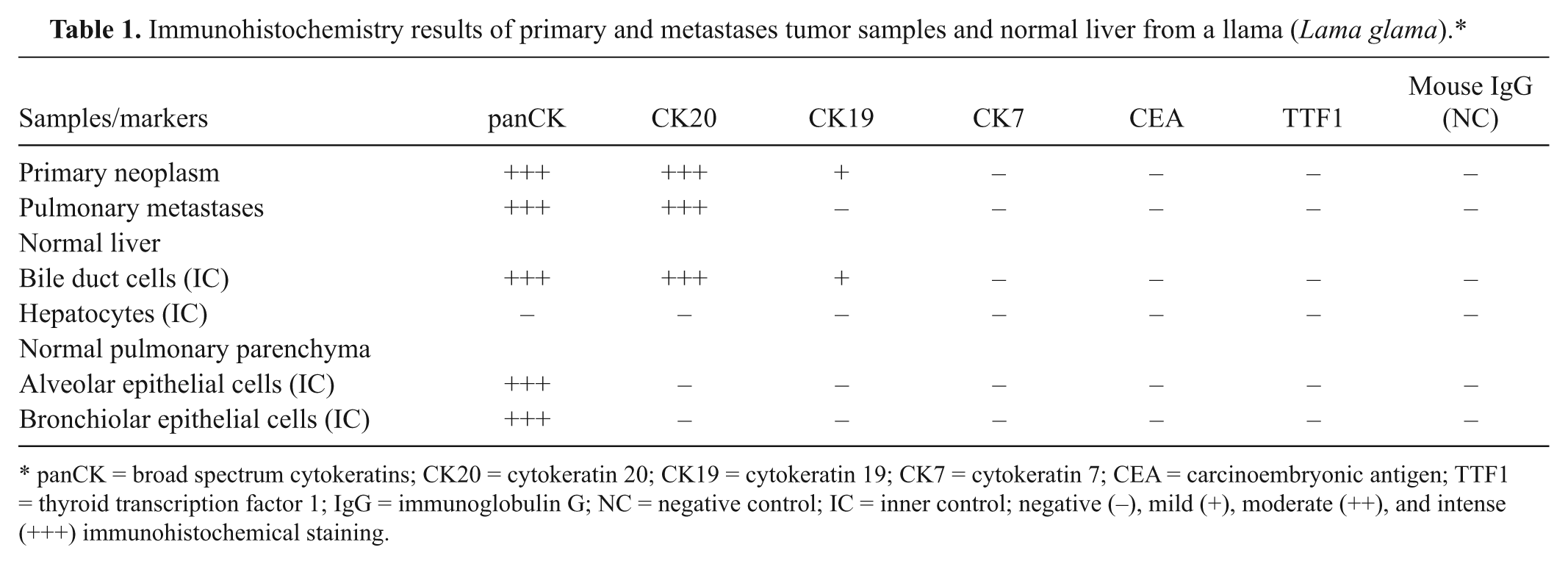
panCK = broad spectrum cytokeratins; CK20 = cytokeratin 20; CK19 = cytokeratin 19; CK7 = cytokeratin 7; CEA = carcinoembryonic antigen; TTF1 = thyroid transcription factor 1; IgG = immunoglobulin G; NC = negative control; IC = inner control; negative (−), mild (+), moderate (++), and intense (+++) immunohistochemical staining.
The current report describes pathological and immunohistochemical findings of metastatic cholangiocarcinoma in a llama. According to the literature reviewed, cholangiocarcinoma in llamas is uncommon, and only 2 cases of disseminated bile duct carcinomas have been reported, but not described, in a retrospective study. 19 In addition to bile duct carcinoma, other tumors that have been reported in llamas include metastatic pulmonary adenocarcinoma, 15 metastatic uterine adenocarcinoma, 9 malignant round cell neoplasia, 12 cutaneous metastases of a mammary carcinoma, 10 gastric squamous carcinoma, 18 retinoblastoma, 3 gemistocytic astrocytoma, 4 and carcinoma in a mixed mammary tumor. 2
Cholangiocellular carcinomas are malignant neoplasms of biliary epithelium, which usually arise from the intrahepatic ducts, but also extrahepatic bile ducts can be affected. Such neoplasms have been recorded in dogs, cats, cattle, sheep, and horses but less frequently than hepatocellular tumors. 6 In human beings, intrahepatic cholangiocarcinoma is much less common than hepatocellular carcinoma.1,7
In human beings, this tumor is frequently associated with liver fluke infestation, primary sclerosing cholangitis, anabolic steroids, ulcerative colitis, intrahepatic lithiasis, Caroli disease, and multiple liver cysts.1,7 In animals, the relationship between bile duct parasitism and bile duct carcinoma is unclear. Mucous gland hyperplasia is common in chronic fascioliasis in cattle and sheep, without bile duct carcinoma formation. 6 There was no evidence of fluke infection in the current case.
The differential diagnosis between cholangiocarcinomas and metastatic tumors of pancreatic origin can be challenging. 8 The presence of mucus secretion, intrasinusoidal and multiple parenchymal invasion by neoplastic cells, a prominent deposition of collagen, abundance of mitotic figures, and multiple foci of hepatic necrosis are distinctive features of bile duct carcinoma.6-8 Cytokeratins are the principal intermediate filament proteins characteristically found in epithelial cells. In the liver, the use of CK7/CK20 immunostaining could help to distinguish cholangiocarcinoma from other primary or metastatic hepatic lesions such as hepatocellular carcinoma, and hepatic metastases of colorectal carcinoma. In human beings, cholangiocarcinoma is positive for CK7 and CK20 in 96% and 70% cases, respectively. 17 In the present case, the negative results for human CK7 in both the neoplasm lesions and the inner control could be due to nonreactivity against llama biliary epithelium. The PAS-positive mucous secretion identified within the neoplastic cells and the comparable immunohistochemical profile of the hepatic and pulmonary neoplasms revealed the epithelial origin of the lesions. Thyroid transcription factor 1 is a marker present in most canine thyroid tumors. 16 Both liver mass and pulmonary metastases were negatively labeled for TTF1 in the current case. CEA, a highly glycosylated cell surface glycoprotein, is considered a colon-specific oncofetal protein 5 and is often found in patients with malignant tumors of the digestive system such as stomach, colon, biliary tract, and pancreas cancer. 11 In the current case, neoplastic cells were negative for CEA, whereas, in human medicine, this marker shows a high sensitivity and specificity for cholangiocarcinoma.13,14 As the inner control was also negative for both CEA and TTF1, it was concluded that these human antibodies do not react with llama biliary and pulmonary epithelium.
To conclude, cholangiocarcinoma is a rare and very aggressive tumor that can affect llamas, and is characterized by a rapid clinical course and by a poor prognosis. Predisposing factors for this neoplasm have not been identified in llamas.
Footnotes
Acknowledgements
Marian Taulescu and Cornel Catoi contributed equally to this work. The authors thank Dr. Beth Valentine, ACVP Diplomate, from the College of Veterinary Medicine, Oregon State University, for reviewing this article.
a.
Dia-Quick Panoptic, Reagent Ltd., Budapest, Hungary.
b.
Phosphate buffer saline (code S3024), carcinoembryonic antigen (rabbit anti-human 1:1), LSAB System-HRP kit (code K0679), mouse IgG1 negative control (code X0931); Dako Denmark A/S, Glostrup, Denmark.
c.
Pancytokeratin (mouse anti-human 1:600), cytokeratin 20 (mouse anti-human 1:100), cytokeratin 19 (mouse anti-human 1:100), cytokeratin 7 (mouse anti-human 1:100), thyroid transcription factor 1 (mouse anti-human 1:200); Novocastra Laboratories, Newcastle, UK.
Declaration of conflicting interests
The author(s) declared no potential conflicts of interest with respect to the research, authorship, and/or publication of this article.
Funding
The author(s) disclosed receipt of the following financial support for the research, authorship, and/or publication of this article: Support provided by the University of Agricultural Sciences and Veterinary Medicine, Cluj-Napoca, Romania.