
Abstract
Avian Influenza A virus (AIV) subtype H5 is divided into American and Eurasian lineages, according to hemagglutinin gene sequences. Although methods for detecting H5 AIVs have been described, no H5 strain–specific detection method has been reported. The purpose of the present study was to develop an antigen-capture enzyme-linked immunosorbent assay (ACE) to detect and differentiate between the American and the Eurasian H5 AIVs. Monoclonal antibodies (mAbs) against the HA fragment of a Eurasian H5N2 AIV were used as the capture antibodies as well as the detector antibodies after labeling with horseradish peroxidase to develop an ACE. One mAb was selected for detecting the American as well as the Eurasian H5 AIVs. The other mAb was used for detecting only the Eurasian H5N2 but not the American H5N2 AIVs, H6N1 AIVs, or Newcastle disease virus. The ACEs developed would be useful for detection and differentiation of H5 AIVs from the Eurasian and the American H5 AIVs.
Avian Influenza A virus (AIV), a member of the Orthomyxoviridae family, is a negative-sense, single-strand RNA virus, and is classified into subtypes based on 2 main surface glycoproteins, hemagglutinin (HA) and neuraminidase (NA). To date, 16 HA subtypes (H1–H16) and 9 NA subtypes (N1–N9) have been identified. HA is the most important antigenic determinant to which neutralizing antibodies are directed.5,10 HA is cleaved into 2 fragments after maturation, hemagglutinin 1 (HA1) and hemagglutinin 2 (HA2), of which HA1 is the main domain of HA to induce antibodies. Chickens immunized with whole HA or HA1 subunit vaccines produce strong humoral immune responses with high titers of HA-specific antibodies. 12
Although both antigen and antibody detection techniques can be used to detect AIV infection, antigen detection is preferred for controlling AIV outbreaks because the production of antibodies lags behind the detection of antigens following infection of birds. 1 Virus isolation and nucleic acid detection by reverse transcription polymerase chain reaction (RT-PCR) can also be used to detect the virus. Several commercial antigen-capture enzyme-linked immunosorbent assay (ACE) kits have been developed based on AIV nucleoprotein detection. They detect all AIV subtypes6,11,13 and are not able to differentiate between subtypes or between different H5 AIV strains. The recent H5N1 AIV infections in Asian and European countries were caused by the Eurasian lineage. 2 The present authors’ laboratory has previously developed monoclonal antibodies (mAbs) against H5 AIVs, 4 which detected only the Eurasian H5 but not the American H5 AIV strains.
Eight American lineage H5N2 AIV strains (A5-1 to -8) and 1 Eurasian lineage H5N2 AIV (E5-1) were used in the present study. Among them, A5-1 was strain A/chicken/Taiwan/1209/03 (H5N2; accession no. AY573917) and E5-1 was strain A/duck/Yunlin/04 (H5N2). The A5-2 to A5-7 strains were H5N2 isolated from chickens similar to the A5-1 strain. The E5-1 strain is a low pathogenic AIV that was isolated from healthy ducks during routine monitoring, 4 and has an HA sequence similar to highly pathogenic Eurasian lineage H5N1 AIV that occurred in Southeast Asian countries. Nine H6N1 AIV strains (H6-1 to -9) from chickens, and 1 Newcastle disease virus (TW-2/00 3 ) were also used in the current study to test the specificity of this assay.
Viruses were propagated by inoculating 9-day-old specific pathogen–free (SPF) embryonated chicken eggs. Allantoic fluid was harvested 3 days after inoculation and stored at −20°C. The 50% virus infectious dose (EID50) of the allantoic fluids was determined using the hemagglutination assay, as previously described. 10 The titers of those viruses ranged from 2.7 × 10⁶ to 4.2 × 10⁹.
The E5-1 H5N2 strain was selected for mAb production because it belongs to the Eurasian lineage as previously reported. 4 The percentage of the nucleic acid sequence identity between A/duck/Yunlin/04 and H5N1 that occurred in Southeast Asian countries (such as A/chicken/Phichit/NIAH600674/08 [H5N1; a Thailand strain, accession no. EU919138]) reached 83.5%, but that between A/chicken/Taiwan/1209/03 and recent H5N1 reached only 75%. Four mAbs, anti–H5-1 (immunoglobulin [Ig]G2a), anti–H5-3 (IgG1), anti–H5-4 (IgG2a), and anti–H5-6 (IgG2b), were previously produced in the authors’ laboratory. 4 Stocked mAb-rich ascetic fluids were purified by 0%–40% ammonium sulfate precipitation and protein A affinity column.
Indirect ELISA was used to evaluate the binding of the 4 mAbs to E5-1. A microtiter plate was coated with 200 ng/well of the formalin-killed E5-1 virus diluted in sodium carbonate buffer b at 37°C for 1 hr. After washing with 340 μl/well of phosphate buffered saline containing 0.1% Tween-80 (PBST), the plate was incubated with 100 μl/well of blocking buffer (PBST + 10% skim milk) at 37°C for 30 min. Washed 3 times with PBST, the plate was incubated with 100 μl/well of 2 ng/μl of mAbs at 37°C for 30 min. A 100 μl/well of goat anti-mouse IgG labeled with horseradish peroxidase (HRP; 1:2,500 dilution) was added after washing 5 times and incubated at 37°C for 30 min. The plate was then washed 5 times and added with 100 μl/well of the 3,3’,5,5’-tetramethylbenzidine (TMB) microwell peroxidase substrate. d The reaction was stopped after 10 min in the dark by adding 100 μl of TMB stop solution d to each well. The optical density (OD) at 450 nm was read with an ELISA plate reader. e The anti–H5-3 and anti–H5-6 mAbs reacted much stronger to the E5-1 virus than the anti–H5-1 and anti–H5-4 mAbs (OD values > 2.5 vs. < 0.5).
The anti–H5-3 and anti–H5-6 mAbs were further tested. The anti–H5-3 mAb reacted with all H5 AIVs tested, whereas the anti–H5-6 mAb reacted only with E5-1 but not A5-1 through A5-7. From different combinations of capture and detection antibodies, the following 2 pairs of mAbs were selected for the ACEs: anti–H5-3/anti–H5-3-mAb-HRP (capture/detector) for detection of all H5N2 AIVs, and anti–H5-6/anti–H5-6-mAb-HRP for the Eurasian lineage H5N2 AIV detection. All the viruses used in the current study as well as an SPF allantoic fluid (negative control) were used for repeatability evaluation by ACE with the anti–H5-3/anti–H5-3 mAb pair. The coefficient of variation of the intra- and interassay variability were 3–5% (n = 33) and 4% (n = 11), respectively.
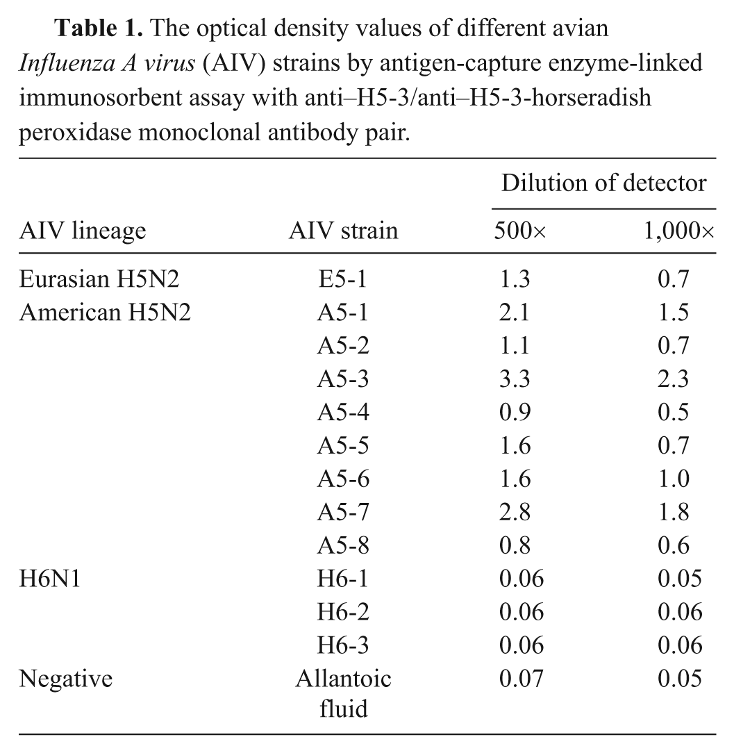
For ACE, the anti–H5-3 and anti–H5-6 mAbs were diluted to 200 ng/well in carbonate buffer and coated 100 μl/well on a microplate as the capture antibodies and incubated at 37°C for 1 hr. After washing with PBST once, the plates were incubated with 100 μl/well of blocking buffer at 37°C for 30 min before washing 3 times and incubating with 100 μl/well of A/duck/Yulin/04 at 37°C for 30 min. After another washing, the plate was added with 100 μl/well of 1:1,000 dilution of anti–H5-3-mAb-HRP or anti–H5-6-mAb-HRP as detector antibodies at 37°C for 30 min. The plate was added with 100 μl/well of the TMB solution after washing and stopping after 10 min in the dark by adding 100 μl of TMB stop solution. The OD at 450 nm was read with an ELISA reader. Fifty tracheal samples, 33 market-aged Taiwan country chickens and 17 adult hens from a broiler breeder farm, known to be negative for AIV by RT-PCR were used to calculate the cutoff value for the assay. The cutoff value was calculated as mean plus 2 standard deviations. Ten-fold serial dilutions of E5-1 allantoic fluid, quantified by EID50, were used as the antigens in the ACE. The detection limit of the ACE was determined as the highest dilution that tested positive. In addition, 301 chicken embryonated eggs were used to test the specificity. Of them, 25 were inoculated with A5-1, 25 with A5-2, and 25 with H6-1, and the rest were uninoculated eggs. The mean OD values plus standard deviation were 0.065 + 0.018 (n = 25), 0.063 + 0.011 (n = 25), 0.064 + 0.015 (n = 25), and 0.062 + 0.008 (n = 226), respectively. The OD values obtained between the tracheal samples and eggs were analyzed using a t-test at the significance level of P < 0.01. 9 The average and standard deviation of the 50 tracheal samples were 0.077 and 0.013, respectively. Thus, the cutoff value was 0.103 (mean + 2 standard deviation). The ACE with the anti–H5-3/anti–H5-3-mAb-HRP pair detected H5 AIV A5-1 through A5-9 as well as E5-1 but not the 3 H6N1 strains (H6-1 to -3) Newcastle disease virus (Table 1). This indicated the absence of false-positive results with H6N1 AIV. No cross-reactivity was observed between the Eurasian H5N2 and the American H5N2 strains.
The optical density values of different avian Influenza A virus (AIV) strains by antigen-capture enzyme-linked immunosorbent assay with anti–H5-3/anti–H5-3-horseradish peroxidase monoclonal antibody pair.
To compare the detection limit between the ACE and RT-PCR, nucleic acid from the allantoic fluid of E5-1–, A5-3–, A5-6–, and A5-7–infected embryonated eggs was extracted by using TRIzol. Reverse transcription was done, followed by amplification of the HA gene by RT-PCR using H5-specific primers H5-155f (5’-ACACATGCYCARGA CATACT-3’) and H5-699r (5’-CTYTGRTTYAGTGTTGA TGT-3’, Y = C/T). Reverse transcription PCR was initiated by reverse transcription at 42°C for 45 min, denatured at 95°C for 3 min, followed by 35 cycles of 95°C for 30 sec, 50°C for 40 sec, and 72°C for 40 sec. The amplified products were further elongated at 72°C for 10 min. The PCR products were run on an agarose gel, with an expected product size of 544 bp.
Because of the recent spread of H5N1 AIV from Asia to Europe and Africa, the Eurasian H5N1 AIV has received considerable attention, with fears that the virus could spread to uninfected countries. 5 Thus, the ability to detect this virus in the early stages is important for controlling its possible spread. Although RT-PCR has been successfully applied to the detection of AIV infection in poultry farms,7,8 the antigen detection assay provides an alternative means for detecting AIV directly. Many farms, like those in Taiwan, own ELISA readers but not a thermocycler for RT-PCR. The present ACE could be readily modified into a convenient diagnostic kit for use on farms for the detection of Eurasian H5 AIV infection.
The present study developed an ACE system that specifically detects the Eurasian lineage H5 AIV, with no cross-reaction with other viruses, including some American lineage H5 AIVs. Many commercial H5 vaccines (such as from the Intervet vaccine) and the H5N2 outbreaks that occurred in Taiwan in 2004 belong to the American H5 lineage. 4 The ability of the new ACE to differentiate between the American and Eurasian lineages of H5 AIV strains would be useful for detection when a new Eurasian H5 AIV intrudes into a country.
AIV-negative tracheas, but not chicken embryonated eggs, were used to determine the cutoff value of the assay because tracheas are the diagnostic sample in the field. A cutoff value with 2 standard deviations (rather than 3) was selected because it provides higher sensitivity in the ACE, which is intended to be a screening assay. The lower OD values obtained from embryonated eggs than from tracheas might be the result of the higher background in the tracheas. However, the lack of field H5 AIV-infected samples in Taiwan means that the assay needs further evaluation.
Footnotes
a.
HiTrap™, GE Healthcare, Uppsala, Sweden.
b.
15 mM Na2CO3, J.T. Baker, Phillipsburg, NJ.
c.
35 mM NaHCO3 (pH 9.6), Sigma-Aldrich, St. Louis, MO.
d.
SureBlue Reserve™, Kirkegaard & Perry Laboratories, Gaithersburg, MD.
e.
EL 312e, Bio-Tek Instruments, Winooski, VT.
f.
Genmedika Biotechnology, Taipei, Taiwan.
Declaration of conflicting interests
The author(s) declared no potential conflicts of interest with respect to the research, authorship, and/or publication of this article.
Funding
The author(s) disclosed receipt of the following financial support for the research, authorship, and/or publication of this article: This work was supported by the Council of Agriculture of Taiwan, and Forward Electronics Co. Ltd., Sanhsia, Taiwan.