
Abstract
Bovine viral diarrhea viruses are economically important pathogens of cattle. Most infections in susceptible animals are acquired from animals persistently infected with the virus. Surveillance programs rely on skin biopsies of persistently infected (PI) cattle to detect the infection. The purpose of this study was to compare antigen capture enzyme-linked immunosorbent assay (ACE) testing results using different types of samples from PI animals. The intent was to determine comparative detection rates in types of samples that are frequently submitted to diagnostic laboratories for evaluation of cases of unknown etiology or samples that could be easily collected for Bovine viral diarrhea virus (BVDV) screening. Eight types of samples were collected from 40 PI animals. The sample types were ear notches, serum, nasal swabs, conjunctival swabs, oral swabs, rectal swabs, vaginal/preputial swabs, and a tail skin fold biopsy. Each type of sample (n = 8) for each animal (n = 40) was evaluated with a commercial ACE kit. When using ACE, tail-skin fold and nasal swab samples were 100% sensitive compared with results using ear notches. Sensitivity using other samples was as follows: serum and vaginal/preputial swabs, 92%; conjunctival swabs, 64%; rectal swabs, 10%; oral swabs, 8%. Testing of tail skin fold biopsies, nasal swabs, and ear notch samples resulted in reliable results. In contrast, other sample types were unreliable for diagnosis of persistent infection in calves.
Exposure to Bovine viral diarrhea virus (BVDV; family Flaviviridae, genus Pestivirus) results in economically important diseases in cattle. 5,6 Infection with BVDV results in both persistent (a result of in utero exposure) and acute (a result of postnatal exposure) infections. 4,7 Although viral shedding from acutely infected animals has the potential to infect other animals, persistently infected (PI) animals can shed high levels of BVDV during their lifetimes and are thought to be the principal source of new infections. An important strategy for controlling the economic impact of BVDV is to test for BVDV and remove PI animals. Several testing strategies have been developed to identify BVDV-infected animals and, more important, differentiate acutely infected from PI animals. Examples of these tests include virus isolation, polymerase chain reaction, and immunohistochemistry (IHC). Antigen capture enzyme-linked immunosorbent assay (ACE) has also been developed to detect viral antigen that is present in either tissues or serum. Antigen capture enzyme-linked immunosorbent assays are robust, economical methods of identifying PI cattle and are the most rapid tests available to date. 2 The purpose of the current study was to investigate the potential of other types of samples for identifying PI cattle. The intent was not to validate alternative samples for BVDV detection but rather to determine diagnostic sensitivity of samples that are frequently submitted to diagnostic laboratories for evaluation of cases of unknown etiology (e.g., cases of respiratory disease outbreaks in cattle) or types of samples that could be easily collected for BVDV screening.
Eight types of samples were collected from 40 animals previously identified as PI based on 2 positive ACEs of ear notch samples collected at least 21 days apart (OIE Manual of Diagnostic Tests and Vaccines for Terrestrial Animals, Chapter 2.10.6; www.oie.int/eng/normes/mmanual/A_00132.htm). Three animals were from a stocker operation in northeastern Oklahoma (Washington County), and 37 were from a stocker operation in south central Oklahoma (Pottawatomie County). All of the animals were segregated from other animals on each premise because of their BVDV infection status. The 8 types of samples collected were as follows: ear notch and caudal tail skin fold biopsies; serum; and swabs from the prepuce or vagina, nasal cavity, ocular conjunctiva, oral cavity, and rectum. Both the ear notch and caudal tail fold samples were obtained with a small ear notcher that was cleaned with dilute chlorhexidine solution and rinsed in clean water between each calf. Serum samples were obtained by collecting whole blood via jugular venipuncture. Vaginal or preputial, nasal, conjunctival, oral, and rectal swabs were obtained by placing a polyester-tipped plastic applicator a in contact with the sampled mucosal surface and rotating the swab rapidly several times. In situations in which bilateral sampling sites were available (conjunctival swab, nasal swab), only 1 side was sampled.
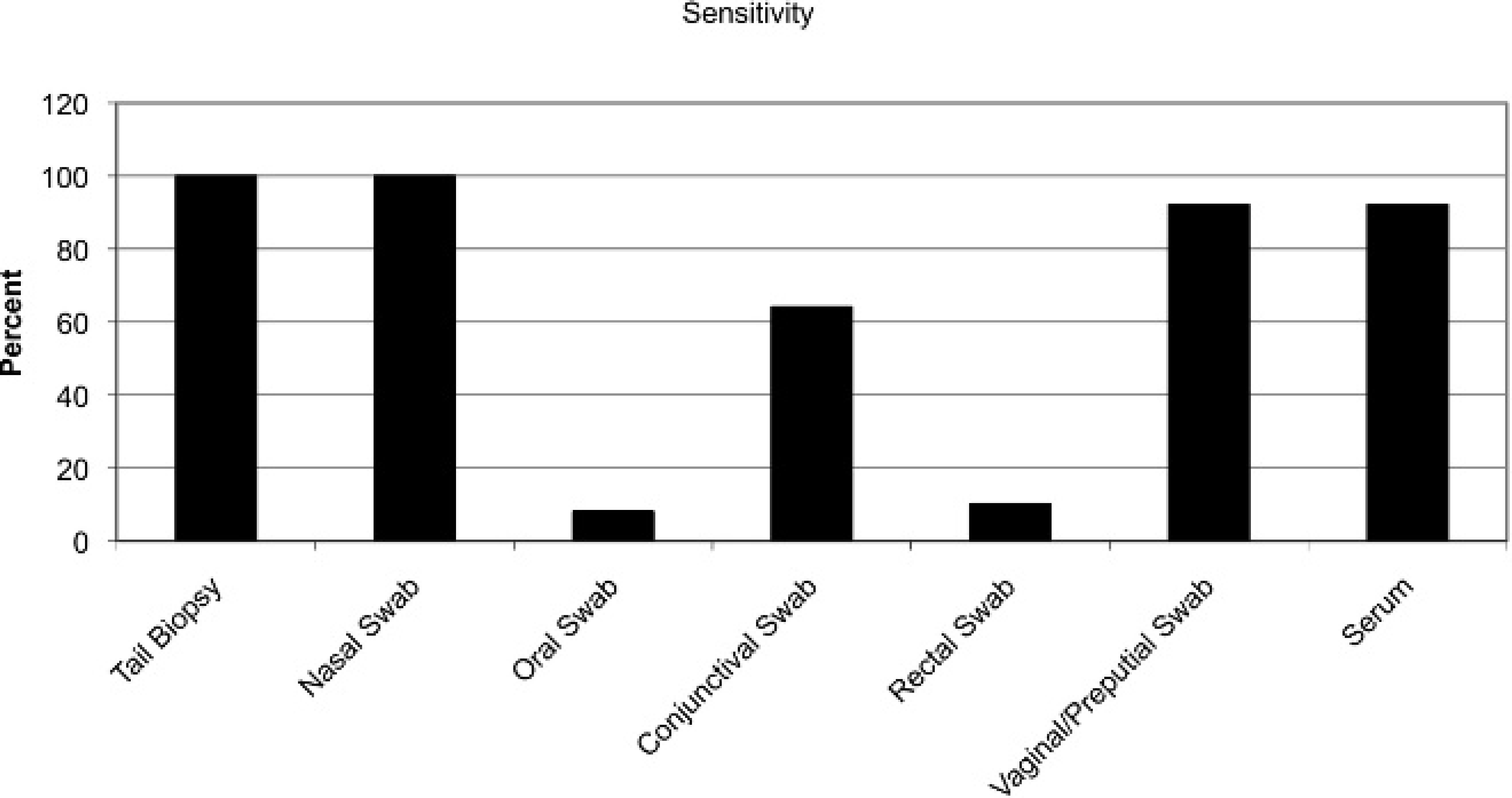
Relative sensitivity of each sample type compared with antigen capture enzyme-linked immunosorbent assay on ear notch samples.
After collection, swabs were immediately submerged in 1 ml of sterile phosphate buffered saline and placed on ice. Ear notch and caudal tail fold samples were placed in dry tubes and held on ice for approximately 2 hr for transport to the laboratory. Both skin and swab samples were frozen at −20°C immediately upon arrival at the lab. The blood collected from each animal was centrifuged at 750 × g for 20 min, and the serum was placed in sterile 15-ml tubes and frozen at −20°C. Twenty-four hr before testing, swab samples were thawed at 4°C. Twenty-four hr prior to testing, 2 ml of phosphate buffered saline was added to each ear notch and caudal tail fold samples. After addition of the saline, samples were held at 4°C until testing. Serum samples were thawed immediately prior to testing.
A commercially available ACE b was used to evaluate the samples for the presence of BVDV Erns (gp48) antigen. 1 The testing was performed according to the manufacturer's directions. Optical density (OD) readings were determined, and the presence or absence of BVDV antigen in each sample was determined by calculating the sample to positive (S/P) ratio for each sample according to the manufacturer's directions. The calculation of the S/P ratio was determined using the following formula:

where Sa is the sample OD, N is the mean OD of the negative control, and Po is the mean OD of the positive control. Guidelines provided with the kit state that an S/P ratio of less than 0.20 indicates a negative BVDV antigen status, while an S/P ratio greater than 0.39 indicates a positive BVDV antigen status. Samples with S/P ratios between 0.20 and 0.39 are graded as “suspect” for BVDV antigen.
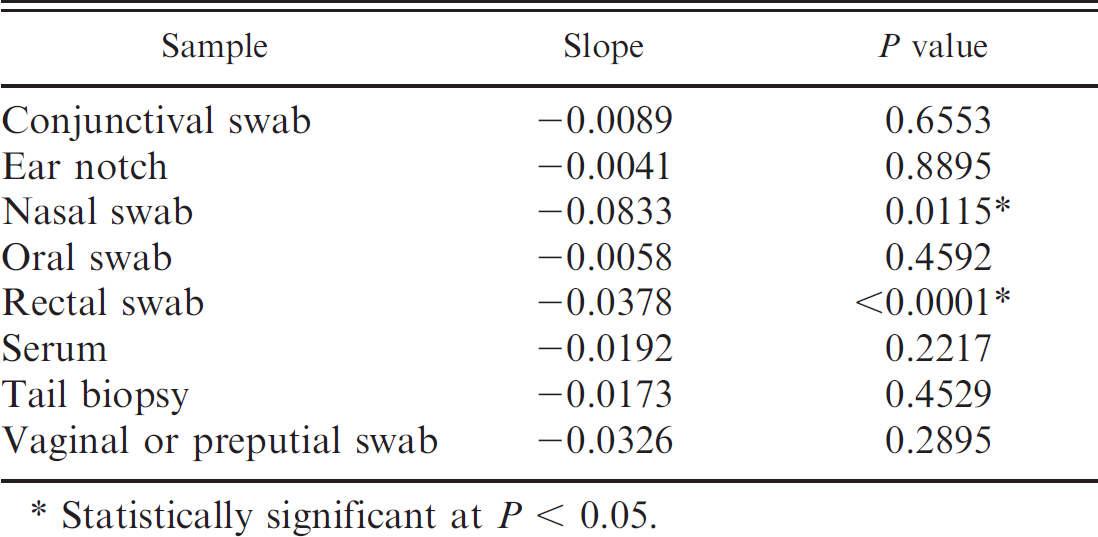
In addition to testing each type of sample by ACE, a second ear notch from each calf was submitted to the Iowa State University Veterinary Diagnostic Lab (Ames, Iowa) for BVDV IHC. 8 Serum virus neutralization assays were completed using BVDV virus from the subgenotypes 1a, 1b, and 2a. The BVDV titers regressed against S/P ratios for each sample type were evaluated using a statistical analysis software package. c
Comparison of effect of virus-neutralizing titer on the sample to positive ratio of each sample type.
Statistically significant at P < 0.05.
Results from testing each sample type were compared with the ACE and IHC results for each of the ear notch samples. Of the 40 calves tested, 39 were positive by ACE and 40 were positive by IHC. Because this investigation is principally concerned with ACE, results will be expressed as sensitivity based on the assumption that calves detected as persistently infected by ACE are true positives (Fig. 1). The ACE results were 100% sensitive using tail biopsies and nasal swabs. The sensitivity of ACE was reduced for the other sample types. The ACE results were 92% sensitive for detecting BVDV using vaginal and/or preputial swabs and serum, 64% sensitive using conjunctival swabs, 10% sensitive using rectal swabs, and 8% sensitive using oral swabs. Average BVDV titers were linearly related to S/P ratio for rectal and nasal swabs. No significant relationship was shown between BVDV titer and any other sample type (Table 1).
Although the prevalence of BVDV PI animals in the general population in North America is less than 1%, studies have shown that 7% of animals dying from infectious diseases in feedlots are PI. 3 Further, mortality rates among PI animals are 10-fold higher than those observed in non-PI animals. 5 Skin biopsies, serum, and buffy coat are the sample types most frequently collected for surveillance efforts, and commercial test kits have been developed based on testing of these sample types. However, collection of skin biopsies is invasive and can be potentially disfiguring. Further, skin biopsies, serum, and buffy coat samples are not always submitted in cases of disease outbreaks of unknown etiology. The purpose of the current study was to investigate the potential benefit of other types of samples for identifying PI cattle. The sample types used in the present study were selected based on similarity to standard sample types (tail fold biopsy), accessibility (nasal, conjunctival, oral, rectal, and vaginal swabs), and comparability (preputial swabs on males vs. vaginal swabs on females) and because samples could otherwise be submitted in respiratory outbreaks of unknown etiology (nasal swabs).
The current study revealed that the test results of skin biopsies from the caudal tail fold and nasal swabs had a high sensitivity compared with the standard ear notch sample. In contrast, testing of vaginal, preputial, conjunctival, oral, and rectal swabs performed at unsatisfactory levels. Although the exact reason for the diminished performance of the vaginal, preputial, conjunctival, oral, and rectal swabs is undetermined, it is suspected that magnitude of viral shedding and bacterial contamination can potentially play a major role in each of the cases. Oral and rectal swabs, both from sites with very high bacterial loads, performed very poorly compared with nasal swabs or ear notches. Vaginal, preputial, and conjunctival swabs were taken from sites that may have had lower levels of viral shedding than other tissues did. Simple linear regression of serum antibody titer versus S/P ratio for each sample type was performed to determine if the presence of antibodies affected detection by ACE (Table 1). Serum antibody titers against BVDV demonstrated a significant linear relationship with S/P ratios from nasal and rectal swab samples. Although nasal samples remained sensitive despite decreasing S/P ratios with increasing titers, sensitivity associated with rectal swabs may be more affected by increased antibody titer. The presence of serum antibody titers did not significantly affect the S/P ratios of other tissues tested. Such observations require further investigation. It should be noted, however, that inclusion of suspect animals substantially increased the sensitivity of several sample types. The result indicates that virus may be successfully detectable at a low level in sample types other than skin and nasal swabs. The use of nasal swab samples has several potential benefits. Nasal swabs are a commonly submitted sample type for screening for a number of respiratory pathogens. Because BVDV often plays a role in the development of respiratory disease, the ability to use 1 sample type for several different tests would be beneficial in terms of both time and economics.
One animal in the current study tested positive by IHC but was negative by all forms of ACE. A previously published report describes a scenario in which 3 acutely infected calves tested positive at initial screening by ACE and by IHC. 1 The calves remained IHC positive for a period after the initial test but were negative by all other tests including, ACE, polymerase chain reaction, and virus isolation. The results from the study indicate that some BVDV antigens may persist in skin tissues after ACE detection is no longer possible. 1 Such a scenario could explain the apparent discrepancy between ACE and IHC in the current study.
As stated above, another advantage to different sample types is the ability to choose a nondisfiguring sample for testing show animals. A significant potential source of BVDV spread is possible if a PI animal is brought to a livestock show and commingled with many other animals. The risk is especially high if pregnant animals are present. Required testing before livestock showing events is becoming more common, but owners are frequently unwilling to have an ear notch collected because the procedure disfigures the ear. Although the collection of a serum sample is not disfiguring, the accuracy of testing serum samples, as determined in the present study, was lower than nasal swabs. A nondisfiguring, noninvasive, more accurate method of sampling, such as nasal swab sampling, would likely increase compliance with testing and reduce the transfer of BVDV at show events.
Footnotes
a.
Puritan Medical Products Co. LLC, Guilford, ME.
b.
IDEXX Laboratories Inc., Westbrook, ME.
c.
SAS Institute Inc., Cary, NC.