
Abstract
The prevalence of Bovine viral diarrhea virus (BVDV) in free-ranging white-tailed deer (WTD, Odocoileus virginianus) in the state of Georgia was evaluated using ear notches collected from hunter-harvested deer during the hunting season of 2010–2011. From September to December 2010, 367 ear samples from WTD were collected from 37 counties in Georgia. The samples were from 178 (48.5%) female deer, 187 (51%) male deer, and 2 (0.5%) of unknown sex. The age of the animals varied from 6 months to 6.5 years. The age was not recorded in 34 animals (9.3%). Of the animals with known ages, 42% were under 2 years. Screening of 367 samples for BVDV using an antigen-capture enzyme-linked immunosorbent assay (AgELISA) resulted in 364 negative samples and 3 suspect samples. The 3 suspect samples were negative for BVDV reverse transcription polymerase chain reaction (RT-PCR), virus isolation, and immunohistochemistry. A subpopulation of samples (n = 89) selected from various geographical regions also tested negative for BVDV RT-PCR. In conclusion, although a few of the samples were suspect for the presence of BVDV by AgELISA, the presence of the virus within the deer population studied could not be confirmed further.
Keywords
Infection with Bovine viral diarrhea virus (BVDV; family Flaviridae, genus Pestivirus)3,16 in cattle can result in respiratory, gastrointestinal, and reproductive tract disease of varying severity, ranging from subclinical to fatal disease. 3 Bovine viral diarrhea virus most commonly infects cattle; however, sheep, goats, pigs, and various wild ruminants may also become infected.4,16
There are 2 types of infection with BVDV, transient and persistent. Depending on the time of infection during gestation, BVDV in cattle can cause early embryonic death, abortion, congenital defects, and persistently infected (PI) carrier calves. Persistently infected animals are the main source of virus transmission as they continuously shed large amounts of virus in the environment. 4 Virus also can be transmitted from acutely infected animals. 4 Other forms of transmission of the virus include semen from both acutely infected or PI bulls and embryo transfer. Transmission can also occur through blood-feeding flies and possibly through air. The possibility of airborne transmission, however, has not been proven and remains controversial. 4
There is some evidence that bovine virus diarrhea (BVD) naturally occurs in free-ranging white-tailed deer (WTD, Odocoileus virginianus) in North America. 1 Surveys for BVDV in wild WTD in other states indicated very low prevalence.1,2,10,11 Bovine viral diarrhea virus 1 (BVDV-1) was isolated from free-ranging WTD. 11 While firm evidence that WTD act as wildlife reservoirs of BVDV is debatable, several experimental studies have evaluated susceptibility of WTD to BVDV infection and transmission of BVDV among WTD, and between WTD and cattle. Initially, it was experimentally confirmed that WTD was susceptible to infection with a BVDV isolate derived from cattle but clinical disease was not reproduced. 15 In later studies, inoculation of pregnant WTD with BVDV resulted in acute infection with death of the does, abortion, fetal resorption, 14 mummified fetuses, 9 and birth of PI fawns,9,14 similar to what is seen in pregnant cows. It was demonstrated that BVDV transmission may occur from cattle to WTD in a model of natural challenge, 8 that BVDV transmission may occur among WTD and that it may be maintained in WTD by congenital infection. 7 More recently, it was experimentally demonstrated that WTD fawns can be experimentally infected with BVDV-1 isolated from free-ranging WTD, 12 that colostrum-deprived calves can be intranasally infected with BDVD-1a strain from free-ranging WTD, 13 and that BVDV transmission from acutely infected WTD fawns to colostrum-deprived calves is apparently possible. 6 The current study was conducted to evaluate the occurrence of BVDV infection in free-ranging WTD in the state of Georgia using ear notches collected from hunter-harvested deer during the hunting season of 2010–2011.
Ear samples were collected at multiple game management areas throughout the state of Georgia. Data on sex, age, and county of origin was recorded. From September to December 2010, 367 ear samples from free-ranging WTD were collected from 37 counties in Georgia, mainly from southern areas (Fig. 1). The samples were from 178 (48.5%) female deer, 187 (51%) male deer, and 2 (0.5%) of unknown sex. Age was estimated based on deer’s teeth. Age varied from 6 months to 6.5 years. The age was not recorded in 34 animals (9.3%). Of the animals with known ages, 42% of the deer sampled were under 2 years. Samples were collected in sterile polyethylene bags and kept refrigerated until transportation to the laboratory.
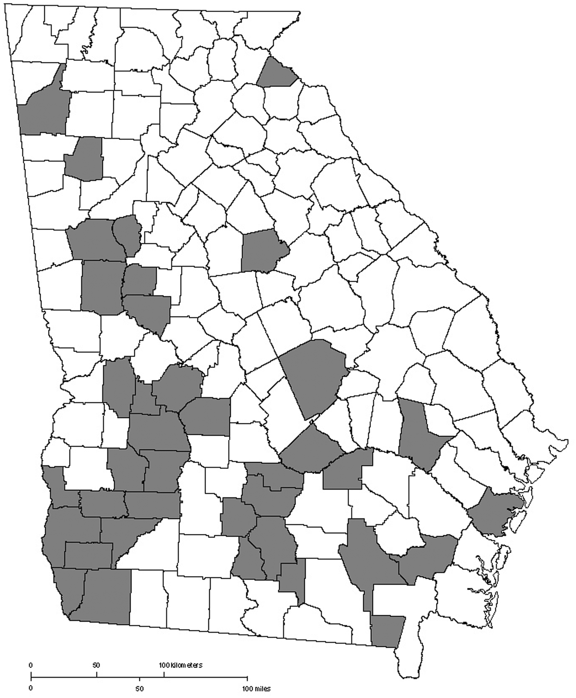
Map of the state of Georgia. The areas in gray depict the counties from where ear samples were collected from hunter-harvested white-tailed deer (Odocoileus virginianus).
At the laboratory, each ear sample was divided into 3 sections. Ear notches of approximately 1 cm2 were placed in 1.5-ml of phosphate buffer solution and processed for antigen-capture enzyme-linked immunosorbent assay (AgELISA). A second sample of similar size was fixed in neutral buffered 10% formalin and embedded in paraffin blocks within 24–48 hr for further immunohistochemistry (IHC) as needed. The remaining sample was frozen at –80°C for further testing including reverse transcription polymerase chain reaction (RT-PCR) and virus isolation (VI). All 367 ear notches were individually tested for BVDV by AgELISA using a commercially available kit a according to the manufacturer’s instructions. Positive and negative controls for AgELISA were controls provided with the manufacture’s kit. Three samples that resulted suspect by AgELISA were processed for IHC using an automated immunostainer b and counterstained with hematoxylin. Immunohistochemistry for BVDV was performed with monoclonal antibody 3.12F1. c A skin sample from a persistently infected calf was used as the IHC positive control.
Out of 367 samples, 89 were tested for BVDV by RT-PCR. The 89 deer samples were selected to equally represent the different geographic areas of sampled population giving preference for younger animals and included samples that resulted in suspect by AgELISA. RNA extraction was performed using a commercially available kit d following the manufacturer’s recommendations. Reverse transcription PCR was carried out with a commercial kit e according to the manufacturer’s instructions. Eight microliters of the sample RNA were added to 17 µl of the RT-PCR master mix. The PCR reaction cycling parameters were: 1 cycle at 45ºC for 10 min, 1 cycle at 95ºC for 10 min, 40 cycles at 95ºC for 15 sec and 60ºC for 45 sec. Positives control for RT-PCR were the positive control provided with the manufacture’s kit and an in-house positive control that consisted of a mixture of BVDV-I and -II virus culture. The negative control was the negative control provided with the manufacture’s kit.
Virus isolation was performed from 3 samples that resulted suspect by AgELISA. For VI, tissue homogenate in Earle minimal essential media was made for each selected ear sample. The homogenate was centrifuged and the supernatant filtered and inoculated onto a preformed monolayer of Madin–Darby bovine kidney (MDBK) and Crandell feline kidney (CRFK) cells. Inoculated cells were incubated in a 5% CO2 atmosphere at 36ºC. Cells were examined daily for virus cytopathic effect. Aliquots of first passage were transferred to a second preformed monolayer of MDBK and CRFK cells on day 7. Chamber slides of MDBK cells were prepared and examined for noncytopathic BVDV using direct fluorescent antibody testing (FAT).
Three ear samples out of 367 resulted in values that fell within the suspect range of the assay by AgELISA. One animal was a 1.5-year-old male deer and the other 2 deer were females 3.5 and 5.5 years of age, respectively. The remaining samples (364) were negative by AgELISA. Seventy-five percent of the selected deer samples tested using RT-PCR was from deer less than 2 years of age (from 6 months to 1.5 years). All suspect samples by AgELISA were tested by RT-PCR. All samples tested with RT-PCR were negative. Immunohistochemistry and VI were performed in all 3 suspect samples for BVDV by AgELISA. All 3 samples were negative by IHC and VI.
Bovine viral diarrhea virus has been isolated from WTD, 1 and studies have suggested that infection, persistent infection, and clinical disease in deer7,9,12,14 and transmission between deer and cattle are possible as a result of experimental infection6,8,13; however, the role of WTD as a reservoir is not fully understood. Previous studies have demonstrated the presence of BVDV in populations of free-ranging WTD in other states in the United States. Two of 745 (0.3%) samples collected from hunter-harvested deer in Indiana tested positive for BVDV with a commercial BVD AgELISA. 11 Skin samples of 406 hunter-harvested WTD collected from deer-processing units in Alabama were tested for BVD antigen using IHC, and only 1 sample (0.24%) was positive. 10 Bovine viral diarrhea virus was detected in 2 deer and, in a follow-up study, 607 samples tested negative in South Dakota. 1 In Colorado, 141 WTD tested negative for BVDV using IHC in sections of skin. 2 The occurrence of BVDV in these populations was very low or nonexistent.
Results from the current preliminary study may not support the hypothesis that WTD could be a potential reservoir for BVDV in the State of Georgia. However, low prevalence of BVDV in populations of WTD in Georgia as seen in other states is possible because BVDV infection has been observed in other species of deer in this state. A study involving 68 hunter-harvested fallow deer (Dama dama) in Little St. Simons Island, Georgia, revealed 1 deer that was positive for BVDV by AgELISA and FAT, but negative by serum neutralization. This deer was considered likely a PI animal. 5
Regarding the population of WTD described herein, the authors believe that age and sex distribution have not influenced the results. Bovine viral diarrhea PI calves typically succumb to BVD under the age of 2. Therefore, surveillance of a hunter-harvested population of deer may not be optimal representation of the general population of deer at risk for infection with BVDV because hunter-harvested deer populations are typically biased toward adult males. The deer population represented in the current study, however, was composed of 42% animals less than 2 years of age and 48.5% of females. In conclusion, although a few of the samples resulted in suspect for the presence of BVDV by AgELISA, the authors could not further confirm the presence of the virus in the deer population studied by RT-PCR, IHC, or VI. The AgELISA used has a sensitivity and specificity of 100%, respectively, for skin samples according to the manufacturer and a designated suspect range of 0.20–0.39. In the present study, 3 out 367 samples fell within the designated suspect range. Although the manufacturer recommends retesting of suspect samples, 3 other tests were used to verify results (i.e., RT-PCR, IHC, and VI). However, all 3 samples were negative by the other tests. Moreover, as no true positives were encountered in this dataset, it is difficult to assess the performance of the AgELISA; it is likely that the AgELISA complies with the manufacturer’s claims. A more detailed validation of the performance of the AgELISA was out of the scope of the paper. However, a similar AgELISA test has been used on samples from WTD in previous survey in which 2 out of 745 samples were positive 11 and, therefore, the test appears suitable to be used in deer samples. The deer population in the State of Georgia is estimated to be approximately 1.2 million (http://www.georgiawildlife.com/DeerFacts). Based on this estimate, approximately 0.03% of the deer population in Georgia was tested in the current study. Future studies targeting a larger population may be necessary to determine if BVDV is present in WTD in Georgia.
Footnotes
Acknowledgements
The authors would like to thank all the volunteers from the Georgia Department of Natural Resources, especially Chris Baumann and Charlie Killmaster. The authors would also like to thank Dr. Doug Waid and his students in the Wildlife Technology Program at the Abraham Baldwin Agricultural College that helped in collecting ear samples, and the staff and technicians from the Tifton Veterinary Diagnostic and Investigational Laboratory that have contributed to this study.
a.
IDEXX Laboratories Inc., Westbrook, ME.
b.
Ventana NextES IHC, Ventana Medical Systems, Inc., Tucson, AZ.
c.
Obtained from Dr. Jeremiah Saliki, Athens Veterinary Diagnostic Laboratory, University of Georgia, College of Veterinary Medicine, Athens, GA.
d.
RNeasy fibrous tissue mini kit, Qiagen Inc., Valencia, CA.
e.
VetMAX-Gold BVDV detection, Applied Biosystems, Foster City, CA.
Declaration of conflicting interests
The author(s) declared no potential conflicts of interest with respect to the research, authorship, and/or publication of this article.
Funding
The author(s) disclosed receipt of the following financial support for the research, authorship, and/or publication of this article: This study was supported by Veterinary Medical Experimental Station, University of Georgia, and U.S. Department of Agriculture.