
Abstract
Endocardial neoplasms are uncommon in veterinary species with most cases restricted to endocardial schwannomas in aged rats. A 15-year-old male rosy-billed pochard (Netta peposaca) was diagnosed following necropsy with numerous, proliferative endocardial masses. Histologically, these masses were composed of interlacing streams and bundles of neoplastic spindle cells supported by a fine fibrovascular stroma. Rare areas of cellular and nuclear palisading were present in the nodules. Approximately 60% of the neoplastic cells were strongly positive for S-100, whereas none of the neoplastic cells was positive for desmin, synaptophysin, neurofilament, and glial fibrillary acidic protein. The histologic features coupled with the S-100 immunoreactivity led to a diagnosis of endocardial neurofibroma.
Keywords
Peripheral nerve sheath neoplasms are a heterogeneous group of neoplasms that include neurofibroma, schwannoma, neurilemmoma, and perineuroma. In veterinary species, peripheral nerve sheath neoplasms are most commonly diagnosed in dogs and cattle. In the latter, such neoplasms are typically encountered as benign masses along the intercostal and epicardial nerves. In contrast, peripheral nerve sheath tumors that originate in the endocardium are extremely uncommon in veterinary species, with most reported cases restricted to proliferative lesions described in the rat.5,9 In the rat, most endocardial neoplasms are classified as schwannomas due to their histologic appearance and immunohistochemical characteristics.5,9 The tumors in rats are typically found in aged animals and are typically incidental findings at necropsy.5,9 Differentiating a schwannoma from a neurofibroma can be difficult and typically relies on a combination of histologic examination, immunohistochemical characterization, and electron microscopic features; however, even after a multitude of diagnostics, differentiation between the 2 entities can be difficult and are often included under the general distinction of peripheral nerve sheath tumor. Schwannomas are best recognized by their variable histologic patterns, which include fusiform to elongate spindle cells enmeshed in a variably dense stroma. The stromal pattern is further defined as densely collagenous and minimal (Antoni A region) or abundantly myxoid (Antoni B region). 7 Neurofibromas typically are composed of more elongate spindle cells with wavy nuclei and very thin collagen fibers that traverse the stroma. Immunohistochemistry can be used to aid in differentiating the 2 neoplasms. Specifically, S-100 immunoreactivity is typically strong and diffuse in cases of schwannoma, whereas in neurofibromas, the immunoreactivity is patchy and is localized to a subset of the neoplastic cells.7,8 This irregular staining in neurofibromas has been purported to result from the different cell populations that typically make up a neurofibroma.
Neurofibromas and schwannomas are only rarely described in birds. Multiple neurofibromas have been described in a red-and-green macaw (or, green-winged macaw; Ara chloropterus) that presented with a large submandibular mass and a glossal mass. 1 The tumors in the macaw were positive for myelin basic protein. 1 A chicken with an ocular and periocular neurofibroma was described in a 2009 report and had approximately 60% immunoreactivity for S-100. 7 Subcutaneous neurofibromas have been described in chickens infected with Avian leukosis virus (Avian leukosis/sarcoma virus subgroup A) and an unusual endocardial proliferative lesion has been reported in chickens experimentally infected with Gallid herpesvirus 2 (previously known as Marek’s disease virus).2,6 Although endocardial and myocardial neurofibromas and schwannomas are well described in rats and cattle, there are no current reports of a similar presentation in any avian species. Herein, the clinical, histopathologic, and immunohistochemical features of multiple endocardial neurofibromas in a rosy-billed pochard are described.
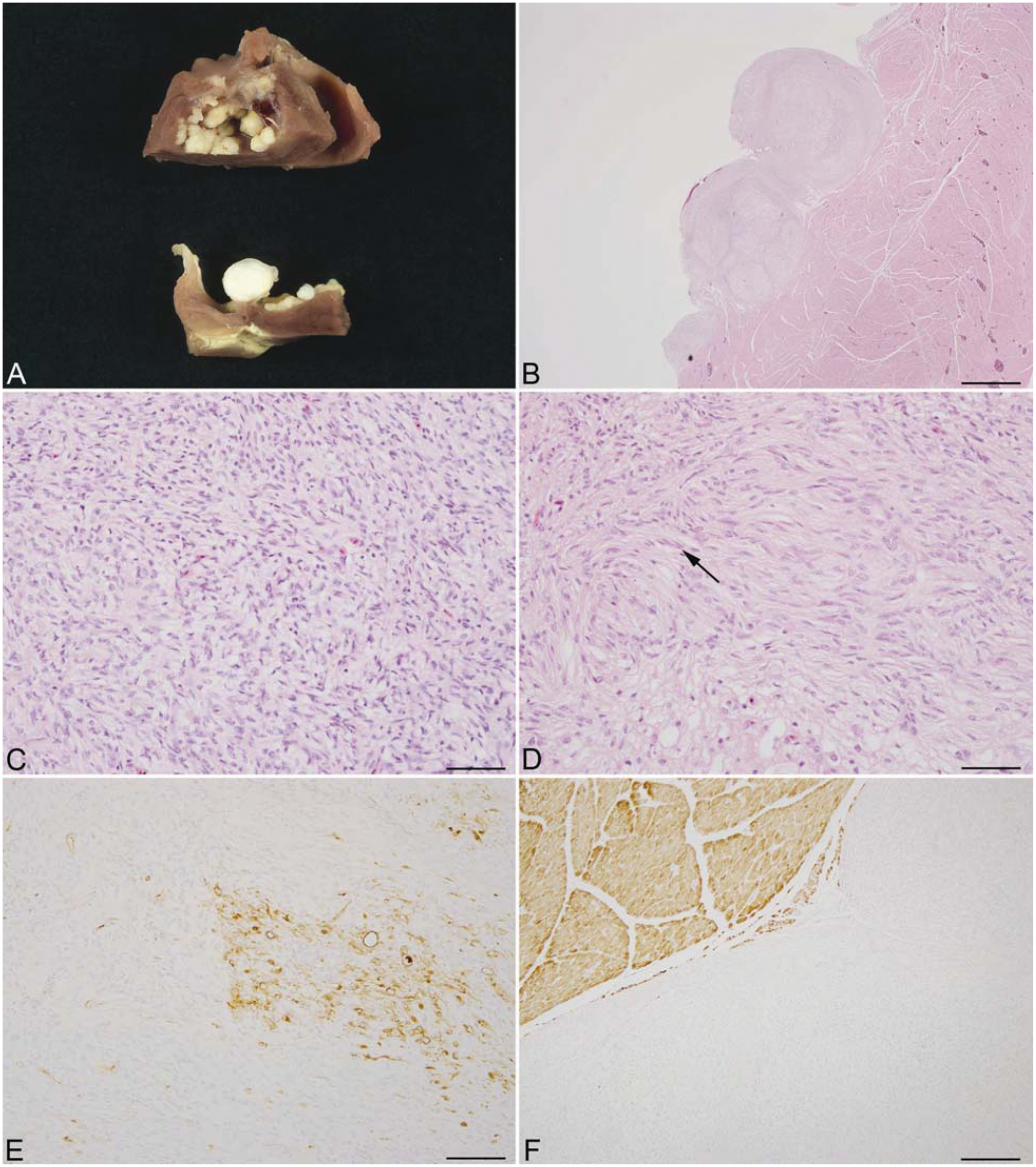
A 15-year-old, male rosy-billed pochard (Netta peposaca) was found dead in its enclosure. No premonitory clinical signs had been noted, and no indication of cardiac disease was recorded. At necropsy, the bird had a moderately enlarged, rounded heart with approximately twelve 0.2–3 cm in diameter, raised, white, firm masses arising from the endocardium. Similar nodules covered the left atrioventricular valve (Fig. 1A). Additional necropsy findings included a focal area of hyperkeratosis around the tarsometatarsal joint and a mildly enlarged liver with rounded edges.
At necropsy, representative sections of all major organs were collected, fixed in 10% neutral buffered formalin, embedded in paraffin, sectioned at 5 µm, and stained using hematoxylin and eosin. To characterize the neoplasm using immunohistochemical methods, standard immunoperoxidase staining for S-100, desmin, glial fibrillary acidic protein (GFAP), neurofilament, and synaptophysin were used. Formalin-fixed, paraffin-embedded sections were deparaffinized, rehydrated, and subsequently blocked with hydrogen peroxide. Pre-treatment for S-100, neurofilament, and synaptophysin involved microwaving for 20 min in 0.01 M citrate buffer followed by 20 min of cooling. There was no pretreatment for desmin and GFAP. All steps were followed by a Tris-buffered saline (TBS) wash. Prior to application of primary antibodies, all slides were treated with protein block a for 10 min. Sections were incubated with anti-human desmin a (monoclonal, 1:50, overnight, 4°C), anti-human S-100 a (polyclonal, 1:3,200, 30 min at room temperature), anti-human synaptophysin a (monoclonal, 1:50, overnight, 4°C), anti-mouse neurofilament b (monoclonal, 1:8,000, overnight, 4ºC), and anti-human GFAP c (monoclonal, 1:1,600, overnight, 4°C). Slides were then incubated with secondary antibody biotinylated goat anti-rabbit d (1:200, 30 min at room temperature) for S-100 and biotinylated horse anti-mouse d (1:200, 30 min at room temperature) for synaptophysin, GFAP, neurofilament, and desmin. This was followed by 30-min incubation at room temperature with ABC Elite d (synaptophysin, S-100, and neurofilament) or ABC standard d (desmin and GFAP). All slides were developed with 3,3′-diaminobenzidine chromogen a and counterstained with Mayer hematoxylin. In all cases, sections were incubated with isotype-specific irrelevant antibodies for negative controls.
Histologic examination of the cardiac mass revealed multiple, pseudoencapsulated endocardial masses that were composed of interlacing streams and whorls of spindle cells embedded in a scant, often myxomatous stroma (Fig. 1B, 1C). Cell density was variable, and there were scattered areas of palisading cells (Fig. 1D). The cells were typically elongate with ill-defined cell borders, abundant pale eosinophilic cytoplasm, and a prominent nucleolus with a single, centrally located nucleolus. Mitotic figures were rare (<1/10 per high power field). Several of the masses were focally mineralized in the center. Immunohistochemically, there was strong, intracytoplasmic immunoreactivity for S-100 in approximately 60% of the neoplastic cells (Fig. 1E). No immunoreactivity was noted for desmin; however, strong immunoreactivity was noted in the unaffected adjacent cardiac muscle (Fig. 1F). No immunoreactivity for synaptophysin was noted; however, strong immunoreactivity was noted in pericardiac nerves. No intratumoral immunoreactivity for neurofilament was noted; however, nerves adjacent to the mass and within the epicardium stained positively. Lastly, no immunoreactivity for GFAP was noted in the neoplastic tissue. Histologic examination of all other organs failed to reveal additional pathologic processes other than mild glycogen accumulation in the hepatocytes.
Although cutaneous schwannomas and neurofibromas are diagnosed with some frequency in most domestic animal species, endocardial schwannomas and neurofibromas are considered rare, with most reported cases restricted to rats. Only 1 case report in a chicken describes proliferative endocardial lesions that had some similarity with the lesions reported herein; however, the lesions in that case report failed to demonstrate immunoreactivity to S-100, and a chronic response to injury was postulated as a potential pathogenesis. 2 In the current case, an adult rosy-billed pochard presented with approximately 12 epicardial masses that histologically and immunohistochemically were most consistent with neurofibromas.
In both human and veterinary medicine, differentiating a schwannoma from a neurofibroma can be difficult and relies on both histologic, immunohistochemical, and electron microscopic features. Schwannomas are benign tumors that are typically composed of variably cellular areas that have fusiform neoplastic cells admixed with areas of myxomatous material (Antoni A and Antoni B).4,8 In the Antoni A pattern, the neoplastic cells are typically aligned in parallel fascicles enmeshed in a collagenous background, whereas the Antoni B regions are typically more mucinous with round to stellate cells. 7 Conversely, neurofibromas are typically more complex than schwannomas due to multiple cell types involved in forming the neoplasm. Neurofibromas have an appearance typified by spindle cells and wavy bundles of collagen intermixed with fibroblasts, eosinophils, mast cells, and numerous blood vessels. 7
Immunohistochemically, S-100 protein is an important differentiating feature. In human medicine, S-100 immunoreactivity in schwannomas is robust and found in a majority of neoplastic cells, both nuclear and cytoplasmic, whereas in neurofibromas, S-100 immunoreactivity is found in a smaller subset of neoplastic cells, sparing a majority of the cells, even though the origin of the neoplastic cells in a neurofibroma are still thought to be Schwann cells. 7 Although a similar expression pattern of S-100 is thought to occur in veterinary species, the majority of these neoplasms are still grouped as peripheral nerve sheath tumors. 8 In the current reported case, approximately 60% of the cells were S-100 positive, thereby favoring a diagnosis of neurofibroma over schwannoma with both GFAP and synaptophysin being non-immunoreactive. Neurofilament staining of intratumoral nerves has historically been used to differentiate schwannomas from neurofibromas; however, this has been shown to not reliably differentiate schwannomas from neurofibromas, and extensive immunoreactivity of the neoplastic cells to S-100 in combination with the clinicopathologic features of the neoplasm remains the most accurate way to differentiate the 2 neoplasms. 3 Lastly, to rule out an unusual presentation of a muscle tumor in the current case, desmin immunohistochemistry was performed. All of the neoplastic cells were negative for desmin, whereas the adjacent cardiac tissue was strongly positive, thereby indicating that the neoplasm was not of cardiac muscle origin. The current case illustrates that proliferative endocardial neoplasms derived from neural tissue occur in birds and should be included as a differential for endocardial masses.
Footnotes
Acknowledgements
The authors wish to thank Kristen Toohey for assistance with photography and Heather Knight for assistance with immunohistochemistry.
a.
Dako North America Inc., Carpinteria, CA.
b.
Sigma-Aldrich, St. Louis, MO.
c.
Lab Vision Corp., Fremont, CA.
d.
Vector Laboratories Inc., Burlingame, CA.
The author(s) declared no potential conflicts of interest with respect to the research, authorship, and/or publication of this article.
The author(s) received no financial support for the research, authorship, and/or publication of this article.