
Abstract
The concentration of GM1 (monosialotetrahexosyl ganglioside) in cerebrospinal fluid (CSF) is markedly increased in dogs with GM1 gangliosidosis due to GM1 accumulation in the central nervous system and leakage to the CSF. The present study established a rapid and simple method for detection of accumulated GM1 in the CSF in dogs with GM1 gangliosidosis using matrix-assisted laser desorption ionization time-of-flight mass spectrometry (MALDI TOF MS) and discusses the usefulness of this method for the rapid diagnosis and/or high-risk screening of this disease in domestic animals. Cerebrospinal fluid was collected from normal dogs and 4- to 11-month-old Shiba dogs with GM1 gangliosidosis. The MALDI TOF MS analysis was carried out in combination with a special sample plate and a simple desalting step on the plate. Specific signs of GM1 could be detected in the standard GM1 solutions at concentrations of 50 nmol/l or more. The signs were also clearly detected in CSF (131–618 nmol/l) in affected dogs, but not in normal canine CSF (12 ± 5 nmol/l, mean ± standard deviation). The results demonstrated that MALDI TOF MS can detect GM1 accumulated in canine CSF even in the early stage of the disease. In conclusion, the rapid detection of increased CSF GM1 using MALDI TOF MS is a useful method for diagnosis and/or screening for canine GM1 gangliosidosis.
Keywords
Lysosomal storage diseases are inherited multi-organ diseases caused by defects in enzymatic acid hydrolytic reactions in lysosomes resulting in accumulation of physiological substrates followed by cellular and organ damages. GM1 gangliosidosis, one of the lysosomal storage diseases, is a neurodegenerative disease caused by an autosomal recessively inherited disorder of catabolism by lysosomal acid β-galactosidase. 25 The disease manifests progressive neurological dysfunction, which results from the intralysosomal accumulation of the specific physiological substrate, GM1 (monosialotetrahexosyl ganglioside), and the subsequent destruction of neurons. The defect of β-galactosidase is caused by deleterious mutations of the GLB1 gene encoding this enzyme. At present, there is no effective treatment.
In domestic animals, naturally occurring GM1 gangliosidosis or β-galactosidase deficiency has been reported in dogs,2,18,19,22,27,30 cats,3-5,7 calves, 8 and sheep.1,20,23 In dogs, the disease has been reported in mixed Beagles, 19 English Springer Spaniels, 2 Portuguese Water Dogs, 22 Siberian Huskies, 18 Shiba Inus, 30 and a mixed-breed dog. 27 In cats, the disease has been reported in Siamese cats, 3 Korat cats, 7 and several families of domestic cats.4,5 GM1 gangliosidosis in ruminants has been reported in Friesian calves, 8 and Suffolk, 1 Coopworth Romny-cross, 23 and Romny sheep. 20 Thus, GM1 gangliosidosis is likely to occur in many animal species and breeds compared to the incidence of other lysosomal diseases in domestic animals.
In veterinary medicine, many lysosomal diseases including GM1 gangliosidosis are not investigated to a definitive diagnosis because a definitive diagnosis of animals in which pathogenic mutations have not been identified requires both identification of the storage materials in the central nervous system and demonstration of the defect of a specific enzyme using fresh tissues or cells from those animals. Therefore, lysosomal diseases are diagnosed by postmortem examination in most cases. Early diagnosis or screening using specimens obtained with minimal invasiveness from living animals allows a quick determination of the prognosis of the affected animals. A previous study conducted by the current authors demonstrated that the concentration of GM1 in cerebrospinal fluid (CSF) is increased in dogs with GM1 gangliosidosis and that this characteristic assists in the antemortem diagnosis of this disease. 21 However, measurement of CSF GM1 using thin-layer chromatography (TLC) immunostaining requires multiple complex processes and a relatively long time (i.e., at least a few days).
Matrix-assisted laser desorption ionization time-of-flight mass spectrometry (MALDI TOF MS) is one of the most advanced soft ionization methods.9,14,26 Advances in this type of ionization method14,26 and electrospray ionization mass spectrometry (ESI MS) 9 have facilitated the rapid ionization of huge biomolecules, such as proteins, heteropolysaccharides, compound lipid, and nucleic acids without complex pretreatment. The current study developed and validated a method of rapidly detecting accumulated GM1 in canine CSF using MALDI TOF MS for the diagnosis and/or screening of GM1 gangliosidosis in domestic animals.
Cerebrospinal fluid was obtained from 2 Shiba dogs (nos. 1 and 2) affected with GM1 gangliosidosis (Table 1). The animals belonged to a breeding colony with this disease in the Graduate School of Veterinary Medicine, Hokkaido University in Sapporo, Hokkaido, Japan. 29 Diagnoses of the dogs were established using a genotyping assay based on pathogenic mutation. 28 The CSF was serially collected from the dogs by cisternal puncture under anesthesia at the ages shown in Table 1, and stored at −80°C until used. In the previous study, 21 normal canine CSF was obtained in the same way from 5 clinically healthy dogs and used as control samples in the present study. All experimental procedures using experimental animals were performed in accordance with the guidelines regulating animal use at the Graduate School of Veterinary Medicine, Hokkaido University.
Concentration of GM1 in cerebrospinal fluid from dogs affected with GM1 gangliosidosis and from unaffected dogs.
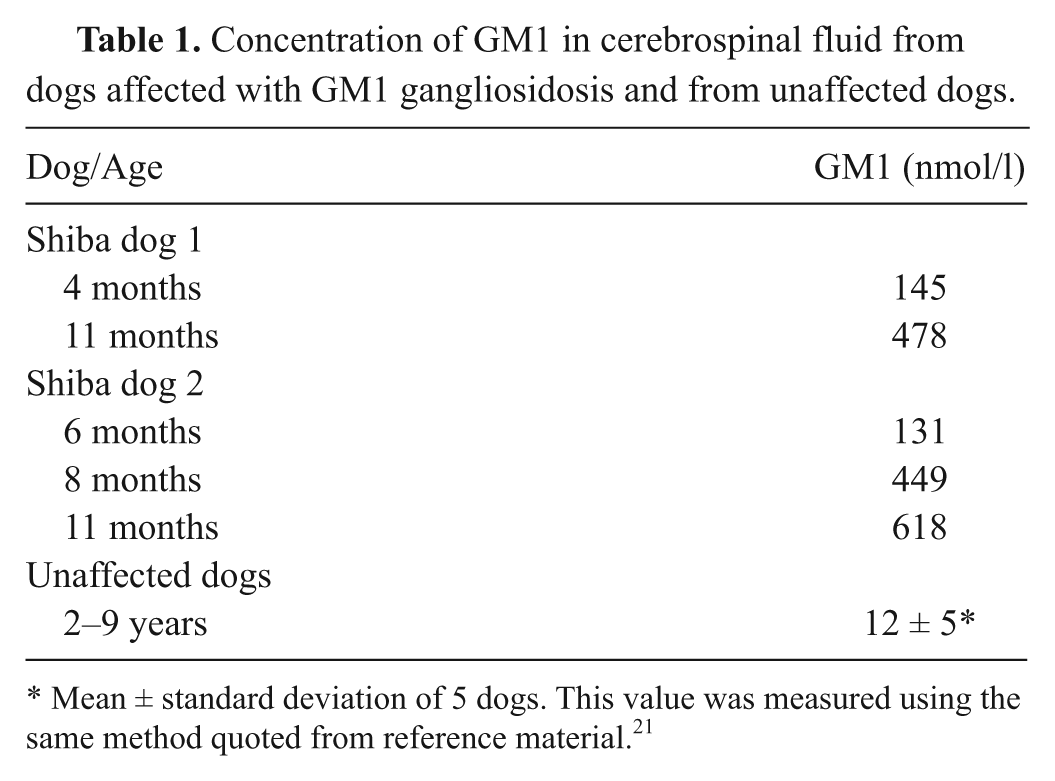
Mean ± standard deviation of 5 dogs. This value was measured using the same method quoted from reference material. 21
An aqueous solution of GM1 was prepared from a commercial GM1 a derived from bovine cerebrum. The concentration of GM1 in this solution was determined by a method reported previously, 13 and adjusted to 20–500 nmol/l as standard solutions of GM1. The concentration of GM1 in canine CSF samples was determined using the TLC enzyme immunostaining method reported previously. 21
As a matrix for MALDI TOF MS, 10 mg/ml of 2,5-dihydroxybenzoic acid b in 10% ethanol was prepared immediately prior to analysis. For application to MALDI TOF MS, 1.5 µl of the matrix solution and 1 µl of sample such as CSF or the standard solution were mixed thoroughly, and 1 µl of this mixture was put on a special sample plate, b referred to as a target plate, followed by ventilation drying. The CSF samples applied to the sample plate were treated for demineralization immediately before MALDI TOF MS analysis. Two microliters of ice-cold 0.1% trifluoroacetic acid c was dropped onto the dried CSF sample on the sample plate, and after 10 sec, the solution was removed by a micropipette. The desalted sample was dried again by ventilation drying.
The MALDI TOF MS analysis was carried out using a mass spectrometer, b coupled with software c for analysis of ion peaks. Ionization was carried out using a N2 laser (wavelength 337 nm), and negative ions were detected using the selective reflector mode of this spectrometer. Commercially available standard peptide solution b was used as external calibrators, and actual calibration was carried out using angiotensin II ([M-H]– = 1044.52725), angiotensin I ([M-H]– = 1294.67025), and adrenocorticotropic hormone amino-acid residues 1–17 ([M-H]– = 2091.07165) among the external calibrators. Laser intensity was regulated to maintain equal irradiation in every experiment, and the laser irradiation was performed 30 times to 4 separate spots on the sample area that could be seen through the magnifying device of the spectrometer. The final data consisted of integrated ion peak spectra obtained by 120 shots of laser irradiation to each sample.
Concentrations of GM1 in CSF in dogs with GM1 gangliosidosis, measured by the TLC enzyme immunostaining method, are shown in Table 1 along with reference values obtained from unaffected dogs. The concentration of CSF GM1 in affected dogs at 4 and 6 months of age was 10 or more times higher than that in unaffected dogs, and the concentration subsequently increased several fold until 11 months of age.
As shown in Figure 1, specific signs of GM1 could be detected in the standard GM1 solutions at concentrations of 50 nmol/l or more. The intensity of the signs was increased according to the concentration of standard solutions. In CSF from Shiba dogs with GM1 gangliosidosis, signs corresponding to those in the standard GM1 were detected (Figs. 2–4), but were not detected in CSF from unaffected dogs (data not shown). The main monoisotopic mass-to-charge (m/z) ratio values of these signals in the canine CSF and the bovine standard GM1 were measured as 1,543.992–1,545.157 at a lower molecular weight and 1,571.892–1,573.176 at a higher molecular weight. However, the lower molecular peak was more intense than the higher molecular peak for canine CSF GM1, while the higher peak was more intense than the lower peak for bovine GM1.
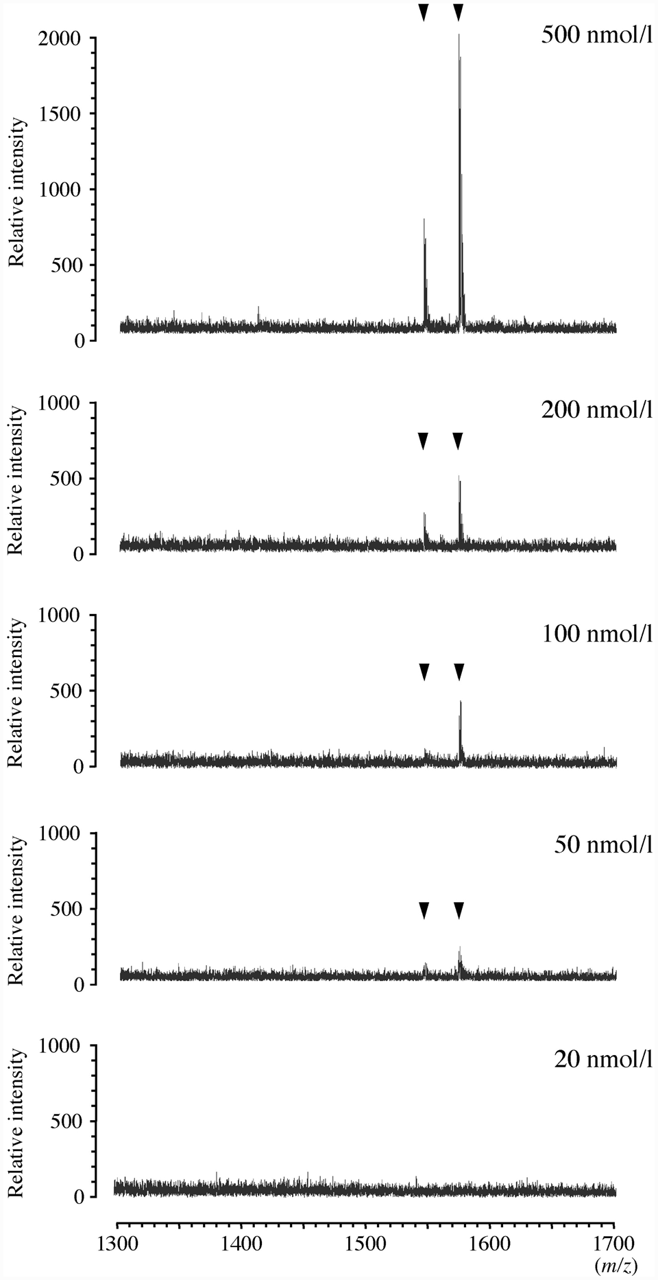
Matrix-assisted laser desorption ionization time-of-flight mass spectrometry spectrum of standard GM1 derived from bovine cerebrum. The concentration of GM1 in the standard solutions is indicated at the upper right of each spectrum. The arrowhead indicates the ion peaks of GM1. m/z = mass-to-charge ratio value.
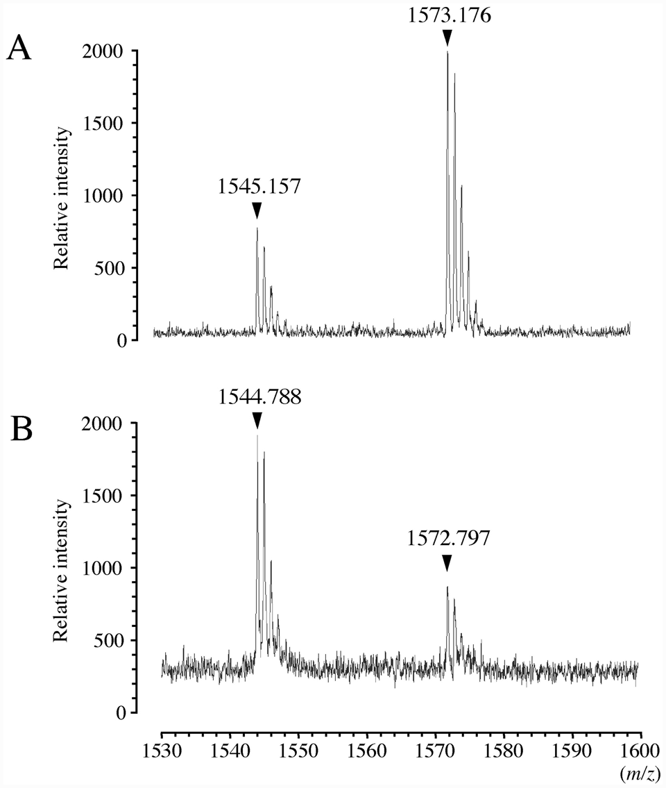
Matrix-assisted laser desorption ionization time-of-flight mass spectrometry enlarged spectrum of GM1 in the standard solution (
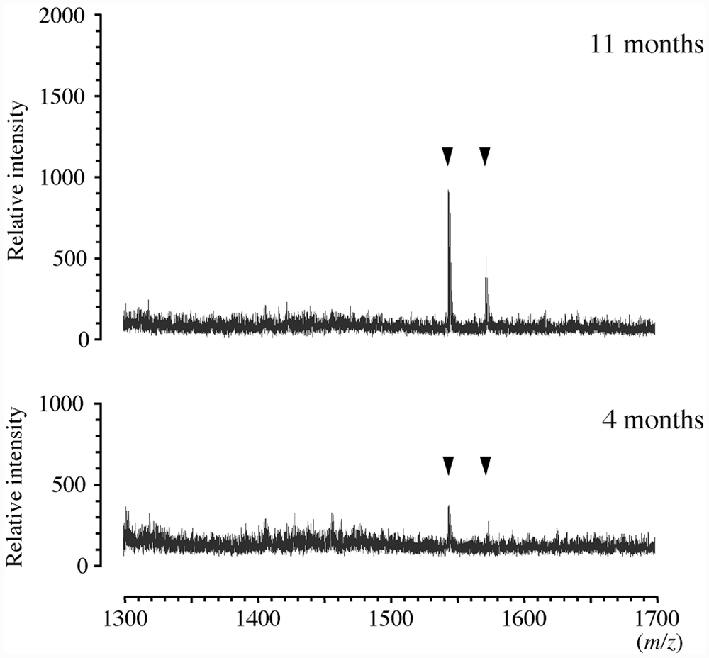
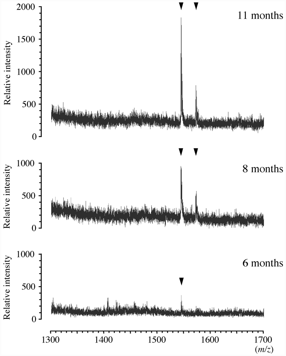
Matrix-assisted laser desorption ionization time-of-flight mass spectrometry spectrum of GM1 in cerebrospinal fluid (CSF) from a Shiba dog (no. 1) affected with GM1 gangliosidosis. The age of the dog when CSF was collected is indicated at the upper right of each spectrum. The arrowhead indicates the ion peaks of GM1. m/z = mass-to-charge ratio value.
Matrix-assisted laser desorption ionization time-of-flight mass spectrometry spectrum of GM1 in cerebrospinal fluid (CSF) from a Shiba dog (no. 2) affected with GM1 gangliosidosis. The age of the dog when CSF was collected is indicated at the upper right of each spectrum. The arrowhead indicates the ion peaks of GM1. m/z = mass-to-charge ratio value.
The signs of GM1 in CSF in affected Shiba dogs were detected slightly at 4–6 months of age and more clearly at 8–11 months of age (Figs. 3, 4). The intensity seemed to be increased according to the age of affected dogs. However, the intensity of canine CSF GM1 tended to be weaker than that of the standard solution at a similar concentration of GM1.
In the past few decades, determination, profiling, quantification, or evaluation of gangliosides including GM1 in tissues, cultured cells, or CSF has been reported by TLC separation coupled with densitometric or immunochemical detection, 21 high performance liquid chromatography, 15 supercritical fluid chromatography, 17 and enzyme-linked immunosorbent assay. 6 These traditional methods have limitations because of their large sample requirement, complex preparation and methodology, and low resolution and sensitivity. To resolve these problems, a 2008 study reported that soft ionization MS–based methods have provided an excellent platform for analysis of gangliosides due to its high sensitivity, precision, accuracy, and resolution. 11 Soft ionization MS–based methodology has been reported to diagnose human patients and a dog with GM1 gangliosidosis using MALDI TOF MS 10 and ESI MS, 27 respectively. However, in these 2 cases, diagnoses were established using postmortem tissue samples. The present study developed the MALDI TOF MS method for detection of GM1 directly by using a tiny amount of CSF from living dogs.
The basic structure of GM1 consists of a ceramide core with an oligosaccharide head including a sialic acid. 12 There are 2 molecular species of GM1 with a different ceramide. One type of ceramide consists of the sphingosine d18:1 and the fatty acid C18:0, and the other type consists of the eicosasphingosine d20:1 and the fatty acid C18:0. Therefore, MALDI TOF MS yields 2 major peaks at m/z 1,544.87 (C73H131N3O31) and m/z 1,572.90 (C75H135N3O31) corresponding to monoisotopic masses of [M-H]– as GM1 (d18:1-C18:0) and GM1 (d20:1-C18:0), respectively. The theoretical m/z values corresponded to those of the 2 main peaks observed in the bovine standard GM1 and canine CSF in affected dogs in the present study (Fig. 2), demonstrating the MALDI TOF MS spectrum of GM1 molecules.
The differing proportions of the 2 main components, GM1 (d18:1-C18:0) and GM1 (d20:1-C18:0), reflect the species and pathology difference. 27 In a mixed-breed dog with GM1 gangliosidosis diagnosed postmortem by ESI MS, the MS spectrum suggested that the ratio of GM1 (d18:1-C18:0) and GM1 (d20:1-C18:0) was approximately 4:1, whereas GM1 (d18:1-C18:0) was less intense than the other (d20:1-C18:0) in the standard GM1 derived from bovine cerebrum. 27 This finding also agreed with the results in the present study.
In Shiba dogs with GM1 gangliosidosis, the increased concentration of GM1 in CSF reflects the storage of GM1 in the central nervous system, and the change is well related with the months of age and clinical course. 21 Clinically, at approximately 5–6 months of age, the affected dog starts to show neurological symptoms recognizable as typically cerebellar signs including ataxia, hypermetria, and intention tremor. 29 Therefore, at this age, high-risk animals first present to a veterinarian and undergo a variety of examinations including a CSF test. In the present study, MALDI TOF MS detected the signs of GM1 molecules in CSF in dogs at 4–6 months of age, suggesting that this method provides a diagnostic support even in the early stage of the disease.
In general, biological samples include numerous minerals such as salts, which inhibit the ionization and analysis of target molecules through the precipitation of these salts, heterogeneity of distributions of matrix and samples, formation of cluster ion of matrix, and other processes. 24 To resolve these problems, there are several methods of washing and desalting the samples on the sample plate.16,24,31 A preliminary examination of CSF samples without desalting did not provide clear signs of GM1 on the MALDI TOF MS spectrum (data not shown). Therefore, a simple desalting method with ice-cold 0.1% trifluoroacetic acid was added to the preparation process and yielded sufficient improvement of the results in the present study. However, the strength of GM1 peaks tended to be weaker than that in the standard GM1 at concentrations similar to the canine CSF. This may be because of an adverse influence from remaining salts in the CSF samples. In addition, the surface of the special sample plate used in the present study is manufactured and designed to concentrate samples into a narrower area in the process of sample drying. This characteristic might also contribute to the detection sensitivity in the present study.
In conclusion, MALDI TOF MS analysis in combination with the special sample plate and the simple desalting step clearly demonstrated GM1 molecules accumulated in only 1 µl of CSF in Shiba dogs with GM1 gangliosidosis. Results suggest that this rapid and simple detection method is useful for the diagnosis and/or high-risk screening of canine GM1 gangliosidosis. This method may also be utilized for antemortem diagnosis of GM1 gangliosidosis in domestic animals other than dogs.
Footnotes
Notes
The author(s) declared no potential conflicts of interest with respect to the research, authorship, and/or publication of this article.
This study was supported financially by grants (nos. 20380173, 20-08112, and 21658109, O.Y.) from the Ministry of Education, Culture, Sports, Science and Technology of Japan.