
Abstract
Molecular screening of GM1 gangliosidosis in Shiba dogs was carried out in northern Japan using blood smear specimens after prolonged storage. Of 125 specimens obtained from 3 veterinary teaching hospitals for this screening, 68 specimens (54%) were adequate for direct amplification in a polymerase chain reaction (PCR)-based DNA test, and the percentage of adequacy was different at each hospital (34%, 73%. and 100%), suggesting that the amount of blood on the smear and the storage condition of specimens may affect adequacy. Of the 68 dogs examined, 2 dogs (2.9%) were heterozygous carriers for this disease and the other dogs were all genotypically normal. The results suggest blood smear specimens can be useful for PCR testing after prolonged storage provided specimens contain a generous amount of blood and have been adequately stored. The study also suggests that GM1 gangliosidosis may be widely prevalent in the Shiba dog population in northern Japan.
GM1 gangliosidosis, a lysosomal storage disease that affects the brain and multiple systemic organs, is due to an autosomal recessively inherited deficiency of acid β-galactosidase coded by the GLB1 gene. 4 GM1 gangliosidosis in Shiba dogs was initially identified in 2000. 12 The homozygous recessive mutation causing GM1 gangliosidosis in Shiba dogs has been identified as a deletion of C nucleotide 1647 in the putative coding region for the canine β-galactosidase gene, 7 allowing diagnosis with a polymerase chain reaction (PCR)-based DNA test, 8,10 as well as an enzyme assay using leukocyte or tissue specimens. 9,10 Affected Shiba dogs manifest neurological symptoms of progressive motor dysfunctions starting from 5 to 6 months of age and finally die by 15 months of age after a clearly defined clinical course, 11,12 which is associated with the progressive accumulation of GM1 ganglioside in the central nervous system 3,12 and cerebrospinal fluid. 2,3
The Shiba dog is one of the original Japanese breeds designated as a protected species in Japan. This canine breed has been one of the most popular breeds for many years, and approximately 40,000 puppies are produced and registered every year in Japan. Shiba dogs have been transported all over the world and are bred and maintained as a standard breed in many countries. Therefore, it is important to control and decrease the prevalence of this fatal inherited disease in Shiba dogs to maintain the quality of this popular breed.
The present study was conducted to investigate the adequacy of blood smear specimens after prolonged storage for molecular screening of canine GM1 gangliosidosis. It also documented the current prevalence of heterozygous carriers in the population of Shiba dogs in northern Japan to facilitate the control of this disease in the near future.
One hundred and twenty-five blood smear specimens from Shiba dogs were collected and examined in 2003 by 3 veterinary teaching hospitals located in northern Japan (Fig. 1). Specimens were stained with Romanowsky-type stains, such as Giemsa staining, and stored at room temperature without mounting with cover glass slips. Twenty-one specimens prepared between 1993 and 2003 were obtained from stored blood smears at Hokkaido University (Sapporo, Hokkaido); 74 specimens prepared between 1993 and 2002 were obtained from Kitasato University (Towada, Aomori prefecture in the Tohoku district); and 30 specimens prepared between 1998 and 2002 were obtained from Iwate University (Morioka, Iwate prefecture in the Tohoku district). Blood smears from Hokkaido University and Iwate University were prepared using a slide film or blood smear technique with a spreader slide slid at an angle. 6 Blood smears from Kitasato University were prepared using a slide-over-slide or squash technique in which the blood film is made by overlapping 2 slide glasses then sliding. 6 The smears from Kitasato University appeared to be thinner and to contain a lower amount of blood than those from the other 2 universities.
After rinsing the surface of the blood smear with pure water followed by xylene, the dry powder of blood was stripped from a part of the blood film using an 18-gauge needle. In the film prepared using the slide film technique, 1 thicker edge of the blood film, approximately 10 × 10 mm. was peeled off and used as the template for the DNA test because this area was thick enough to obtain a useful amount of blood powder but was not essential for observing blood cells microscopically. In the film prepared using the slide-over-slide technique, one-third to half of the area of the film had to be peeled off for the DNA test because this type of film is typically very thin and does not contain a generous amount of blood.
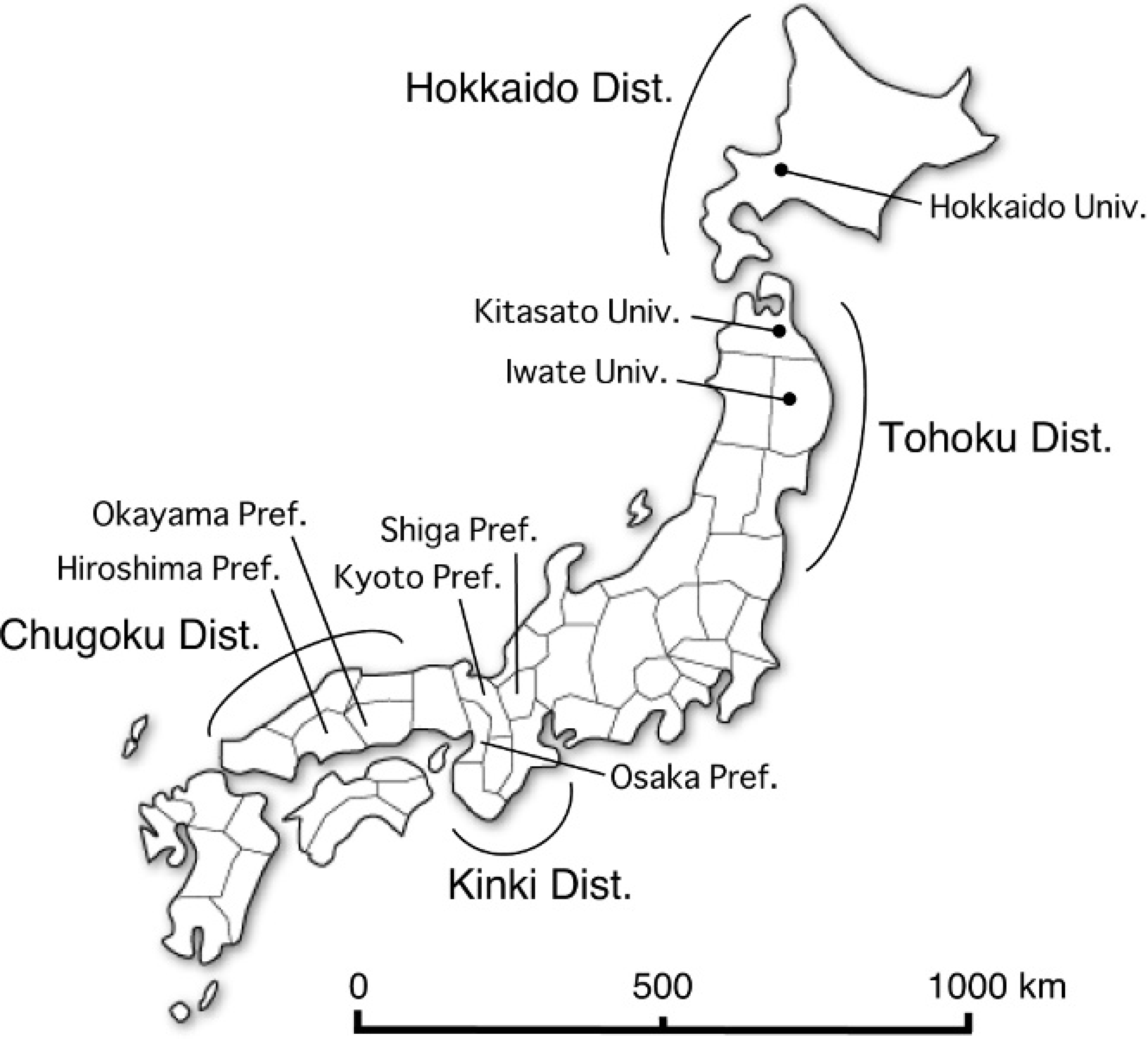
Map of Japan indicating locations of universities (Univ.), districts (Dist), and prefectures (Pref.) involved in the study.
The genotyping test was performed according to the direct PCR amplification method reported previously 8 in which the DNA extraction process can be eliminated. Briefly, PCR was carried out in a 25-μl reaction mixture containing 1× Ampdirect-A, a 0.6 units of Taq polymerase, b 0.2 mM deoxynucleoside 5′-triphosphate, and 12.5 pmol of specific primers (forward: 5′-AAC ACT GAG GAT GCA GTA CGC AGC-3′, reverse: 5′-TCC AGG AAA CTG GAT AAA GGT GTC-3′). The dry powder of blood was first put into an empty PCR tube and spun down on the tube bottom, and the reaction mixture was layered over the specimen without mixing. After preheating at 80°C for 15 minutes and the first denaturation at 94°C for 4.5 minutes, 40 cycles of amplification were carried out at a denaturing temperature of 94°C for 30 seconds, an annealing temperature of 60°C for 1 minute, and an extension temperature of 72°C for 1 minute. Extension during the last cycle was carried out for 7 minutes. The amplification product (8 μl) was added together with 5–20 units of the restriction endonuclease NciI c to a 10-μl reaction mixture containing 1× restriction endonuclease buffer and digested for 90 minutes at 37°C. Both the amplification and digested products were analyzed by 3% agarose gel electrophoresis, d stained with ethidium bromide, and detected using an ultraviolet trasilluminator.
For examinations of a carrier dog identified by the survey, blood was collected from the dog (with the owner's permission) and pedigree analysis was performed using the dog's pedigree paper. The genotype was determined by PCR using whole blood as the template for direct PCR amplification. 8
The results are shown in Table 1. Of 125 specimens obtained from 3 veterinary teaching hospitals in northern Japan, only 68 specimens were adequate for direct PCR amplification. The overall adequacy rate was 54% and differed considerably among hospitals (34%, 73%, and 100%).
Of the 68 specimens adequate for PCR, 2 dogs from Hokkaido and Aomori were heterozygous carriers. The total prevalence of carriers was 2.9%. One carrier dog from Hokkaido was still living in 2003 and was examined further with the owner's permission. The carrier status of the dog was confirmed with PCR using whole blood as the test sample. The pedigree analysis using the dog's pedigree paper showed that the carrier dog had a forefather in common with affected dogs reported previously (Fig. 2). By contrast, information about the other carrier dog identified in Aomori prefecture was not available. Based on the identification of 2 carrier Shiba dogs in the present study in Hokkaido and Tohoku districts, the prevalence of carriers was 2.9% using simple arithmetic, although the number of samples examined in this survey was not sufficient to calculate an accurate prevalence.
The prevalence of human GM1 gangliosidosis is not known. 4 However, people of certain ethnic origins show a higher prevalence of this disease. Among these ethnic groups, a high prevalence of human patients with infantile GM1 gangliosidosis has been reported in the Maltese Islands, 1 where the prevalence of the disease was calculated as 1/3,700 live births, indicating a carrier prevalence of about 1/30 (approximately 3%) according to the Hardy-Weinberg principle. Therefore, the prevalence of carriers in Shiba dogs in the present study corresponds approximately to the highest prevalence shown in the human ethnic populations.
Genotype screening of GM1 gangliosidosis in Shiba dogs in northern Japan.
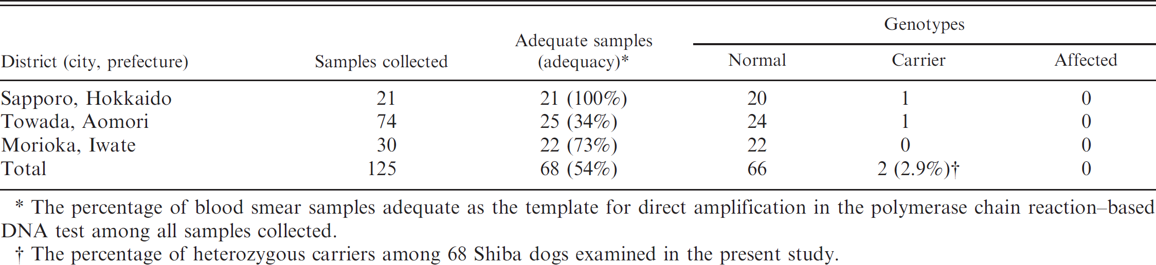
The percentage of blood smear samples adequate as the template for direct amplification in the polymerase chain reaction-based DNA test among all samples collected.
The percentage of heterozygous carriers among 68 Shiba dogs examined in the present study.
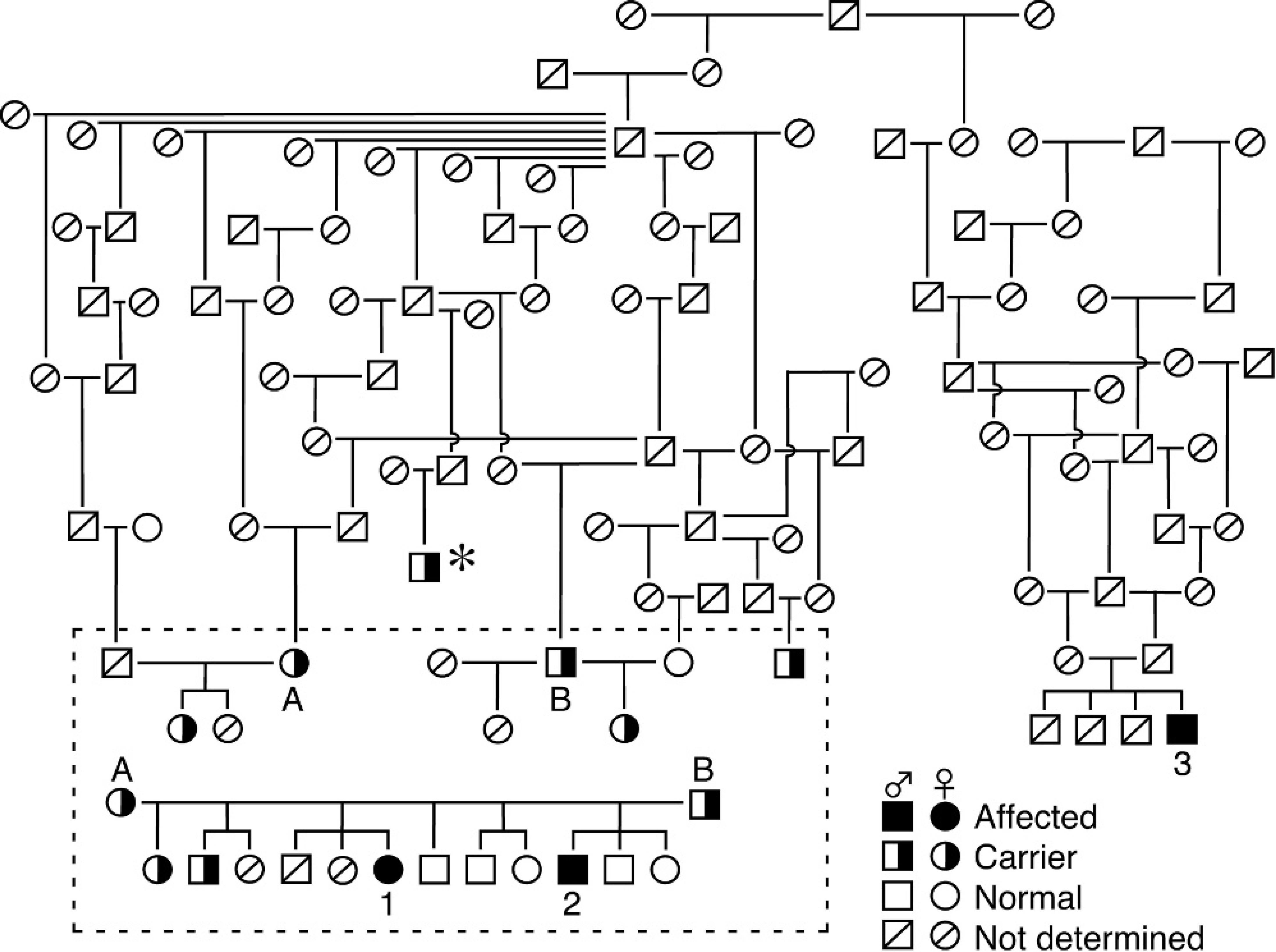
In Shiba dogs, 2 affected cases (dogs 1 and 2 in Fig. 2) were first identified in a breeding colony in Hokkaido, 12 and then 1 sporadic case (dog 3 in Fig. 2) was identified in Shiga prefecture in the western area of Japan (Fig. 1). 5 Recently, 5 sporadic cases possessing a distant common forefather with previous cases studied were diagnosed with a DNA test in Hiroshima, Okayama, Osaka, and Kyoto prefectures in Chugoku and Kinki districts in western Japan (Osamu Yamato, unpublished data). This suggests that the mutant allele is distributed widely in breeding colonies of Shiba dogs all over Japan. The data in the present study were limited to that of northern Japan, but there may be a high prevalence of carriers all over Japan. Therefore, preventive measures should be taken against GM1 gangliosidosis in Shiba dogs to eradicate this disease or at least to reduce the prevalence of carriers.
In the present study, the usefulness of blood smears after long-term storage for PCR testing was investigated. As a result, the overall adequacy was not very high (54%; 68 of 125 specimens). However, the adequacy differed considerably among hospitals (34%, 73%, and 100%). The lowest rate at Kitasato University seems to be due to the lower amount of blood on the smear, resulting from the use of a slide-over-slide technique for preparing the blood film. The specimens at Iwate University might have been damaged by light or heat during the storage period. In conclusion, the PCR assay using blood smear specimens is useful for preliminary screening of small populations, but the specimens should be prepared using a slide smear technique that maintains a good amount of blood, and they must be stored without damage in order to prevent DNA fragmentation.
Acknowledgements. This study was supported financially by grants (14560258 and 16380210, O.Y.) from the Ministry of Education, Culture, Sports, Science and Technology of Japan (MEXT) and by the project “Development of Preventive Veterinary Medicine using Genome and Proteome Analyses” of Nippon Veterinary and Life Science University (T.A.), which was also funded by MEXT.
Footnotes
a.
Ampdirect for human blood, Shimadzu Corp., Kyoto, Japan.
b.
Takara EX Taq, Takara Shuzo, Tokyo, Japan.
c.
NciI, Nippon Gene, Tokyo, Japan.
d.
Agarose 21, Nippon Gene, Tokyo, Japan.