
Abstract
Severe ventriculitis and emaciation caused by the infestation of the nematode Hadjelia truncata occurred in meat-type breeder rock pigeons (Columba livia) in southern and central California. Hadjelia truncata can infest several species of birds, although it has only been reported as pathogenic in pigeons. The factors that contribute to H. truncata pathogenicity are not known. The gross and microscopic pathology caused by the infestation of H. truncata in the ventriculus of pigeons and its morphological identification are presented.
Hadjelia truncata (order Spirurida, superfamily Habronematoidea, family Habronematidae, genus Hadjelia 2 ) is a nematode found in the digestive system of several species of birds. 3 Hadjelia truncata has been reported to be pathogenic only in commercial pigeons from a few countries, including Egypt, Iran, Saudi Arabia, and Cyprus.1,3,4,8 The current study describes a severe parasitic ventriculitis in meat-type rock pigeons (Columba livia) diagnosed in southern and central California.
During 2009–2010, a total of 20 live and 3 dead, adult meat-type breeder pigeons were received as 4 submissions at the California Animal Health and Food Safety Laboratory System (CAHFS) for diagnostic examination. Three submissions were received at the CAHFS, Turlock Branch, and 1 submission was received at the CAHFS, San Bernardino Branch. The submissions came from 3 different producers. All the submissions had a common clinical history of birds not doing well, losing weight, or experiencing poor weight gain, poor food consumption, diarrhea, and increased mortality. In one of the flocks, the percent of affected birds was approximately 10%. Percent of mortality of the other flocks was unavailable. The affected pigeons were housed in wooden, chicken wire–fenced lofts. On one property, ducks were kept in the area surrounding the lofts.
All the submitted worm-infested pigeons had crops filled with yellow corn and were moderately to severely emaciated. Ventriculi were enlarged approximately 2–3 times the normal size due to thickening of the smooth muscle layer (Fig. 1). The surface of the koilin layer of the cranial and caudal areas of the ventriculus had a moderate to severe irregular and rough appearance with either a greenish or a dark brownish discoloration and multiple rounded cavities and erosions (Fig. 2). Additionally, the koilin layer was thick, soft, fragile, and easily detachable from the ventricular glandular epithelium. Underneath the koilin layer, there were too numerous to count (>100) thread-like worms approximately 1–2 cm long (Fig. 3). Some birds had a whitish film on the proventricular mucosa. Whole corn kernels were present in the duodenal lumen. Moderately enlarged, dilated, and flaccid hearts were seen in some birds.
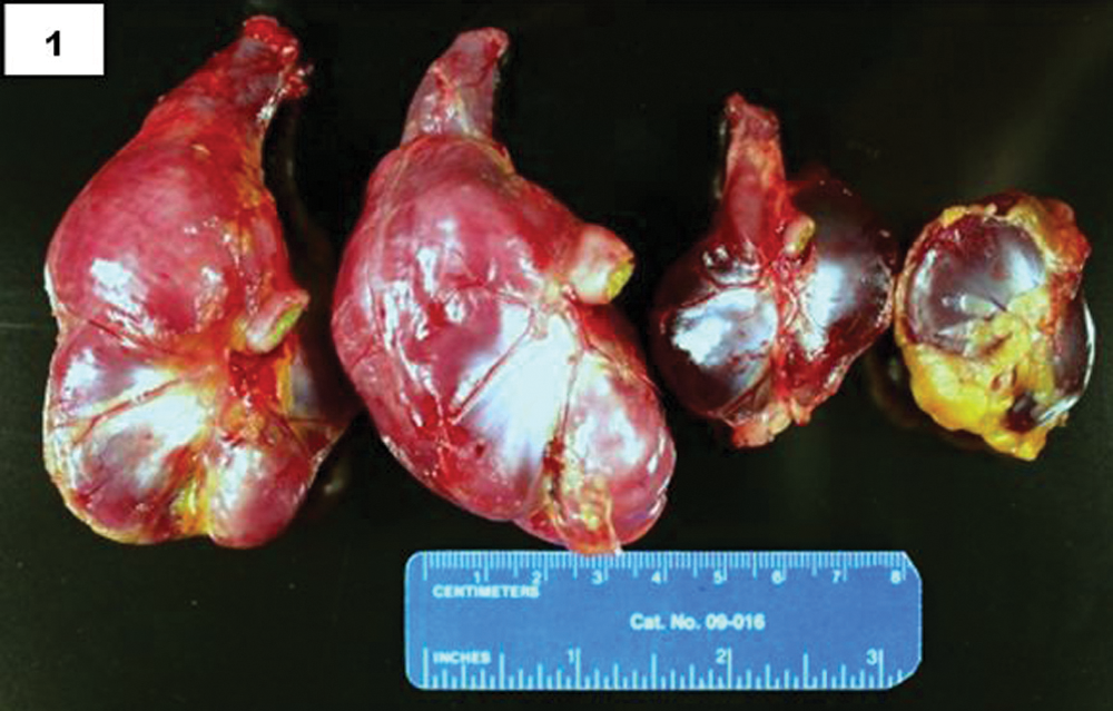
Rock pigeon (Columba livia). Severe hypertrophy of the 2 ventriculi on the left side compared to 2 normal-sized ventriculi on the right side.
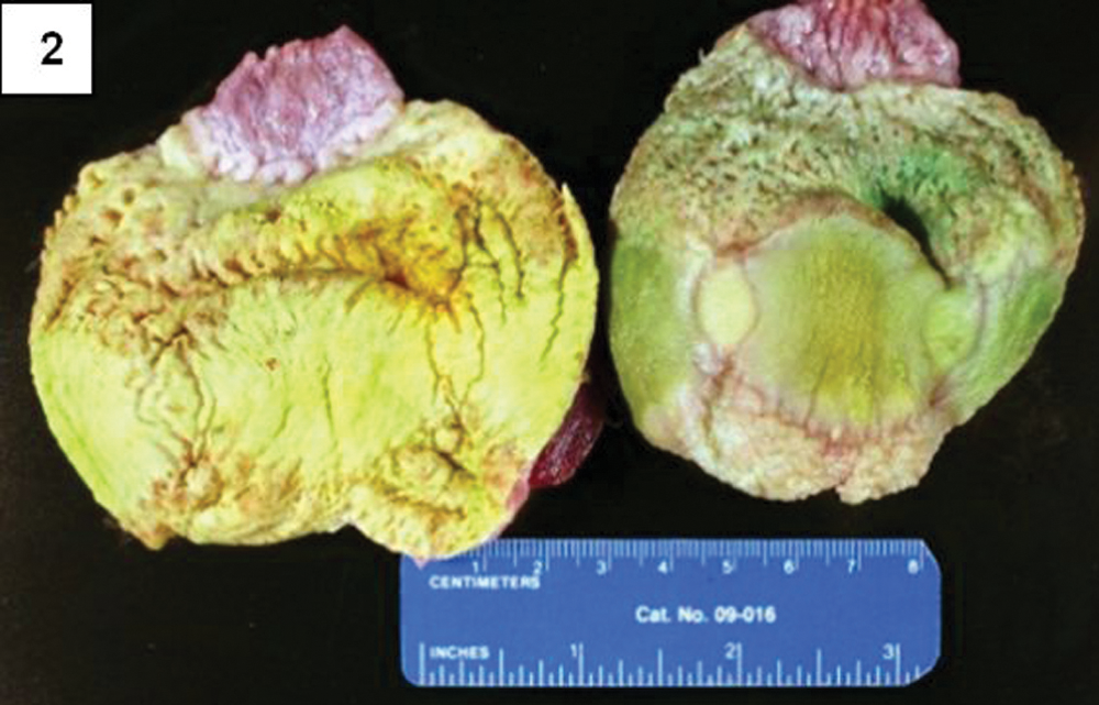
Rock pigeon (Columba livia). Rough appearance and ulcerations of the koilin layer on the proximal portion of the ventriculus.
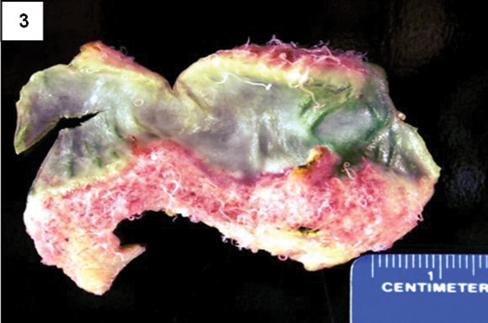
Rock pigeon (Columba livia). Large numbers of thread-like worms located between the koilin and glandular epithelium of the ventriculus.
The histological examination of ventriculus sections revealed that the koilin layer had marked and extensive disruption with uneven staining and numerous clear spaces. In some sections, there were moderate numbers of bacterial clusters mixed within the koilin layers (Fig. 4). Between the koilin and glandular epithelium layers there were large numbers of nematode cross sections. Histologically, H. truncata exhibits an ornamented thick cuticle containing regularly spaced ridges and lateral wing alae. The musculature had coelomyarian and ciromyarian arrangements along the body and around the esophagus, respectively. Females had large numbers of oval, thick-shelled, embryonated eggs at different stages of development in their oviducts (Fig. 5). The egg morphology of H. truncata matched the microscopic characteristics for spirurid eggs reported previously. 5
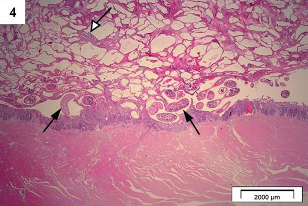
Rock pigeon (Columba livia). Ventriculus histological section exhibiting nematode cross sections located between the glandular epithelium and koilin layers (black arrows). The koilin layer is disrupted and fragmented, with multiple clear spaces. Bacterial clusters are also noted in the koilin layer (white arrow). Bar = 2,000 µm.
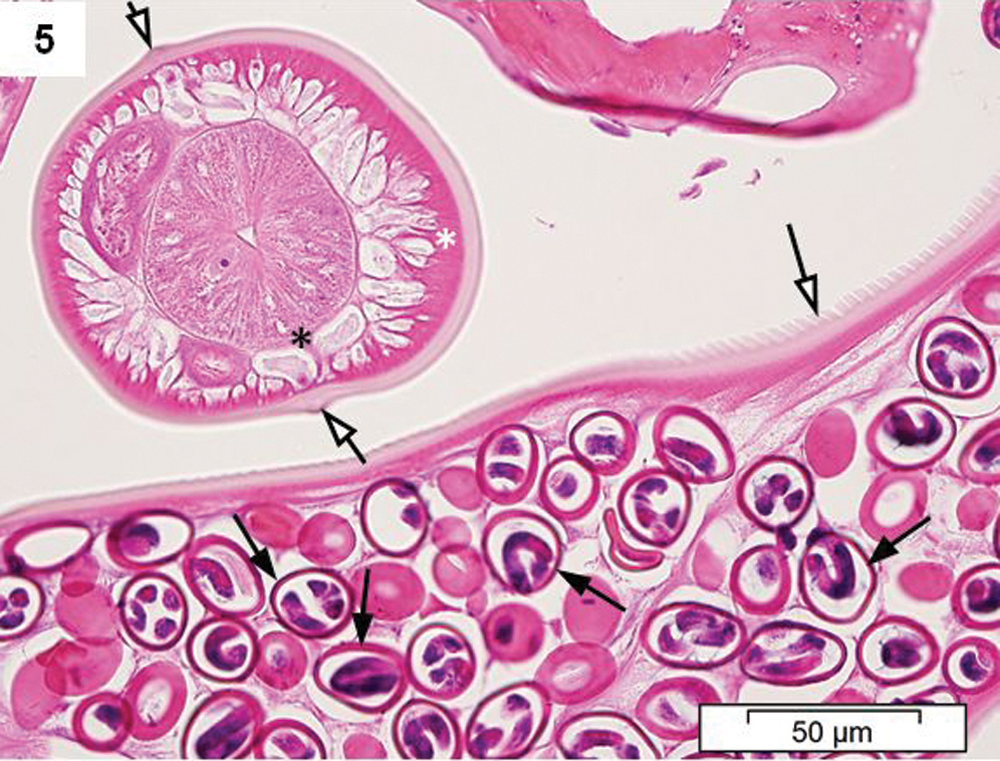
Hadjelia truncata cross sections located between the glandular epithelium and koilin layers of the ventriculus exhibiting large numbers of embryonated eggs (black arrows), the ornaments of the cuticle (white arrows), and the coelomyarian (white asterisk) and ciromyarian (black asterisk) musculatures. Bar = 50 µm.
The ventricular epithelium showed extensive cystic glandular hyperplasia with accumulation of a homogeneous eosinophilic proteinaceous material in the dilated epithelial glands. In the lamina propria, there were moderate lymphocytic and heterophilic infiltrations, although in some sections the inflammatory infiltration was predominantly eosinophilic. Small numbers of lymphoid aggregates were also seen in the lamina propria. The smooth muscle layer was hypertrophied approximately twice the normal thickness.
Ventriculus worm identification was carried out by light microscopy examination. The length of males and females were 6.5–9 mm and 12–16.5 mm, respectively (Figs. 6, 7). The bodies were straight with moderate uniform thickness in both males and females, narrowing at the anterior end. The mouth was surrounded by 2 lateral lips that were trilobed and had a cylindrical pharynx (Fig. 8). There were 6 caudal papillae including 4 pairs of pre-anal and 2 pairs of post-anal papillae (Fig. 9). The male posterior end was spirally coiled at the region of the caudal alae (Fig. 10). Spicules were unequal and dissimilar, one measured 1.27 mm and the other 0.35 mm long. In females, the anal pore was situated at 0.1 mm from the posterior end. The morphology of the parasites examined was consistent with key identification characteristics for H. truncata reported previously.3,6-8
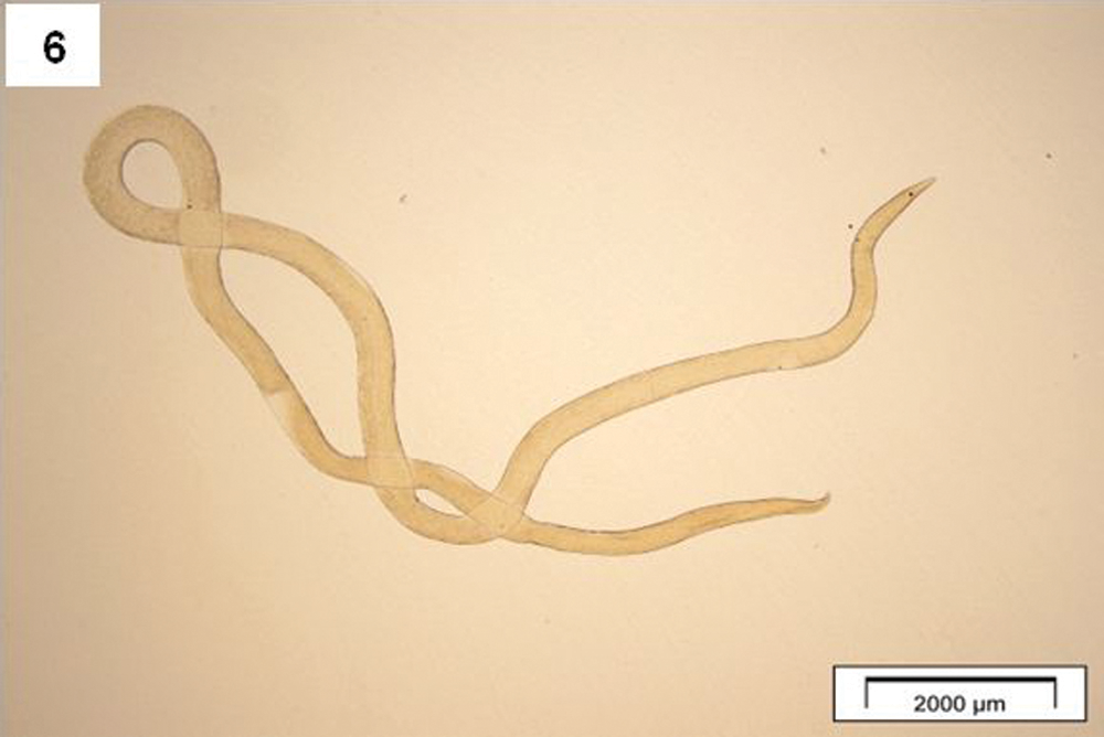
Image of the whole body of a Hadjelia truncata female. Bar = 2,000 µm.
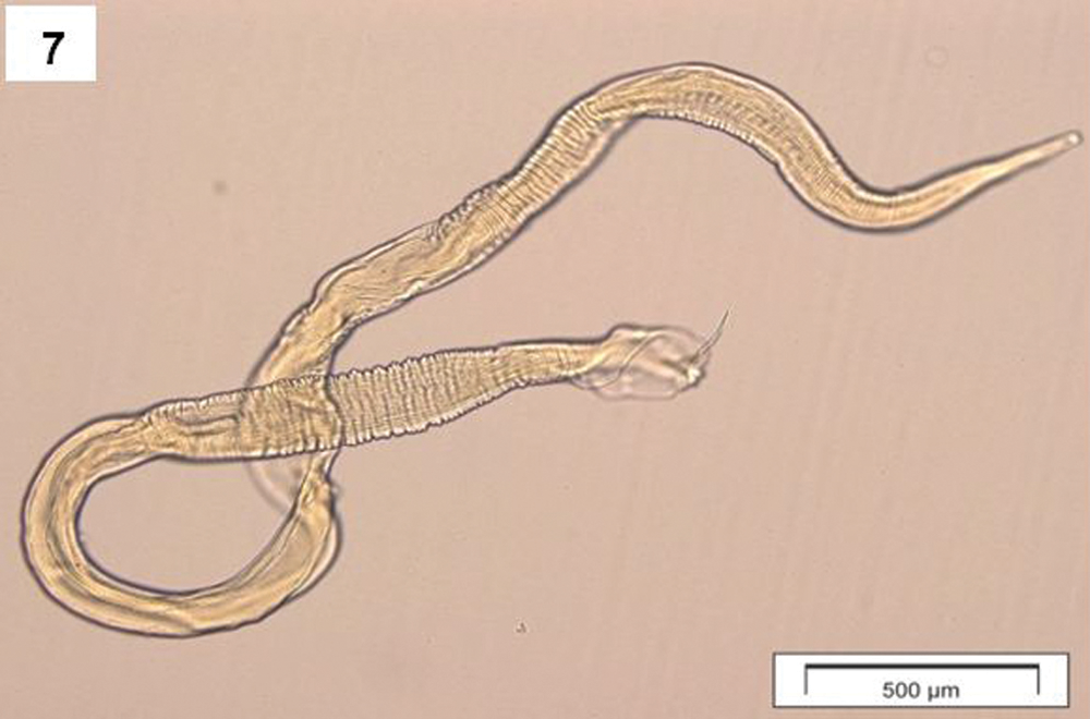
Image of the whole body of a Hadjelia truncata male. Bar = 500 µm.
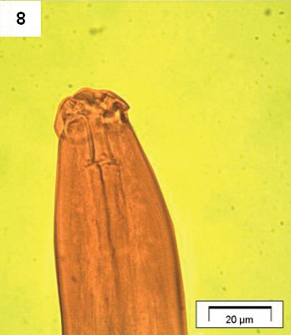
Image of the cephalic end of Hadjelia truncata exhibiting 2 trilobed lateral lips and a cylindrical pharynx. Bar = 100 µm.
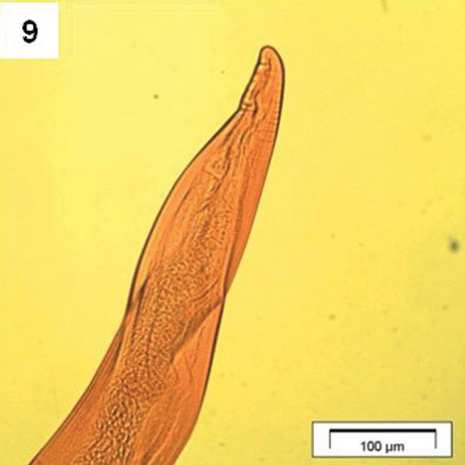
Image of Hadjelia truncata exhibiting the caudal end of the female. Bar = 20 µm.
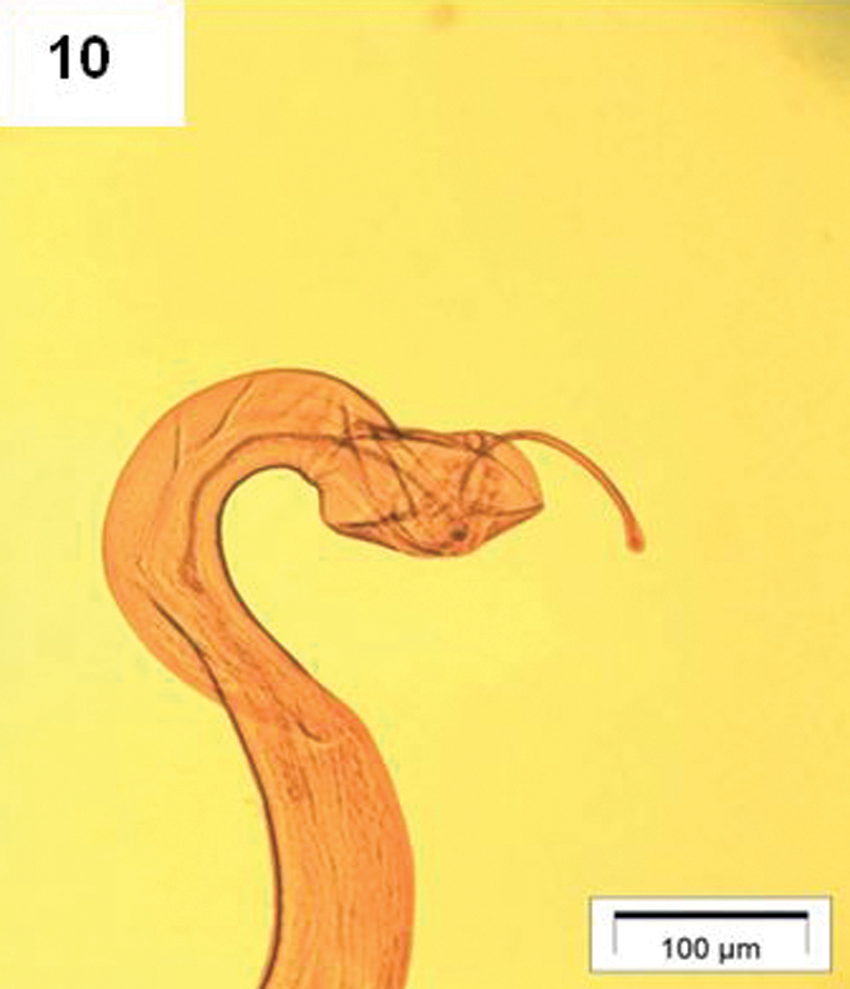
Image of Hadjelia truncata exhibiting the caudal end of the male spirally coiled at the region of the caudal alae. Bar = 100 µm.
A previous study reported findings of H. truncata in the digestive system of several wild birds including magpie, cuckoo, blackcap, hoopoe, nightjar, roller, shrike, and other avian species from Asian and European countries2,3; however, no pathology associated with the infestation of H. truncata in the birds was reported. 3 It is interesting that despite many avian species being able to host H. truncata, it has only been reported to be pathogenic in pigeons in a few countries of the Middle East and Egypt, and now in the United States. It is unknown how or when H. truncata infestation may have occurred in California rock pigeons. Possibly, international trade of this avian species may have facilitated importation to the United States of this parasite, although transmission by other birds cannot be ruled out. It is also unknown if diet, environmental and management conditions, any probable intermediate host, degree of infestation, or other undetermined factors may be associated with the pathogenic manifestation of this nematode.
Clinical signs and pathological findings were similar to those reported previously, 3 including loss of weight, severe hypertrophy of the ventricular muscle layer, severe damage of the koilin layer, and moderate inflammation of the mucosa; however, the number of parasites found underneath the koilin layer was larger in the cases reported herein. Ventricular smooth muscle hypertrophy may have occurred to compensate for ventriculus dysfunctional activity caused by the H. truncata infestation.
A previous study indicates that heteroxeny, a parasite’s utilization of more than one species to complete its life cycle, has been adopted by the entire Spirurida order. 2 Considering the lifecycle characteristics of the Spirurida order, susceptible pigeons may have been infested with H. truncata by ingesting the intermediate host harboring the infective stage. The intermediate host possibly is an arthropod as suggested previously. 4 After ingestion, the infective stage may be released in the ventriculus lumen for burrowing into the koilin layer to develop into the adult stage and stay in the ventriculus’s glandular epithelium. Cheilospirura sp., another nematode of the Spirurida order, causes severe ventriculitis in chickens, turkey, Guinea fowl, grouse, pheasant, and quail, but not in pigeons. 9
Footnotes
Acknowledgements
The authors acknowledge Alicia Gutierrez, Julie Reader, Monica Harris, and Jamie Hall for their excellent technical support.
The author(s) declared no potential conflicts of interest with respect to the research, authorship, and/or publication of this article.
The author(s) received no financial support for the research, authorship, and/or publication of this article.