
Abstract
A competitive liquid-phase–blocking concanavalin A enzyme-linked immunosorbent assay (LPB-ConA-ELISA) was developed in the current study. The assay used ConA as a capture reagent, and the sera of specific pathogen–free chickens immunized with nonpurified Newcastle disease virus (NDV) suspension as detector antibodies, to detect and quantify specific antiviral antibodies in serum samples from free-ranging pigeons. The comparison between the LPB-ConA-ELISA and the hemagglutination inhibition (HI) test for the detection of antibodies in serum samples from 107 pigeons showed significant correlation between the assays (r = 0.875), a high sensitivity (100%), specificity (95.8%), accuracy (96.3%) for the ELISA, and good agreement (κ = 0.83) between the 2 assays. The results of this study suggest that the LPB-ConA-ELISA could be a useful alternative to HI test in the serodiagnosis of NDV in pigeons, or other species of birds.
Keywords
Introduction
Newcastle disease virus (NDV; order Mononegavirales, family Paramyxoviridae, subfamily Paramyxovirinae, genus Avulavirus), an Avian paramyxovirus type 1 (APMV-1), is the etiologic agent of an important infectious poultry disease, characterized by its highly contagious nature and lethality, resulting in significant economic losses, either directly, or due to trade embargoes. 1 Highly virulent NDV has been frequently isolated from chickens and other species of birds, both domestic and wild, with these other bird species being important in the epidemiology and control of the disease in chickens.1,8
Serological tests have been used for both monitoring postvaccination antibody responses and for the indirect diagnosis of NDV infection through the detection of antivirus antibodies. The presence of seropositive results in nonvaccinated birds, or seroconversion in vaccinated birds, is associated with the occurrence of an NDV infection at some past time. 21 The hemagglutination inhibition (HI) test is the reference and most widely applied test to detect anti-NDV antibodies in a number of bird species. 21 In addition, commercial kits based on an indirect enzyme-linked immunosorbent assay (ELISA) have been used for the detection of antibodies in chicken and turkeys. Despite the advantages of such kits, indirect ELISAs require a specific conjugate for the detection of antibodies in species of birds other than chicken and turkey.9,11,17,21
To overcome this limitation, competitive and blocking ELISAs (cELISA and bELISA, respectively), using polyclonal or monoclonal antibodies as detector reagents were developed. Such ELISAs have been successfully applied for the detection and titration of antibodies in serum samples from chickens and other species.5,9,11,17 However, a problem in using the cELISA is that purified whole virus particles are required as target antigens. The NDV purification process requires the propagation of virus in specific pathogen–free (SPF) embryonated eggs and the use of ultracentrifugation. The purified NDV antigen is required to be attached to the solid phase of these cELISAs, or in the preparation of capture or detector antibodies.5,11,17 Additionally, the cELISAs, when using a given anti-NDV monoclonal antibody, may not block antibodies from test sera against all strains of APMV-1, because the monoclonal antibody reacts with a single epitope of 1 NDV antigen. 21
Interactions between mannose-binding lectins, such as concanavalin A (ConA), and RNA viruses, including paramyxoviruses, have been demonstrated and applied to the purification of these viruses. 3 Additionally, ConA was reported to bind efficiently with NDV, blocking virus hemagglutination. 10 The lectin has been used as a capture reagent for Infectious bronchitis virus (IBV; Avian coronavirus) in a sandwich ELISA method. 4 However, to the authors’ knowledge, lectins, including ConA, have not been used to capture NDV using ELISA methods. The replacement of the capture NDV antibodies by the lectin ConA and monoclonal anti-NDV detector antibodies by polyclonal antibodies from SPF chickens immunized with nonpurified NDV suspension could facilitate and improve the execution of cELISAs. There would be no need for virus purification, or any restrictions due to epitope specificity of monoclonal anti-NDV antibodies.
Thus, an alternative method for the detection of anti-NDV antibodies in sera samples from pigeons, using the format of a liquid-phase–blocking ConA ELISA (LPB-ConA-ELISA) was developed in the current study. The new assay was compared with the HI test.
Materials and methods
Propagation of Newcastle disease virus
The LaSota strain of NDV was grown by inoculation into the chorioallantoic cavity of SPF embryonated chicken eggs, as recommended.11,21 The allantoic fluid was harvested from the embryonated chicken eggs 48 hr postinoculation, and titered for infectivity in embryonated chicken eggs and used as the NDV antigen suspension to immunize chickens and pigeons, and as the antigen in the LPB-ConA-ELISA.
Pigeons
A total of 107 free-ranging rock pigeons (Columba livia) were captured in Jaboticabal, São Paulo, Brazil between January and May 2012. Blood samples were collected from the brachial vein of the birds. The serum samples were obtained after blood clotting and stored at −20°C. All experimental procedures with these birds, and the others described below, were approved by the Ethics and Animal Welfare Commission of the Faculty of Agriculture and Veterinary Sciences, Universidade Estadual Paulista–Jaboticabal (protocol no. 021719/11).
Detector anti–Newcastle disease virus antibodies
Anti-NDV polyclonal antibodies were prepared by hyperimmunization of SPF White Leghorn chickens. A group of six 28-day-old chickens were placed into positive pressure isolators and received, at an interval of 2 weeks, 2 immunizations with non-purified attenuated LaSota strain of NDV containing 104.0 50% embryonic infectious doses (EID50), via the intraocular route. Two weeks after the last immunization, the chickens were boosted, at an interval of 2 weeks, by the intramuscular route, with 2 doses of inactivated LaSota strain of NDV (106.0 EID50/dose) emulsified in Freund incomplete adjuvant. a Blood samplings were carried out 2 weeks later, and the serum was separated and stored at −20°C.
Newcastle disease virus–positive and –negative pigeon sera
A group of 12 free-ranging pigeons were captured in Jaboticabal, São Paulo, Brazil. The birds were placed in positive pressure isolators for a period of 60 days in order to adapt to captivity conditions and for the collection of 2 blood samples, at first and fiftieth day of housing, to test as paired serum samples in HI test. Negative HI titers (<3 [Log2]) were detected for all serum samples, confirming that the pigeons were seronegative for NDV. The pigeons were separated into 2 groups of 6 pigeons and placed into 2 positive pressure isolators. The first group received 4 doses of LaSota strain of NDV with 104.0 EID50 per bird via intraocular route, at intervals of 10 days. Blood samples were collected 10 days after the last immunization, and the sera were obtained and stored at −20°C. A pool of the sera from the last bleeding was prepared and used as NDV-positive pigeon serum. The second group of 6 pigeons remained nonimmunized for the same period of immunization. These birds were bled similarly to the pigeons from the immunized group, to prepare the NDV-negative pigeon sera.
Western blotting
The reactivity of polyclonal antibodies from chickens that were hyperimmunized with LaSota strain of NDV was characterized by Western blot analysis. 19 In brief, the polyethylene glycol–concentrated NDV antigen suspension, prepared as previously described, 11 was separated by electrophoresis in a 12% polyacrylamide gel with sodium dodecyl sulfate; the polypeptides were then electrotransferred from the gel to a polyvinylidene difluoride membrane. b This step was followed by the addition of anti-NDV chicken serum (1:100) and rabbit anti-chicken immunoglobulin G (IgG)-peroxidase conjugate (1:1,000). c Each of these reagents was incubated for 1 hr at 37°C. The color reaction was developed by adding a commercial chromogen–substrate mixture (3,3′-diaminobenzidine plus urea). d
LPB-ConA-ELISA
The LPB-ConA-ELISA was performed according to a previously described methodology, 11 except that ConA e was used as the capture reagent, polyclonal antibodies from chickens hyperimmunized with non-purified NDV were used as the detector reagent, and 10 instead of 22 wells were used to measure the maximum reactivity (maximum optical density [OD]) of detector antibodies with NDV antigen in the absence of competitor antibodies from test serum. Before the application of the LPB-ConA-ELISA to detect anti-NDV antibodies in pigeon sera, optimum concentrations of the capture reagent, ConA, in phosphate buffered saline (PBS; pH 7.4), optimum dilutions of nonpurified antigen preparation, and chicken anti-NDV sera (detector antibody) were determined by checkerboard titration. Reagents were added in volumes of 50 µl per well, and the microplates f were washed 4 times between each step with PBS plus 0.05% Tween-20 (PBST). The microplates were coated with ConA e diluted in PBS, and incubated for 18 hr at 4°C. After washing, the microplates were blocked with a solution containing PBS supplemented with 10% skimmed milk (SM) and incubated for 45 min at 37°C. The liquid phase of the reaction was carried out in the wells of a separate carrier microplate, and a mixture of equal volumes (50 µl) of nonpurified NDV LaSota at a constant predetermined dilution (1:2) in PBS and duplicates of 1:8-diluted test sera in PBS were used. After incubation at 37°C for 45 min, 50-µl volumes of the test serum–virus mixtures were transferred from the carrier plates to the ELISA plates and incubated at 37°C for 1 hr. Thereafter, the ELISA plates were washed, and a pretitrated optimal dilution (1:1,000) of the anti-NDV chicken hyperimmune serum in PBST was added. The plates were incubated for 1 hr at 37°C and then washed. Then, rabbit anti-chicken IgG horseradish peroxidase conjugate, g diluted in PBST-SM, was added to each well, and the reaction was incubated for 1 hr at 37°C. After washing, the plates received the substrate solution containing 0.4 mg/ml of ortho-phenylenediamine h and hydrogen peroxide (0.006%) diluted in 0.1 M Na2PO4 and 0.1 M citric acid buffer (pH 5.0). After incubation for 15 min in the dark and at room temperature, the reaction was stopped by the addition of 2 M HCl. Noninfected tissue or allantoic fluid samples were used as a negative control antigen in each plate. Plates were read spectrophotometrically at 490 nm. The mean ODs obtained for each test sample were converted, as reported previously, 11 to percent inhibition (PI) values, which was calculated using the formula: PI = [(maximum OD − sample OD)/(maximum OD)] × 100.
Cutoff point of LPB-ConA-ELISA
The two-graph receiver operating characteristic (TG-ROC) analysis method 7 was used to determine the cutoff point of LPB-ConA-ELISA. The sensitivity and specificity values were plotted against the PI values. The equivalence point between sensitivity and specificity was at the intersection between the curves of these 2 parameters and it is related to a PI value that is considered the cutoff point of LPB-ConA-ELISA.
Hemagglutination inhibition test
The HI test was performed using 4 hemagglutinating units of the LaSota strain of NDV and chicken red blood cell suspension at 0.5%, as recommended previously. 2 Aliquots of pigeon sera were pretreated at 56°C for 15 min and with 5% kaolin (weight/volume) for 30 min, to eliminate nonspecific inhibitors of hemagglutination. 20 Serum samples were 2-fold diluted (1:2–1:2,048), and the titer was expressed as Log2 of the reciprocal of the highest serum dilution showing inhibition of hemagglutination. The HI titers ≥3 were regarded as NDV-positive as previously described. 11
Statistical analysis
The PI values of samples obtained in the LPB-ConA-ELISA were compared to their HI titers by linear regression analysis, and the Pearson correlation coefficient (r) was established. i The relative sensitivity and specificity, predictive values, and agreement (kappa [κ] index) were determined. 11
Results
Specificity of chicken polyclonal anti–Newcastle disease virus antibodies
The anti-NDV hyperimmune chicken sera, used as a detector polyclonal antibody in the LPB-ConA-ELISA, reacted in a Western blot specifically with the following major viral antigens: hemagglutinin–neuraminidase (HN), nucleoprotein (NP), and matrix (M) protein as shown by bands of 72, approximately 66, and 42 kDa, respectively. No reactivity with other antigenic components from allantoic fluid was observed (Fig. 1).
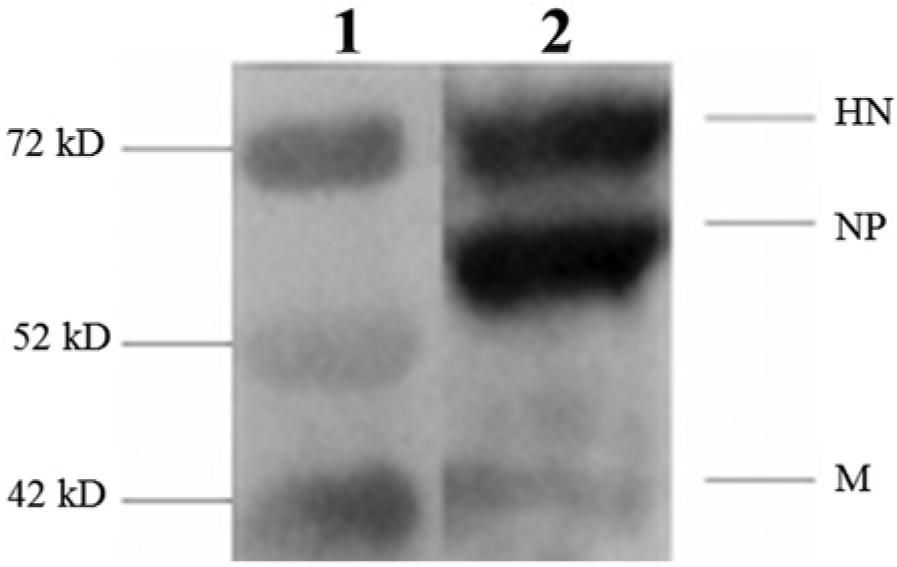
Western blot analysis of anti–Newcastle disease virus (NDV) chicken hyperimmune serum. Lane 1: molecular weight marker j ; lane 2: polyethylene glycol-concentrate NDV antigen suspension. The horizontal lines indicate the major structural proteins of NDV detected: hemagglutinin-neuraminidase (HN), nucleoprotein (NP), and matrix (M) protein.
Standardization of the LPB-ConA-ELISA
The checkerboard titration of the reagents of LPB-ConA-ELISA showed that the maximum discrimination between reactions (PI) with NDV-positive and -negative pigeon sera were detected at coating concentrations of 1 mg/ml of ConA, NDV antigen dilution of 1:2, detector antibody dilution of 1:1,000, and pigeon serum dilution of 1:8. The cutoff point for the LPB-ConA-ELISA of a PI value of 19.73% was determined by TG-ROC analysis (Fig. 2). This cutoff point corresponded to the point of equivalence of 95.3% for the relative sensitivity and specificity. Based on this analysis, pigeon serum samples were classified as positive if the PI value was ≥19.73%, and as negative if the PI value was <19.73%.
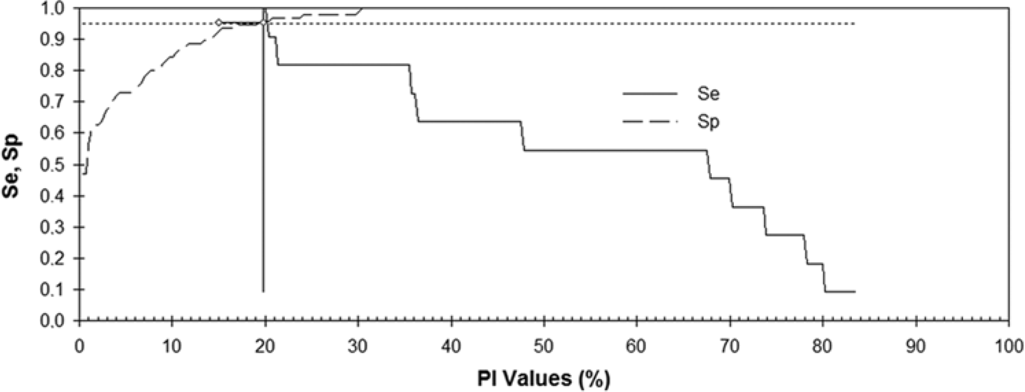
Profiles of the curves obtained by two-graph receiver operating characteristic analysis by plotting the relative sensitivity (Se) and specificity (Sp) values against the percent inhibition (PI) values (see text for details of calculation) generated by the liquid-phase–blocking concanavalin A enzyme-linked immunosorbent assay. The intersection point of the 2 curves corresponded to the cutoff point (PI = 19.73%) at which Se is equivalent to Sp (0.95; dotted and dashed horizontal line). The level of accuracy adopted was 95%.
Titration of Newcastle disease virus–positive and –negative pigeon sera
Distinct profiles of reactivity were shown by the LPB-ConA-ELISA (Fig. 3) in the titration of the pools of NDV-positive and -negative pigeon serum samples. The positive pigeon serum with a HI titer of 7 inhibited, in a dose-dependent manner, the reactivity of the detector anti-NDV antibody with the NDV antigens in LPB-ConA-ELISA. The reactivity of the NDV-positive pigeon serum started with a PI value of 97.78%, and declined down to a PI value of 0.80%. Negative pigeon sera with a HI titer ≤3, showed low inhibition activities on the reactivity of the detector (PI values ≤0.95%) in all dilutions tested.
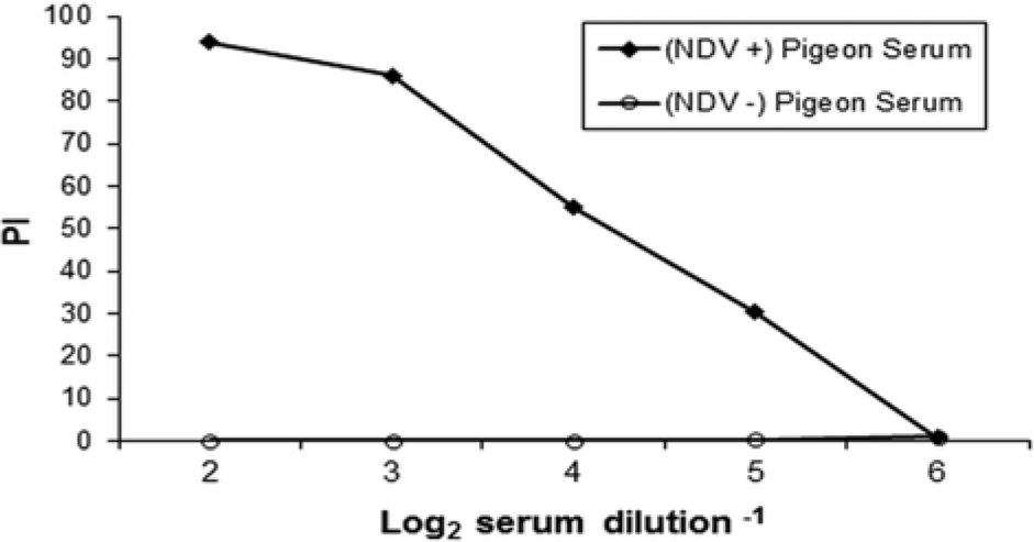
Titration in the liquid-phase–blocking concanavalin A enzyme-linked immunosorbent assay (LPB-ConA-ELISA) of Newcastle disease virus (NDV)-specific antibodies in pools of sera from experimentally immunized [(NDV+) pigeon serum], or nonimmune/noninfected [(NDV−) pigeon serum] pigeons. The LPB-ConA-ELISA results are expressed as the percent inhibition (PI) values (see text for details of calculation).
Comparison between the LPB-ConA-ELISA and HI
Based on the testing of the 107 free-ranging pigeon sera, the sensitivity of the LPB-ConA-ELISA was 100%, the specificity was 95.8%, and the accuracy was 96.3%. The positive predictive value was 73.3% and negative predictive value was 100%. A total of 11 serum samples were positive and 92 were negative for the presence of anti-NDV antibodies in both serological tests (Table 1). Four sera were classified as positive only in the LPB-ConA-ELISA (3.74% of false-positive), and no serum was positive in the HI assay and negative in the LPB-ConA-ELISA (0% of false-negative rate; Table 1). There was a good agreement (κ = 0.83) and also a high correlation (r = 0.875 and R2 = 0.766) between the LPB-ConA-ELISA and HI (Fig. 4).
Comparison between the hemagglutination inhibition (HI) test and liquid-phase–blocking concanavalin A enzyme-linked immunosorbent assay (LPB-ConA-ELISA) for detection of antibodies to Newcastle disease virus in pigeon sera.*.
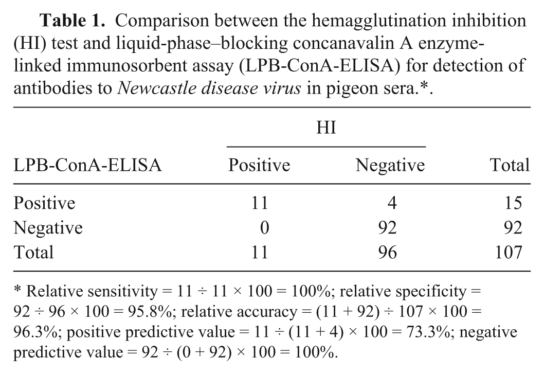
Relative sensitivity = 11 ÷ 11 × 100 = 100%; relative specificity = 92 ÷ 96 × 100 = 95.8%; relative accuracy = (11 + 92) ÷ 107 × 100 = 96.3%; positive predictive value = 11 ÷ (11 + 4) × 100 = 73.3%; negative predictive value = 92 ÷ (0 + 92) × 100 = 100%.
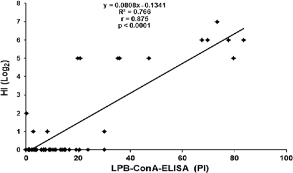
Linear regression analysis and correlation between antibody levels as measured by the liquid-phase–blocking concanavalin A enzyme-linked immunosorbent assay (LPB-ConA-ELISA; expressed as percent inhibition [PI] values) and hemagglutination inhibition (HI) titers (expressed as Log2 serum dilution−1).
Discussion
The bELISA developed in the current study demonstrated that ConA, when used as capture reagent, was able to bind to the NDV antigens present in nonpurified allantoic fluid. Furthermore, ConA was able to immobilize these antigens onto a solid phase, leaving their epitopes free to react with detector antibodies or the NDV-specific antibodies present in test serum samples. In fact, ConA has also been reported to efficiently trap IBV on the microplate surface, which was fundamental for the development of another ELISA based on the sandwich format (S-ConA-ELISA). This S-ConA-ELISA was successfully used for the detection of both IBV and IBV-specific antibodies in chickens. 4 Additionally, the anti-NDV chicken serum used as detector antibody in the LPB-ConA-ELISA, may have contributed in providing a high specificity in the interaction with NDV antigens in this test. This inference is supported by the results of Western blot analysis, which showed specific reactions of this antiserum with major NDV antigens. All these properties of the ELISA reagents permitted an effective determination of the competition between the detector and the antibodies from pigeon serum for NDV antigens in the LPB-ConA-ELISA; consequently, a more accurate determination of the anti-NDV antibody levels, calculated as PI values, was achieved. Another advantage of the LPB-ConA-ELISA with polyclonal anti-NDV antibodies is the potential of this technique to detect antibodies raised by variant NDV strains, especially those classified as pigeon APMV-1. The antibody responses to NDV are polyclonal in the natural hosts, chickens or wild birds, so that a wide spectrum of reactivity for the epitopes of APMV-1 antigens is displayed by the anti-NDV antibodies. 13
The results of the LPB-ConA-ELISA for sera from pigeons were in close agreement with those obtained in the HI test, as a high correlation (r = 0.875) and agreement (κ = 0.83) were observed when comparing these assays. In a previous study, 9 bELISAs, based on the use of purified NDV particles adsorbed directly to the microplate with the use of 2 different monoclonal antibodies as detector, were found to have slightly higher (r = 0.90) or lower (r = 0.71) correlation coefficients with HI test than those found in the current study. A high coefficient of correlation (r = 0.875) and a high agreement (κ = 0.82) were recorded in the measuring of anti-NDV antibodies in ratite sera by another LPB-ELISA method. 11 This demonstrates that the replacement of capture anti-NDV antibodies by ConA as done in the present study, did not negatively affect the specific trapping and epitope availability of NDV antigens present in allantoic fluid to react in the LPB-ELISA format. Similarly, the use of nonpurified NDV suspension, as adopted in the current study, in place of the purified viral antigen suspension used by others 11 to prepare detector anti-NDV antibodies in chickens, caused no relevant negative effects on the ELISA results.
The sensitivity and specificity values obtained by comparing LPB-ConA-ELISA with HI did not markedly differ from those values found for other bELISAs used for the detection of anti-NDV antibodies in chickens or other species.5,9,11,17 The sensitivity values varied in those studies from 90.91% to 100%, and the specificity values from 91.18% to 100%, while the sensitivity and specificity found for the current LPB-ConA-ELISA were 100% and 95.8%, respectively. However, results of 100% for sensitivity or specificity should be treated with caution because a serological test rarely reaches such a value for these parameters. 6
Part of the differences in correlation, agreement, sensitivity, and specificity found in the bELISAs described above could be because of differences in the ELISA reagents used (use of capture antibody versus ConA; use of purified versus nonpurified NDV antigen as capture reagent, and use of monoclonal versus polyclonal anti-NDV as detector antibodies), and in the cutoff point determination of each test. The TG-ROC analysis used in the current study, as well as a previous study, 11 may have favored the determination of a more appropriate cutoff point for this bELISA method, because it can provide a better balance between sensitivity and specificity of immunodiagnostic tests.7,11 Another difference is that the LPB-ConA-ELISA tested a single dilution (1:8) of each serum sample, as conducted in a previous study. 11 The use of a single dilution was adopted for the current study to save time, reduce the number of microplates, simplify the handling of the assay, and reduce the amount of reagents required to test sera.
The pigeons experimentally immunized with NDV in the current study reached a maximum mean HI titer of 6 (range: 5–7), while the PI values detected for the birds in LPB-ConA-ELISA reached the maximum mean of 86.1% (range: 78.5–93.72%). Moderate HI titers (≤4.36) were found in another study after vaccination of pigeons with LaSota NDV. 15 However, higher HI titers (6.23) were observed in pigeons vaccinated with Ulster NDV when the serum samples were tested by HI with this homologous virus. 15 In addition, higher mean HI titers (8.25; range: 4.67–8.25) were detected in pigeons experimentally infected with a chicken NDV isolate of a virulent pathotype. 12 Unfortunately, no ELISA antibody levels determined for experimentally infected or vaccinated pigeons with NDV are available to compare with the results from the LPB-ConA-ELISA.
In the present study, 10.28% seropositivity for NDV was found among the sampled pigeons by both serological tests, whereas a further 3.74% of the sera were positive by the LPB-ConA-ELISA. Seropositivity rates for NDV are highly variable among free-ranging birds. Frequencies of 5.5% of NDV seropositivity were reported for 109 free-ranging pigeons, using HI, in the same region of São Paulo State, Brazil. 18 Additionally, HI NDV-specific antibodies were detected in 2.91% of sparrows sampled in Pernambuco State, Brazil, 16 while a serosurvey in Switzerland, using a commercial kit based on a bELISA, found 10.2% of NDV positivity among wild birds. 14
The LPB-ConA-ELISA developed in the current study for the detection of anti-NDV antibodies achieves comparable results with those from the reference serological test (HI test). The results of the new assay matched the reports in the literature for other bELISAs, particularly the high correlation and agreement with HI test. Overall, the LPB-ConA-ELISA offers potential for use in the diagnosis of NDV in pigeons, or other species of nongalliforme birds.
Footnotes
a.
Sigma-Aldrich, St. Louis, MO.
b.
GE Healthcare, Buckinghamshire, United Kingdom.
c.
Sigma-Aldrich, Steinheim, Germany.
d.
Sigma-Aldrich, St. Louis, MO.
e.
Sigma-Aldrich, Steinheim, Germany.
f.
Costar, Corning, NY
g.
Sigma-Aldrich, St. Louis, MO
h.
Sigma-Aldrich, St. Louis, MO
i.
Excel 2007, Microsoft Corp., Bellevue, WA.
j.
Thermo Scientific, Rockford, IL.
Declaration of conflicting interests
The author(s) declared no potential conflicts of interest with respect to the research, authorship, and/or publication of this article.
Funding
This study was funded by the Coordenação de Aperfeiçoamento de Pessoal de Nível Superior (CAPES), and the Conselho Nacional de Desenvolvimento Científico e Tecnológico (CNPq), Brazil (grant 578453/2008-8).