
Abstract
Histologic and immunohistochemical features of a malignant mesothelioma of the testicular tunica vaginalis in a 9-year-old male mixed-breed dog are reported. The dog had a large scrotal swelling determined by ultrasonography to be a heterogeneous irregular mass characterized by a mixed echogenic pattern and located in the left vaginal canal of the testis. Histologically, the neoplasm was nodular, was poorly demarcated, and consisted of pleomorphic cells that were associated with a significant desmoplastic reaction. By immunohistochemistry, neoplastic cells stained positively for cytokeratin and vimentin and were immunoreactive with an antimesothelial cell monoclonal antibody. Malignant mesotheliomas of tunica vaginalis testis are rare in mammalian species, including dogs.
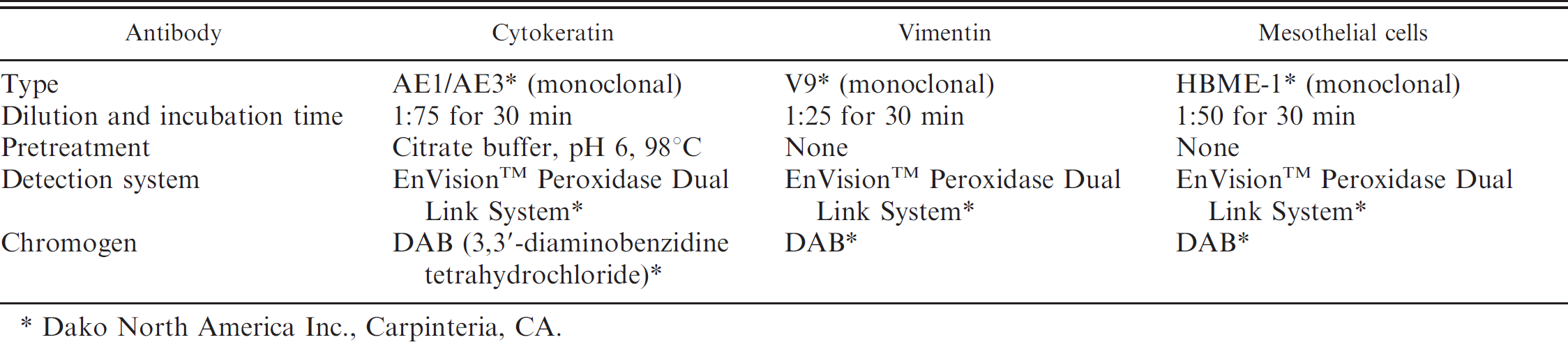
A 9-year-old male mixed-breed dog was examined by the referring veterinarian for a large swelling of the scrotum. Inguinal exploration and ultrasonography revealed a heterogeneous and irregular mass located in the left testicular vaginal canal that was characterized by a mixed echogenic pattern (Fig. 1). Ultrasonography also revealed abundant anechoic effusion surrounding the left testis that was morphologically consistent with hydrocele. A color Doppler study revealed reduction of blood flow to the affected testis compared with the contralateral side. No significant echographic alteration of the parenchymal pattern was observed. The medial iliac and hypogastric lymph nodes were slightly enlarged, but other lymph nodes were unremarkable after additional physical examination, biochemical profile, and survey chest radiographs. A number of scrotal nodules and a firm subcutaneous mass approximately 3 cm in diameter were surgically removed. However, the mass involving the testicular vaginal canal that protruded into the abdominal cavity was not completely removed (Fig. 2). Radical surgery was postponed until the histopathologic findings were available. Both testicles and the excised tissue masses were fixed in 10% buffered formalin, routinely processed, embedded in paraffin, sectioned at 4 μm, and stained with hematoxylin and eosin for histologic evaluation. Immunohistochemical staining with cytokeratin AE1/AE3, a vimentin V9, a and mesothelial cell HBME-1 a (Hector Battifora mesothelial epitope) antibodies was done using an automated immunostainer, b and appropriate positive and negative controls were included (Table 1).
Histologically, the mass was nodular, poorly demarcated, and composed of neoplastic epithelial cells arranged in solid irregular tubular structures associated with an intense desmoplastic reaction consisting of intricate bundles of ovoid-shaped cells intermingled with stromal connective tissue (Fig. 3). The neoplastic cells were cuboidal and had distinct cell borders, a low to moderate nuclear-to-cytoplasmic ratio, abundant granular eosino-philic cytoplasm, and pleomorphic round-to-oval–shaped nuclei. One to 2 mitoses, including a few bizarre mitotic figures, were seen per high-power field. Superficially, the mesothelial cell layer of the tunica vaginalis testis was evident. Neither the testis nor the epididymis was affected by the neoplastic process, and the seminiferous tubules were well preserved. The neoplastic cells were strongly positive for cytokeratin (Fig. 4), weakly positive for vimentin (Fig. 5), and extensively immunoreactive for HBME-1 antigen (Fig. 6). On the basis of these findings, the morphological diagnosis was malignant mesothelioma (MM) of the tunica vaginalis testis.
After surgery, the dog was treated with prednisone (1 mg/kg twice a day) for 1 month. Postoperative recovery was uneventful, and ultrasonography of the thoracic and abdominal cavities did not show evidence of metastatic disease. Four months after surgery, however, the dog was euthanized as a result of relapse of the scrotal neoplasm associated with involvement of the inguinal skin and subcutaneous tissues, pain, and worsening of general health condition. Necropsy revealed 3 nodular metastatic lesions in the peritoneum and the mesentery. Apart from mild edema, no histologic evidence of metastatic disease was seen in sections of the regional lymph node.
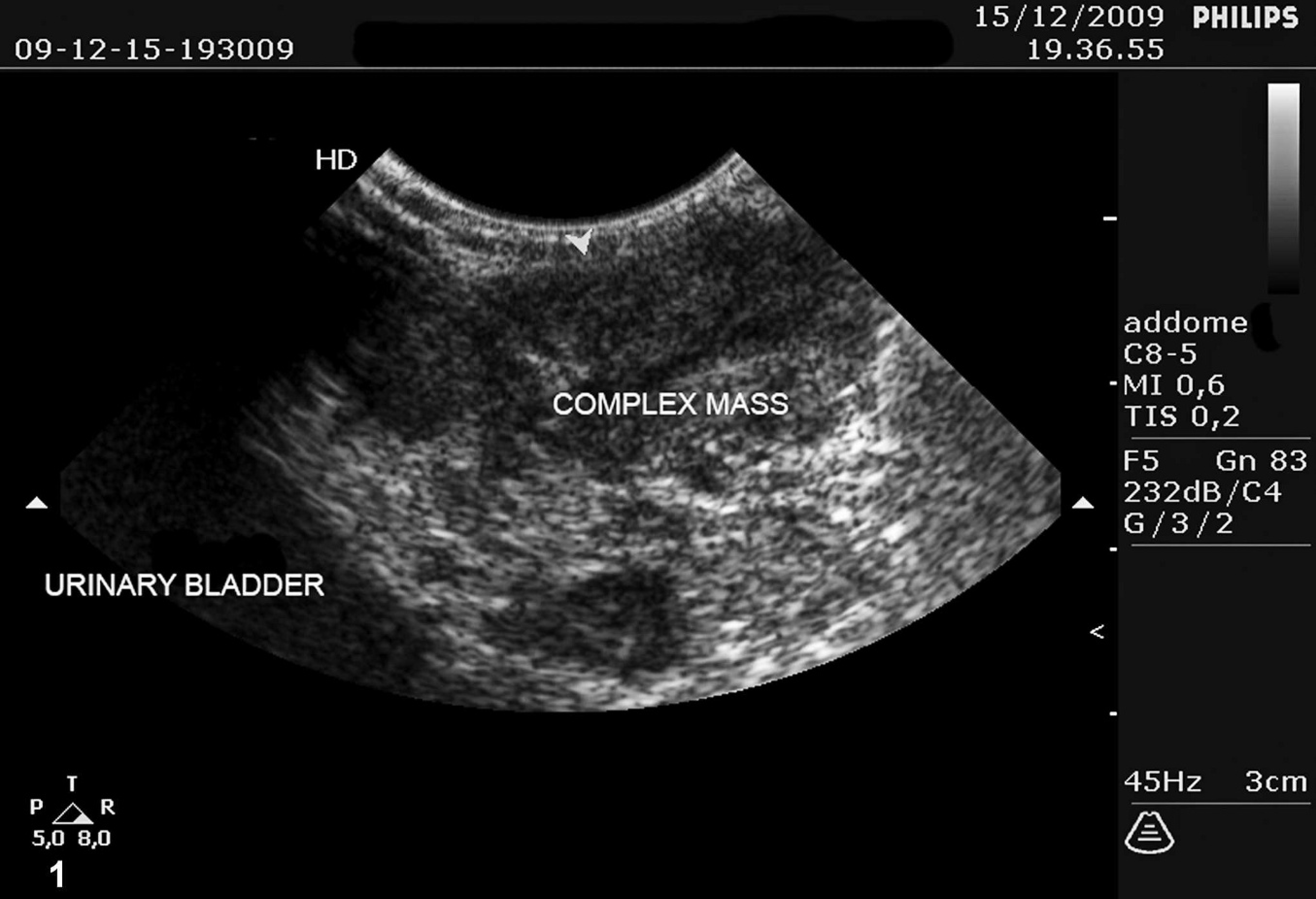
Preoperative inguinal ultrasonography revealed the presence of a complex mass located within the left tunica vaginalis testis of a 9-year-old male mixed-breed dog. The neoplasm was characterized by a mixed echogenic pattern.
Malignant mesothelioma is often associated with a poor clinical prognosis and occurs most frequently in the pleura, peritoneum, and pericardium. Generally, MM is uncommon in mammalian species, including dogs and human beings. Tunica vaginalis testis is an extension of the peritoneum normally lined by a single layer of mesothelial cells. MMs of the tunica vaginalis testis are rare. In dogs, only one other instance of MM of the tunica vaginalis has been reported in a 12-year-old male Scottish Terrier dog. 3 In that case, however, the diagnosis was established by histologic examination without adjunct immunohistochemical and/or ultrastructural characterization of the neoplasm. The dog of that report died 3 months after surgery. Histologic and ultrastructural features of a case of MM of tunica vaginalis testis were also described in an 18-month-old bull. 13 In human beings, MMs of the tunica vaginalis testis are also rare, accounting for only 0.3–5.0% of all MMs. 7 Although previous reports indicate that 34.2–41.0% of MMs of the tunica vaginalis testis correlate with asbestos exposure, 8 previous testicular trauma or hernial repair are also considered risk factors. 1 In the present case, the dog was an indoor pet, and no member of the family was similarly affected. Furthermore, no history of asbestos exposure was found, and the etiology of the neoplasm remains undetermined. In human beings, MMs have been subclassified into 3 groups on the basis of histopathologic features: the epithelial (tubular papillary) type, most often seen in the peritoneal cavity and the tunica vaginalis testis; the mesenchymal or sarcomatous type, commonly found in the pleural cavity; and the biphasic or mixed type, which occurs in and/or on serosal membranes. 9
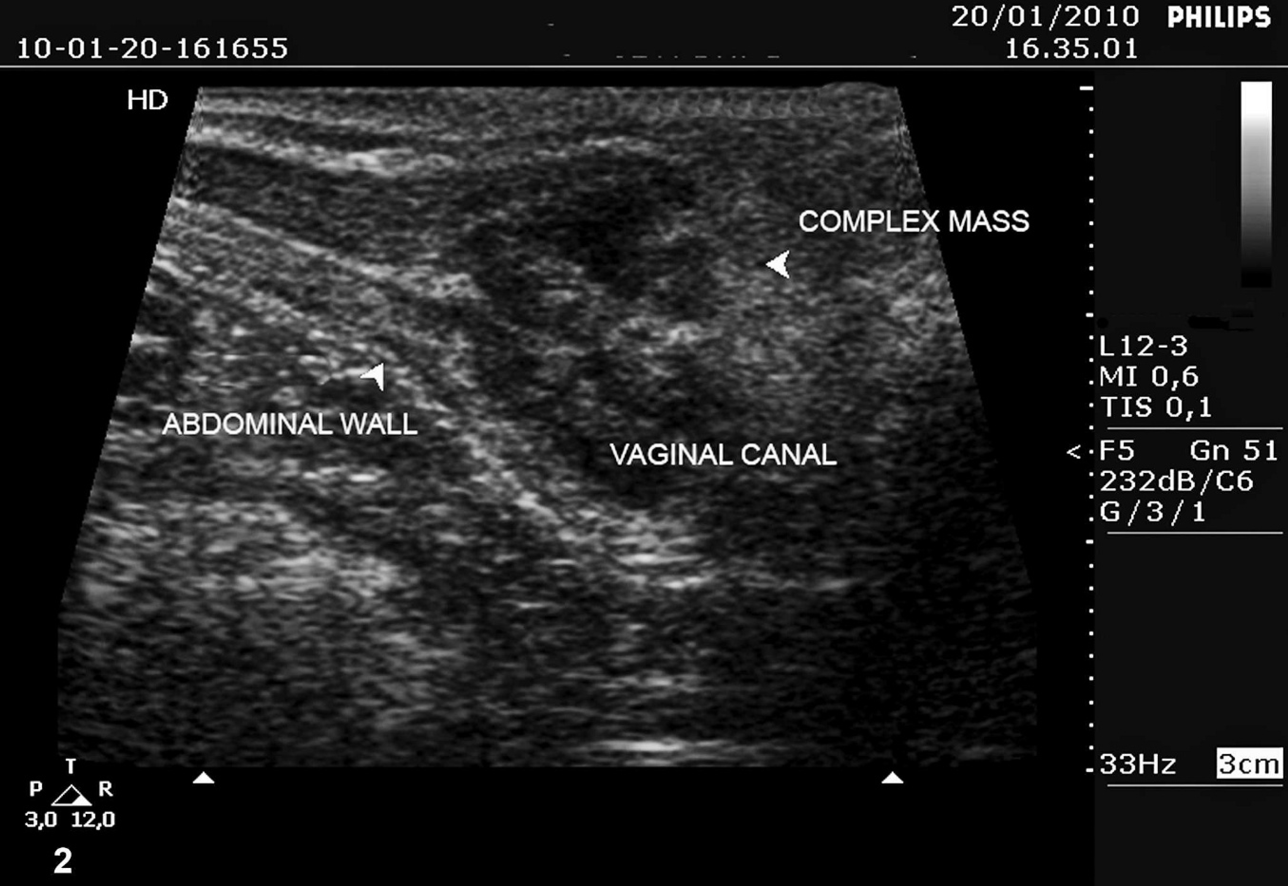
Postoperative inguinal ultrasonography revealed a residual complex mass occupying the left tunica vaginalis testis of a 9-year-old male mixed-breed dog.
The neoplasm described in the present report had malignant histologic features characterized by a tubular pattern that was partially adenomatoid in appearance. In the previous report of canine MM of the tunica vaginalis testis, 3 the neoplasm had a tubulopapillary pattern, and neoplastic cells had a low to moderate nuclear-to-cytoplasmic ratio along with cytoplasmic vacuoles. In that case, special stains failed to reveal epithelial mucin, but a few psammoma bodies were present. The authors considered mesothelial hyperplasia and metastatic adenocarcinoma as possible differential diagnoses. In the neoplasm of the present report, mesothelial hyperplasia was excluded on the basis of the clinical course of disease as well as by gross and microscopic findings. Metastatic adenocarcinoma was also considered unlikely because of the absence of a primary tumor site as well as the neoplasm's histologic and cytologic characteristics. According to the human medical literature, immunohistochemistry is required for a definitive classification of MM. 2 In the present case, tumor cells stained positively for cytokeratin, vimentin, and HBME-1 antigen. These findings strongly suggest that canine MM of the tunica vaginalis testis shares immunophenotypic characteristics with a similar neoplasm reported in human beings. 10 Although the outcome of MM of the tunica vaginalis testis is not known for every reported case in human beings, the overall clinical prognosis for mesothelioma is poor, 4 and, as such, radical orchiectomy should be the first-line therapy. 11 The efficacy of adjuvant chemotherapy and radiotherapy has not yet been clearly determined. 8 Because MM of the tunica vaginalis testis has been infrequently reported in pets, 5 management has been limited to palliation with corticosteroids, repeated drainage of malignant effusion, or intracavitary platinum compound injection. 6 In a recent study, it was reported that a combination of piroxicam and intracavitary platinum-based chemotherapy as a treatment of advanced mesothelioma had remarkable efficacy at controlling the malignant effusion in 3 patients. 12 The sporadic cases of MM of the tunica vaginalis reported in dogs do not permit any etiologic speculation. In addition, future reports of MM are encouraged to enhance the understanding of the etiology and biological behavior of this neoplasm. In particular, MMs should be considered when patients are clinically diagnosed with hydrocele of the testicle. The accurate preoperative diagnosis could be helpful in planning the appropriate therapy. The present case enhances understanding of the biological behavior and management of this neoplasm in dogs.
Antibodies, dilutions, pretreatments, detection system, and chromogen used for the immunohistochemical characterization of malignant mesothelioma of the tunica vaginalis testis in a 9-year-old male mixed-breed dog.
Dako North America Inc., Carpinteria, CA.
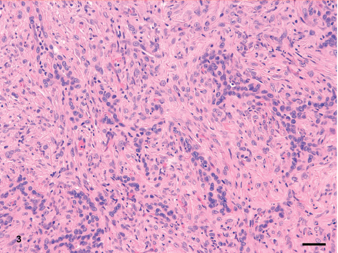
Malignant mesothelioma characterized by a poorly demarcated proliferation of epithelial cells arranged in solid and irregular pseudotubular structures and intermingled with a desmoplastic reaction. Hematoxylin and eosin. Bar = 50 μm.

Immunohistochemistry for cytokeratin shows diffuse positive intracytoplasmic staining of the epithelial cells. Note that components of the desmoplastic reaction are cytokeratin-negative. Immunohistochemistry, Mayer hematoxylin counterstain. Bar = 50 μm.

Immunostaining for vimentin shows strong positive staining of the neoplastic epithelial cells and the surrounding connective tissue stroma. Immunohistochemistry, Mayer hematoxylin counterstain. Bar = 50 μm.

Immunohistochemistry for HBME-1 antigen showing marked cytoplasmic-positive staining of the neoplastic cells. Immunohistochemistry, Mayer hematoxylin counterstain. Bar = 100 μm.
Footnotes
a.
Dako North America Inc., Carpinteria, CA.
b.
Bond-maX™, Leica Microsystems, Menarini, Italy.