
Abstract
A 9-year-old Haflinger mare presented to the Liphook Equine Hospital with a history of weight loss, azotemia, and repeated episodes of ascites over a period of 10 days. The horse was euthanized after exploratory laparotomy revealed large numbers of variably sized masses distributed throughout the peritoneal cavity. Macroscopically, some masses were papillary, while others were nodular. Histologically, the masses were comprised of large to giant, variably shaped, and occasionally multinucleated neoplastic cells with marked anisokaryosis and anisocytosis and a high mitotic rate. Small to moderate numbers of neoplastic cells were swollen by 1 to several, moderately sized to large, clear, circular or ovoid vacuoles, which stained positive with oil red O. Immunohistochemically, the neoplastic cells co-expressed vimentin and cytokeratin. Electron microscopy demonstrated tumor cells with tight junctions, microvilli, and numerous intracytoplasmic lipid droplets. These findings are consistent with a lipid-rich form of mesothelioma, which should be considered as a differential diagnosis if lipid vacuoles are present in potentially neoplastic cells in equine abdominocentesis samples.
Mesotheliomas are malignant neoplasms arising from the mesothelial cells that in normal circumstances form a single-cell lining covering peritoneal, pericardial, and pleural surfaces. Neoplastic transformation of these cells is an infrequent occurrence in domestic animals. 3,8 There have been a small number of reports of mesothelioma in horses, including tumors arising within the pleural cavity, 6,7 peritoneal cavity, 4,10 and pericardium, 12 and those that had spread to involve more than one of these sites. 6,13,14 The ages of the horses ranged from 2 to 27 years. The clinical presentation depended on the site affected; for example, tumors arising in the peritoneal cavity typically produced marked abdominal distension due to ascites, as in other species. 4,8,10
Mesotheliomas have been classified into 3 main histological types: epithelioid, biphasic, and sarcomatoid. 8 Additional epithelioid variants include tubopapillary, acinar, polygonal, microcystic, clear cell, deciduoid, desmoplastic, and lymphohistiocytic mesotheliomas. 3,8 Ultrastructurally, mesotheliomas are composed of cohesive cells that are connected by tight junctions, and have numerous apical microvilli of variable length. Lipid-rich mesotheliomas are an unusual morphological variant previously reported in human beings 2,5,9 and 1 dog. 1 These tumors are epithelioid, express both vimentin and cytokeratin, and contain large numbers of lipid-filled, variably sized cytoplasmic vacuoles that stain positively with oil red O. This report describes an unusual lipid-rich variant of mesothelioma in a horse.
A 9-year-old Haflinger mare presented to the Liphook Equine Hospital (Hampshire, United Kingdom) with a history of weight loss, ascites, and azotemia, and underwent exploratory laparotomy following repeated draining of abdominal fluid. Approximately 60 liters of turbid fluid was removed on the day of presentation; this fluid had a total nucleated cell count of 5.3 × 10⁹/l (normal value is typically less than 9 × 10⁹/l in mature horses 11 ) and a protein concentration of 40 g/l (normal level is less than 25 g/l). The next day, a further 75 liters of similar fluid was drained immediately prior to surgery. At surgery, hundreds of nodular and papillary masses measuring from 2 to 50 mm in diameter were distributed throughout the peritoneal cavity (Fig. 1). The horse was euthanized due to repeated accumulations of very large amounts of abdominal fluid and the resultant poor prognosis.
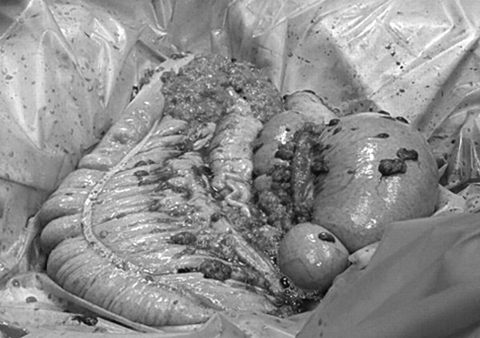
Disseminated abdominal mesotheliomas, horse. Intraoperative photograph of large numbers of variably sized masses coating the mesentery and intestinal serosa.
Multiple representative samples of the masses were fixed in 10% neutral buffered formalin and submitted to the Veterinary Diagnostic Services Unit (School of Veterinary Medicine, University of Glasgow, Glasgow, Scotland) for histologic examination. The fixed specimens were embedded in paraffin and routinely processed according to accepted histologic technique. For light microscopy, 5-µm thick sections were stained with hematoxylin and eosin (HE). Selected sections were immunohistologically labeled for vimentin a and pancytokeratin. a Cryosections of snap-frozen, formalin-fixed tissue were stained with oil red O.
Processing for electron microscopy was carried out in a routine fashion in an automatic, programmable processor. b Briefly, the tissue was post-fixed in 1% osmium tetroxide in a sodium cacodylate buffer, dehydrated through increasing concentrations of ethanol, followed by propylene oxide, and propylene oxide:resin mixtures. Tissue specimens were then embedded in araldite resin. Initial sections were stained with 1% azure II and 1% methylene blue in 1% borax for light microscopy. Ultrathin sections were then cut from appropriate areas and stained with a saturated solution of uranyl acetate followed by contrast staining with lead citrate. Sections were examined using a transmission electron microscope. c Photographs were taken on cut plate films. d
Histologically, the nodular masses were comprised of sheets and streams of tumor cells supported by a sparse to moderately abundant fibrovascular stroma. In sections from papillary masses, the variably thick and often branching projections were comprised of neoplastic cells in sheets that extended down from the surface into the core stromal tissue. In both nodular and papillary masses (Fig. 2A and 2B, respectively), the neoplastic cells were polygonal, ovoid, or elongate, with a moderate to large amount of brightly eosinophilic cytoplasm that had well-defined margins. Neoplastic cells often contained 1 to several, small to large (up to 80 µm in diameter), clear, circular or ovoid, intracytoplasmic vacuoles, some of which stained positively with oil red O, consistent with lipid. Cells containing these lipid droplets were more numerous in the nodular masses than the papillary ones, and numbers varied throughout the masses. The eccentric or paracentral nuclei were large or giant and round, ovoid, or irregularly shaped, with variable amounts of stippled chromatin and 1–6 small to large, deeply eosinophilic nucleoli. There was marked anisocytosis and anisokaryosis, with small to moderate numbers of mononuclear or multinucleate neoplastic giant cells. Mitotic figures, some of which had a bizarre appearance, varied from 1–6 per high power field (400×). Tumor cells were intermixed with various other cell populations, including reactive fibroblasts, adipocytes, neutrophils, lymphocytes, and plasma cells. Scattered throughout the section were numerous necrotic tumor cells and small to large coalescing areas of hemorrhage.
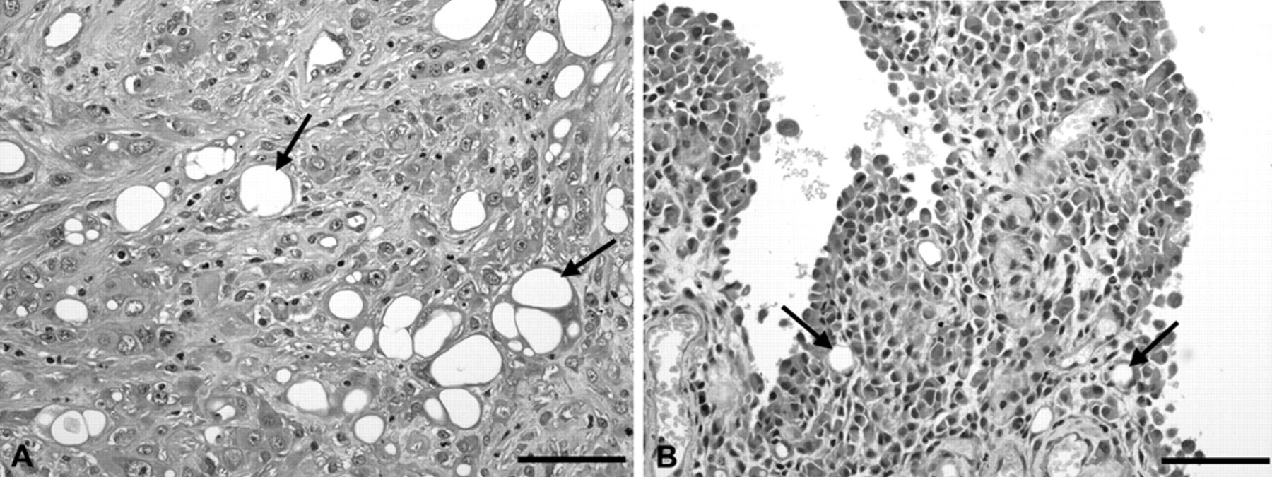
Abdominal mesothelioma, horse.
Immunohistochemically, there was strong positive cytoplasmic labeling of tumor cells for both pancytokeratin and vimentin, nonspecific markers known to be co-expressed by mesothelial cells. Ultrastructurally, the neoplastic cells often had numerous characteristic long microvilli, visible intercellular tight junctions, and frequent intracytoplasmic vacuoles of variable size and number, in addition to numerous intracytoplasmic lipid droplets (Fig. 3). These findings were consistent with the diagnosis of a lipid-rich mesothelioma.
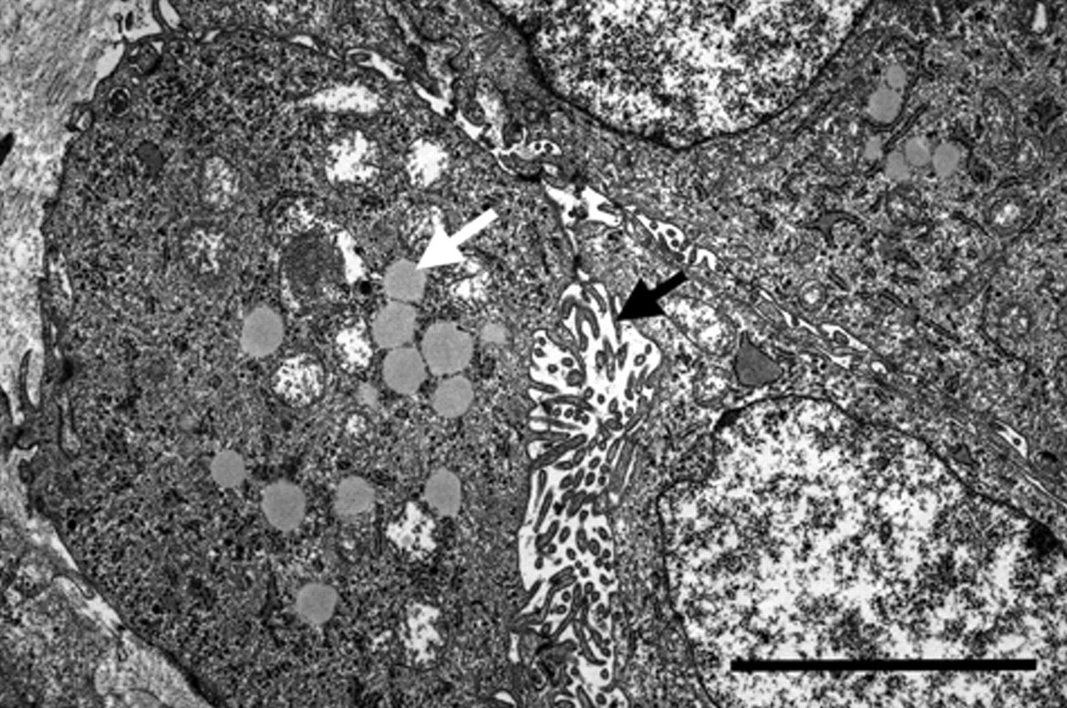
Abdominal mesothelioma, horse. Transmission electron micrograph showing neoplastic cells with numerous intracytoplasmic lipid droplets (white arrow) and surface microvilli (black arrow). Bar = 3 µm.
The mesothelial origin of the neoplasm in the present study was diagnosed based on gross, histological, immunohistochemical, and ultrastructural features. Additionally, the specific characteristics of intracytoplasmic lipid droplets within neoplastic cells were demonstrated by both electron microscopy and histochemical staining with oil red O. These features were similar to those previously reported in human patients and 1 dog. 1,5,9 One of the human cases involved a diffuse, lipid-rich, epithelioid tumor in the pleural cavity where tumor cells contained large intracytoplasmic vacuoles. 5 In the second report, foam cells, with large numbers of lipid droplets in the cytoplasm and a macroscopic appearance similar to “codfish roe,” were described. 9 In a 2008 case of a lipid-rich mesothelioma in a dog, the tumor cells contained cytoplasmic vacuoles some of which stained positive with oil red O. The neoplastic cells expressed vimentin, pancytokeratin, and S-100 protein in the surrounding cytoplasm, and had a similar ultrastructural appearance to the current case. The 2008 report did not contain an HE photomicrograph with which to compare the current case. 1
To the authors’ knowledge, the lipid-rich variant of mesothelioma has not previously been reported in the horse. Cells from which lipid-containing tumor cells should be differentiated cytologically include macrophages with intracytoplasmic lipid droplets seen in chylous effusions, tumor cells exfoliating from interstitial cell tumors involving cryptorchid testes, or metastatic carcinoma cells containing mucin vacuoles. 2 If lipid-containing tumor cells are noted, lipid-rich mesothelioma should be considered as a potential differential diagnosis when examining cytological aspirates from horses with abdominal, pleural, or pericardial effusions.
Footnotes
Acknowledgements
The authors wish to thank Lynn Stevenson, Iain MacMillan, Lynn Oxford and Liz Strachan in the Histopathology Laboratory, Veterinary Diagnostic Services, School of Veterinary Medicine, University of Glasgow, and Mailis McCulloch from the Veterinary Biosciences Division, School of Veterinary Medicine, University of Glasgow for their technical assistance.
a.
Dako Denmark A/S, Glostrup, Denmark.
b.
Leica Lynx Processor™, Leica Microsystems GmbH, Saarland, Germany.
c.
JEOL CX-100, JEOL Ltd., Tokyo, Japan.
d.
Kodak 4469, Eastman Kodak, Rochester, NY.
The authors declared that they had no conflicts of interest with respect to their authorship or the publication of this article.
The authors declared that they received no financial support for their research and/or authorship of this article.