
Abstract
Two unrelated bovine beef calves, aged 2 mo and 3 mo, were presented to The Ohio State University Veterinary Medical Center because of scrotal swelling and abdominal distension. On postmortem examination, there was abundant peritoneal fluid and numerous small friable masses covering all peritoneal surfaces and extending into the scrotum via the tunica vaginalis, with no identifiable primary neoplasm. Based on light microscopy, differential diagnoses included malignant mesothelioma and anaplastic carcinoma. Immunohistochemically, the neoplasms labeled positive for cytokeratin, and negative for vimentin and calretinin. Neoplastic cells contained periodic acid–Schiff-positive, diastase-resistant cytoplasmic granules, and lacked Alcian blue–positive, hyaluronidase-negative cytoplasmic vacuoles. Ultrastructurally, the cells had features of carcinoma, including secretory granules, and lacked typical features of mesothelioma, such as long slender microvilli. Our final diagnosis was carcinoma in both calves, despite the equivocal gross and light microscopic findings. We propose that a presumptive diagnosis of peritoneal mesothelioma in bovine calves should be avoided without corroboration by a combination of histology, histochemistry, immunohistochemistry, and, if possible, electron microscopy.
Juvenile neoplasia is uncommon in all species, including cattle. In calves, juvenile lymphoma is the most frequently reported neoplasm, followed by mesothelioma.11,13 Tumors of embryonal or mesenchymal origin are even less common; carcinomas are rarely reported except as epizootic outbreaks of viral or genetic origin.11,12,17 Mesotheliomas are difficult to distinguish from carcinomas on light microscopy depending on differentiation, and a diagnosis is often reached based on a combination of gross and light microscopic findings as well as signalment.2,4,8,11 When histologic findings are equivocal, mesothelioma is typically the presumptive diagnosis, given the rate of occurrence reported in the literature compared to carcinomas.11,17 Herein, we report 2 cases of carcinomas of unknown origin in bovine beef calves, initially diagnosed as mesotheliomas using conventional methods, and then reclassified as carcinomas following histochemical and ultrastructural examination.
Calf 1 was a 3-mo-old mixed-breed beef bull calf that was presented to The Ohio State University Veterinary Medical Center (OSU-VMC; Columbus, OH) because of bilateral scrotal swelling. The calf was thin and had a pot-bellied appearance, but was otherwise healthy with no previous medical history. Physical examination revealed a distended abdomen and scrotum, a fluid wave on ballottement of the abdomen, and scrotal fluid distension.
Complete blood count and serum biochemical profile were unremarkable. Scrotal ultrasound revealed diffuse irregular thickening of the tunica vaginalis, with similar proliferative lesions affecting the greater omentum associated with marked peritoneal effusion. An aspirate of the abdominal fluid was taken prior to abdominal exploratory surgery. Cytologically, the fluid was dark-red and cloudy, and small numbers of neutrophils were present. There were a few markedly atypical, irregularly rounded cells individually or in small clusters, with marked anisocytosis and anisokaryosis. Atypical cells had distinct cellular borders, pale-to-moderately basophilic cytoplasm with a few variably sized distinct colorless vacuoles, and occasionally phagocytized neutrophils and erythrocytes. Nuclei were round, oval, or irregular, with finely to coarsely stippled chromatin and up to 3 nucleoli. There were occasional multinucleate or binucleate cells as well as bizarre mitotic figures. Given the extent of the gross surgical findings within the abdomen, the calf was euthanized on the surgical table.
A partial autopsy was performed on calf 1. Upon opening the abdomen, there was a large volume of serosanguineous fluid in the peritoneal cavity and omental sling. The omentum, mesentery, and peritoneal wall were diffusely and irregularly thickened, nodular, and red. Grossly, the proliferative lesion affected all peritoneal and serosal visceral surfaces and extended into the scrotum via the tunica vaginalis (Fig. 1). A presumptive diagnosis of mesothelioma was made based on reported cytologic features and gross morphology.
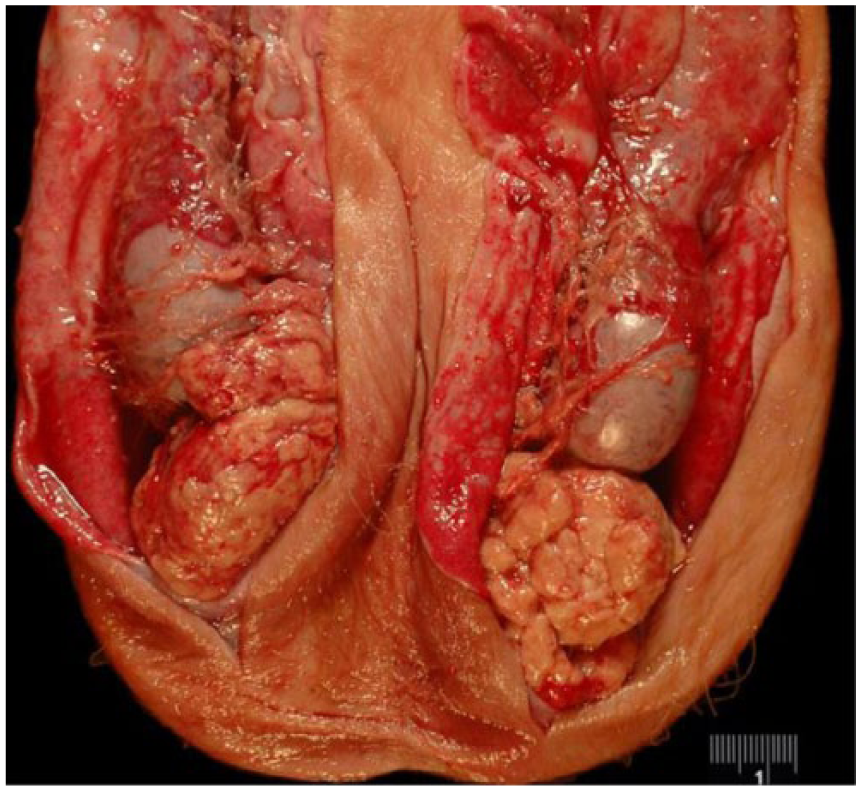
Carcinoma of unknown origin in the scrotal sac of calf 1.
Calf 2 was a 2-mo-old Charolais bull calf that was also presented to the OSU-VMC because of scrotal swelling and abdominal distension. The owner reported that the scrotum had been swollen since birth. The calf was otherwise healthy with no previous medical history. Physical exam revealed a fluid-filled abdomen and scrotum, which was confirmed on ultrasound. Given the poor prognosis, no further testing was performed, and the calf was euthanized and submitted to the OSU Applied Pathology service for postmortem examination.
At autopsy, there were ~72 L of serosanguineous fluid in the peritoneal cavity, with an additional 4–5 L retained in the omental sling and 25 mL of similar fluid in the scrotum. The peritoneal surfaces, serosal surfaces of the gastrointestinal tract, serosal surface of the bladder, visceral surface of the liver, and omentum were covered in coalescing, friable, adherent, raised, white-to-brown nodules, ranging from pinpoint to 1 cm diameter (Fig. 2). There were similar masses in the vaginal cavity, with some adherent to the visceral and parietal vaginal tunics and some free-floating. A primary neoplasm could not be identified grossly in either calf, and again a presumptive gross diagnosis of mesothelioma was made.
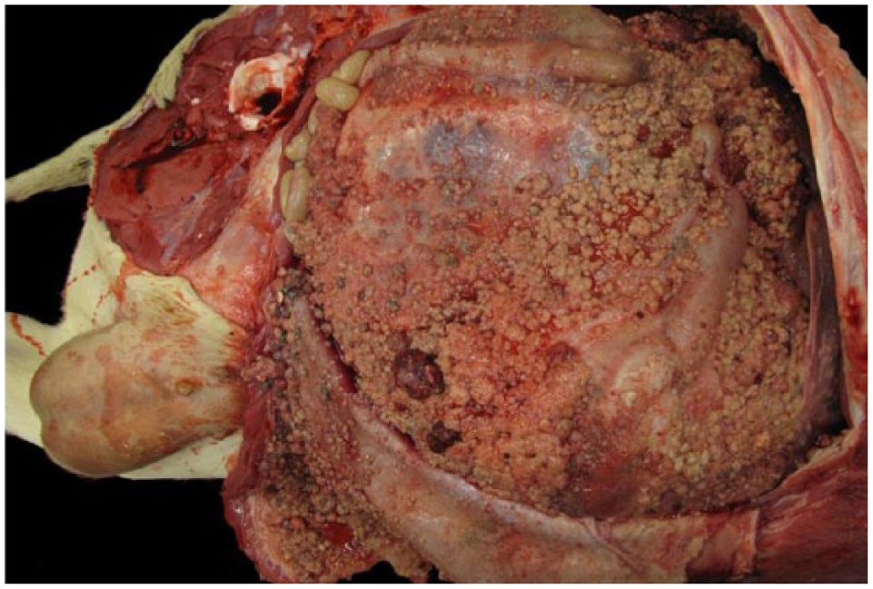
Carcinomatosis on the omental sling, peritoneal wall, and peritoneal serosal surfaces of calf 2. Note the scrotal swelling.
Sections of masses from both cases were fixed in 10% neutral-buffered formalin. Tissues were routinely processed for histologic evaluation, and were stained with hematoxylin and eosin, periodic acid–Schiff (PAS) with and without diastase, and Alcian blue with and without hyaluronidase. Immunohistochemistry was performed using monoclonal mouse antibodies for cytokeratin AE1/AE3 (Dako, Santa Clara, CA), vimentin clone V9 (Dako), and calretinin (Santa Cruz Biotechnology, Dallas, TX) utilizing horseradish peroxidase for detection. Additional sections from masses of both calves were fixed in 3% buffered glutaraldehyde, routinely processed for evaluation by electron microscopy (EM), embedded in epoxy resin, and sectioned at 70–90 nm on copper 200-mesh EM grids.
Histologically, the masses from calf 1 were encapsulated and densely cellular, and composed of highly pleomorphic cells often arranged in papillary projections along fibrous trabeculae. Cells were large and polygonal, with abundant eosinophilic cytoplasm, large round-to-irregularly shaped nuclei with stippled chromatin, and prominent, often multiple, nucleoli. There were occasional binucleate to multinucleate cells. Occasionally, cells were tall, columnar, with vacuolated cytoplasm. There was marked anisocytosis and anisokaryosis with marked atypia. Mitoses were 15 per ten 400× fields.
The masses from calf 2 were well-demarcated and partially encapsulated, composed of sheets and clusters of large, loosely arranged, neoplastic polygonal cells. These cells occasionally formed small tubular structures. Neoplastic cells had abundant pale eosinophilic cytoplasm, often containing 1–4 large clear distinct vacuoles. There was marked anisocytosis and anisokaryosis with marked atypia. Neoplastic cells were often binucleate and had 1–4 round-to-oval nuclei per cell. There were 1–3 prominent nucleoli per nucleus; nucleoli were often irregularly shaped. There were 17 mitotic figures per ten 400× fields, and mitotic figures were often bizarre. Admixed throughout the neoplasm were regions of necrosis and mineralization, as well as minimal lymphocytic inflammation. Neoplastic cells were also found as emboli in sections of lung. Based on the light microscopic findings in both cases, additional techniques were pursued to confirm the presumptive diagnosis of mesothelioma.
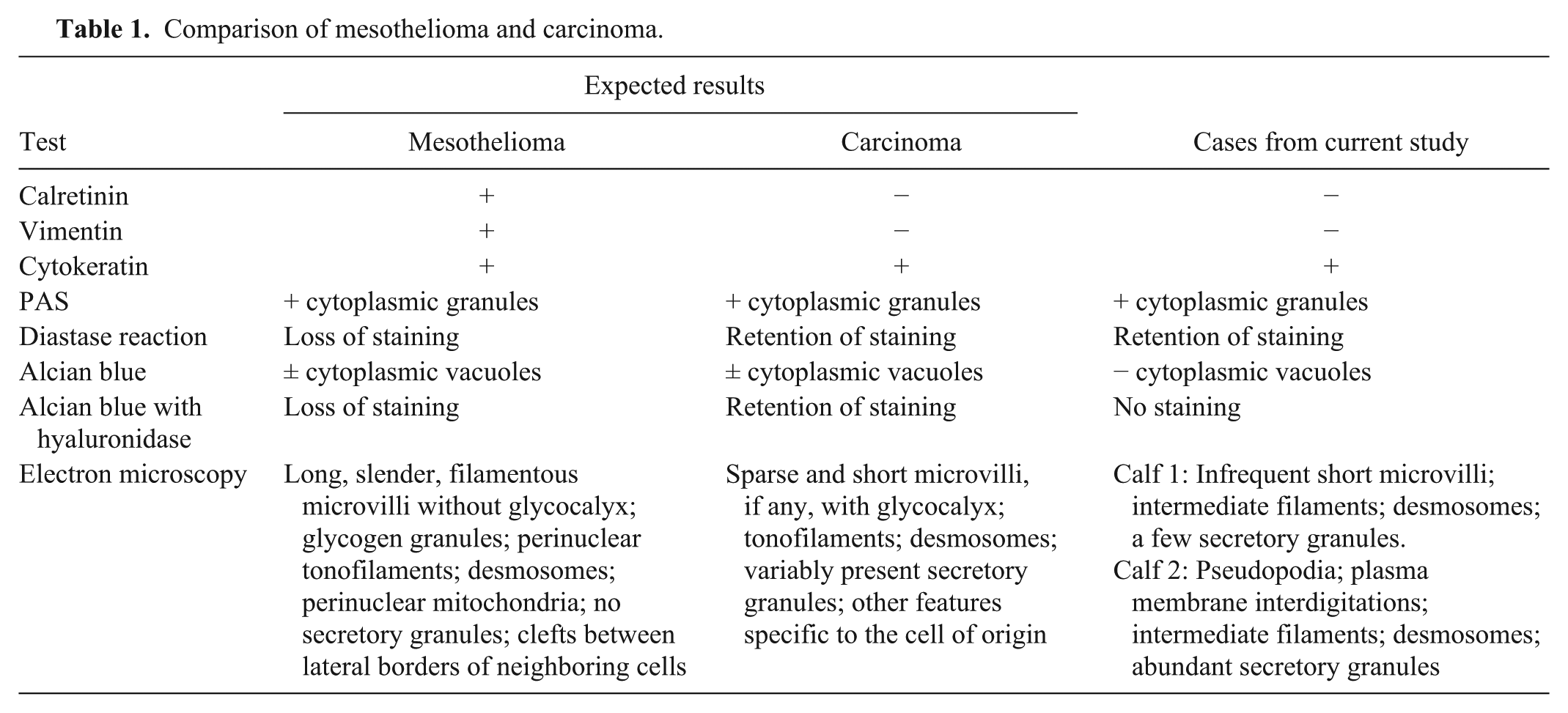
Expected results of additional testing in diagnosis of carcinoma versus mesothelioma are presented in Table 1, along with a summary of findings in our cases. Neoplastic cells from both cases had strong intracytoplasmic labeling for cytokeratin, and did not label for vimentin or calretinin. Neoplastic cells from both cases contained numerous PAS-positive, diastase-resistant granules. Slides were also stained with Alcian blue and hyaluronidase, but no Alcian blue–positive cytoplasmic vacuoles could be identified.
Comparison of mesothelioma and carcinoma.
Based on the gross, cytologic, and initial histologic appearance, a presumptive diagnosis of mesothelioma was made in both cases. However, given the immunohistochemical findings that were not consistent with mesothelioma, EM was pursued. Both cases showed features consistent with carcinoma ultrastructurally. In calf 1, all cells present were epithelioid, characterized by prominent intercellular tight junctions (desmosomes), cytokeratin intermediate filaments, and infrequent short apical microvilli (Fig. 3). Nuclei were predominantly round with perimembranous clumped heterochromatin. The endoplasmic reticulum was multifocally swollen. Minimal secretory granules were noted, and there were a few lysosomes. There was no apparent basal lamina. In calf 2, all cells present were epithelioid, characterized by desmosomes and cytokeratin intermediate filaments, as well as abundant secretory granules (Fig. 4). Frequently, the plasma membrane was thrown into projections that interdigitated with adjacent cells, and there were occasional pseudopodia, but no microvilli. Neither neoplasm had significant supporting stroma or vasculature present. The specific tissue of origin was not apparent given the lack of characteristic morphologic ultrastructural features in the cells examined; however, it was determined that both tumors were carcinomas, and were likely of different tissue origin.
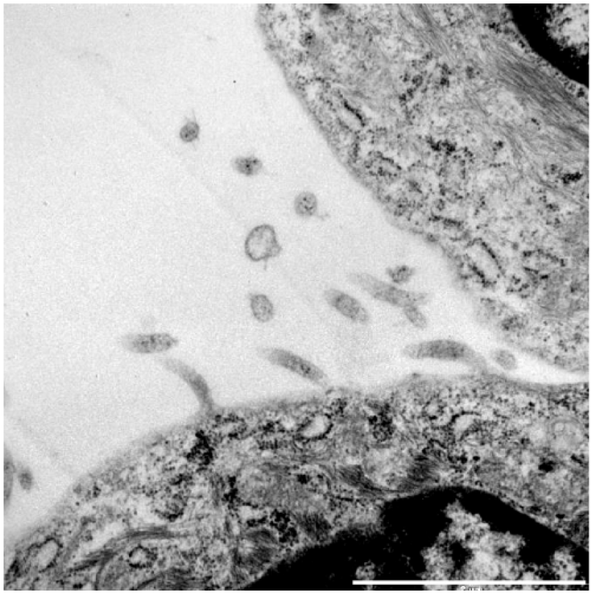
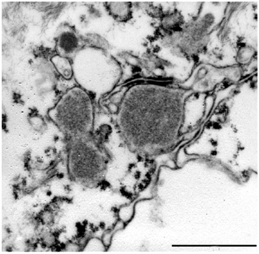
Ultrastructural features of a carcinoma of unknown origin in calf 1. A few neoplastic cells had occasional short microvilli. Bar = 2 µm.
Ultrastructural features of a carcinoma of unknown origin in calf 2. Neoplastic cells contained numerous secretory granules of variable electron density. Bar = 2 µm.
Despite ruling out a diagnosis of mesothelioma based on extensive testing, the tissue of origin of either neoplasm was not apparent based on morphology. Additionally, there was no primary neoplasm noted grossly or histologically, resulting in a challenging final diagnosis of carcinoma, based on histochemical staining and EM. Classically, carcinomas are positive for cytokeratin and negative for vimentin and calretinin, as in our case, whereas mesotheliomas are positive for vimentin, calretinin, and cytokeratin,3,19 although mesotheliomas in cattle positive for only vimentin have been reported. 16 Calretinin has a high sensitivity and specificity when used to distinguish mesotheliomas from metastatic carcinomas in human oncology, and is a standard component of a diagnostic panel.6,9 Mesotheliomas should not have diastase-resistant, PAS-positive cytoplasmic granules, whereas the granules can be present in carcinomas, as in our cases. 6 Mesotheliomas can contain Alcian blue-hyaluronidase–negative cytoplasmic vacuoles; this feature should be absent in carcinomas. 6 Ultrastructural characteristics of mesotheliomas include the presence of long, slender, filamentous microvilli, glycogen granules, tonofilaments, desmosomes, and clefts between borders of adjacent cells; cells lack secretory granules. 10 Ultrastructural characteristics of carcinomas are variable depending on the cell of origin, but typically microvilli are sparser and shorter than in mesotheliomas, if any microvilli are present, and cells often contain secretory granules. 10
Carcinomas are rarely reported in calves. 11 A thymic carcinoma has been reported in a calf, 1 as well as ethmoid carcinoma, 12 bronchoalveolar carcinoma, 20 and anaplastic small cell carcinoma of the lung. 15 Two undifferentiated carcinomas in adult cows were examined ultrastructurally in one report, and were found to be squamous cell carcinoma and adenocarcinoma of the lung. 7 Our cases lacked macroscopic primary thoracic tumors, making any of these origins unlikely. A case of peritoneal carcinomatosis has been reported in a calf, with liver suspected as the tissue of origin, 13 but the livers in our cases were grossly and histologically normal.
EM is the gold standard for diagnosis of mesothelioma in humans.10,14 Light microscopy combined with a panel of immunohistochemical stains is also acceptable, but histochemical stains alone, such as PAS with diastase or Alcian blue with hyaluronidase, are considered insufficient given the variability of glycogen and mucin production in both mesotheliomas and adenocarcinomas. 14 In a review of the literature, few cases of bovine mesothelioma, either in juvenile or adult cows, are corroborated with additional histochemistry, such as PAS or Alcian blue stains,2,3,5,18 or immunohistochemistry.3,16,19 We found no reports of suspected mesotheliomas in calves that had been examined ultrastructurally, although 2 cases of mesothelioma in a 6-y-old and a 4-y-old cow were confirmed via EM. 5 In the literature, a mesothelioma examined ultrastructurally in the youngest bovid was in the scrotum of an 18-mo-old bull. 18 Carcinomas are rarely reported in calves, and herein we report 2 cases. Given that both of our cases were initially diagnosed as mesothelioma using standard clinical and pathology modalities, it is possible that mesotheliomas have been misdiagnosed in juvenile bovids and the prevalence of carcinomas in young bovids is higher than previously thought.
Footnotes
Acknowledgements
We thank Edward Calomeni for technical assistance with EM analysis, and Dr. Krista LaPerle for assistance with EM image selection.
Declaration of conflicting interests
The authors declared no potential conflicts of interest with respect to the research, authorship, and/or publication of this article.
Funding
The authors received no financial support for the research, authorship, and/or publication of this article.