
Abstract
The current study describes the clinical, gross, histopathologic, and immunohistochemical findings of a T-cell lymphoma in a captive porcupine (Coendou prehensilis), a species typically seen in the tropical forests of Brazil. At necropsy, extensive neoplastic involvement was observed in the cervical lymph nodes, with extension into the salivary gland. The spleen was mildly enlarged, and neoplastic nodules were grossly evident in the liver and right kidney. Histologically, sheets of large and markedly pleomorphic round cells were observed in the cervical lymph nodes, lung, liver, spleen, and kidney. The neoplastic cells were positive for cluster of differentiation (CD)3 and negative for CD79a by immunohistochemistry.
The porcupine (Coendou prehensilis), an animal typically seen in the tropical forests of Brazil, is an arboreal, nocturnal, and frugivorous rodent of the family Erethizontidae. The upper parts of the body are covered with strong and dense erectile spines. 4 Little is known about diseases in this species, including neoplastic conditions. Tumors of lymphoid cells are common in domestic animals and can be classified according to anatomical location (e.g., multicentric, digestive, mediastinal or thymic, cutaneous) and phenotypic characteristics (B cells or T cells). 1 The current study describes the clinical, gross, histologic, and phenotypic characteristics of a lymphoid neoplasm in a porcupine.
An adult male porcupine with a body length of approximately 60.0 cm and housed in a zoologic garden in the state of Minas Gerais, Brazil was referred to the attending veterinarian for clinical care several times over a period of 2 months (September and October 2006). The animal presented with lethargy, anorexia, and progressive weight loss (1 kg in 7 months). At the beginning of September 2006, the animal presented with mild dyspnea and mucopurulent discharge from both nostrils. The body temperature was 36.9°C. Physical examination revealed a moderate increase in volume of the ventral region of the mandibular and cervical regions. A complete blood cell count at this time revealed mild leukocytosis caused by mild lymphocytosis and monocytosis. Clinical chemistry revealed increased blood urea nitrogen (BUN) and creatinine concentrations suggestive of renal or prerenal azotemia (Table 1). The animal was treated with antibiotics (because of suspicion of sinusitis on the basis of clinical signs of dyspnea and nasal discharge) and probiotics, but there was no clinical improvement. The animal was submitted for re-examination and treatment in mid-October 2006. One week later, the animal died.
Hematology and clinical chemistry data of a Brazilian porcupine (Coendou prehensilis) with disseminated T-cell lymphoma. *
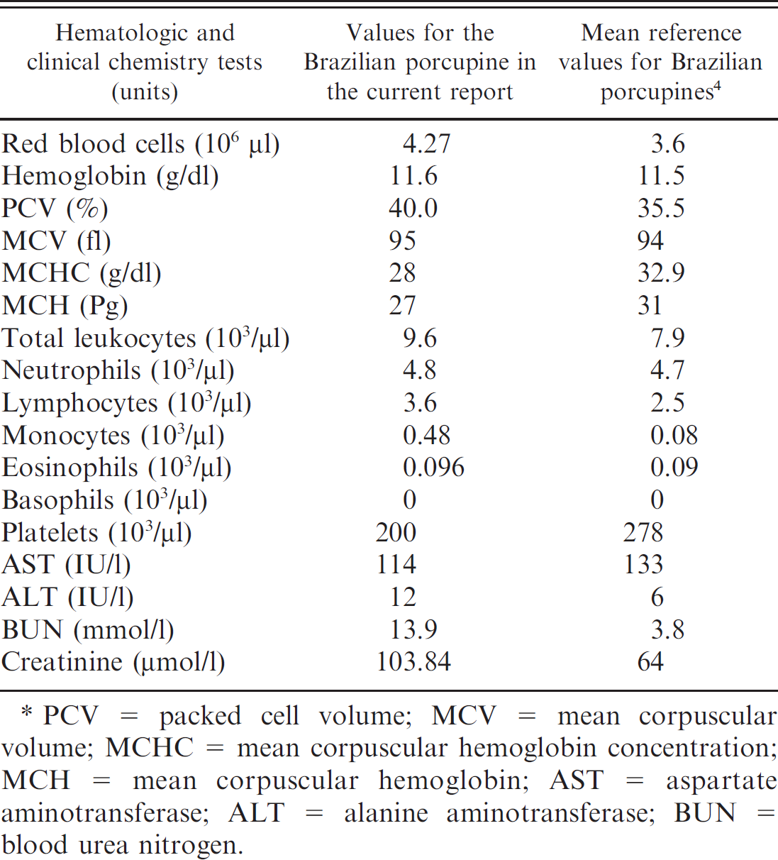
PCV = packed cell volume; MCV = mean corpuscular volume; MCHC = mean corpuscular hemoglobin concentration; MCH = mean corpuscular hemoglobin; AST = aspartate aminotransferase; ALT = alanine aminotransferase; BUN = blood urea nitrogen.
At necropsy, the cadaver was in poor body condition. The mucosal membranes were pale, and there was crusting mucopurulent exudate at the nares. Moderate subcutaneous edema was observed in the mandibular and cervical regions. An extensive soft, white, nodular mass (Fig. 1) with multifocal friable and red areas (interpreted as necrosis and hemorrhage) was present beneath this edematous tissue. The cervical lymph nodes and salivary glands were not discernible from the mass. The cranial lung lobes were consolidated and red, whereas the caudal lung lobes were slightly firm. The lumen of the trachea contained moderate amount of yellowish viscous material, and the mucosa had numerous petechiae. The liver was congested and moderately enlarged, with scattered, white, poorly delineated areas on the capsular and cut surfaces. The spleen was slightly enlarged, friable, and pale. The cortices of the mesenteric lymph nodes were more prominent than expected. The right kidney was pale and had one nodule measuring 1.5 cm in diameter embedded in the parenchyma (Fig. 2). Representative tissue samples were fixed in 10% buffered formalin and were submitted to the Veterinary School of the Universidade Federal de Minas Gerais (Belo Horizonte, Minas Gerais, Brazil), where they were processed routinely, embedded in paraffin, sectioned at 5 μm, and stained with hematoxylin and eosin.
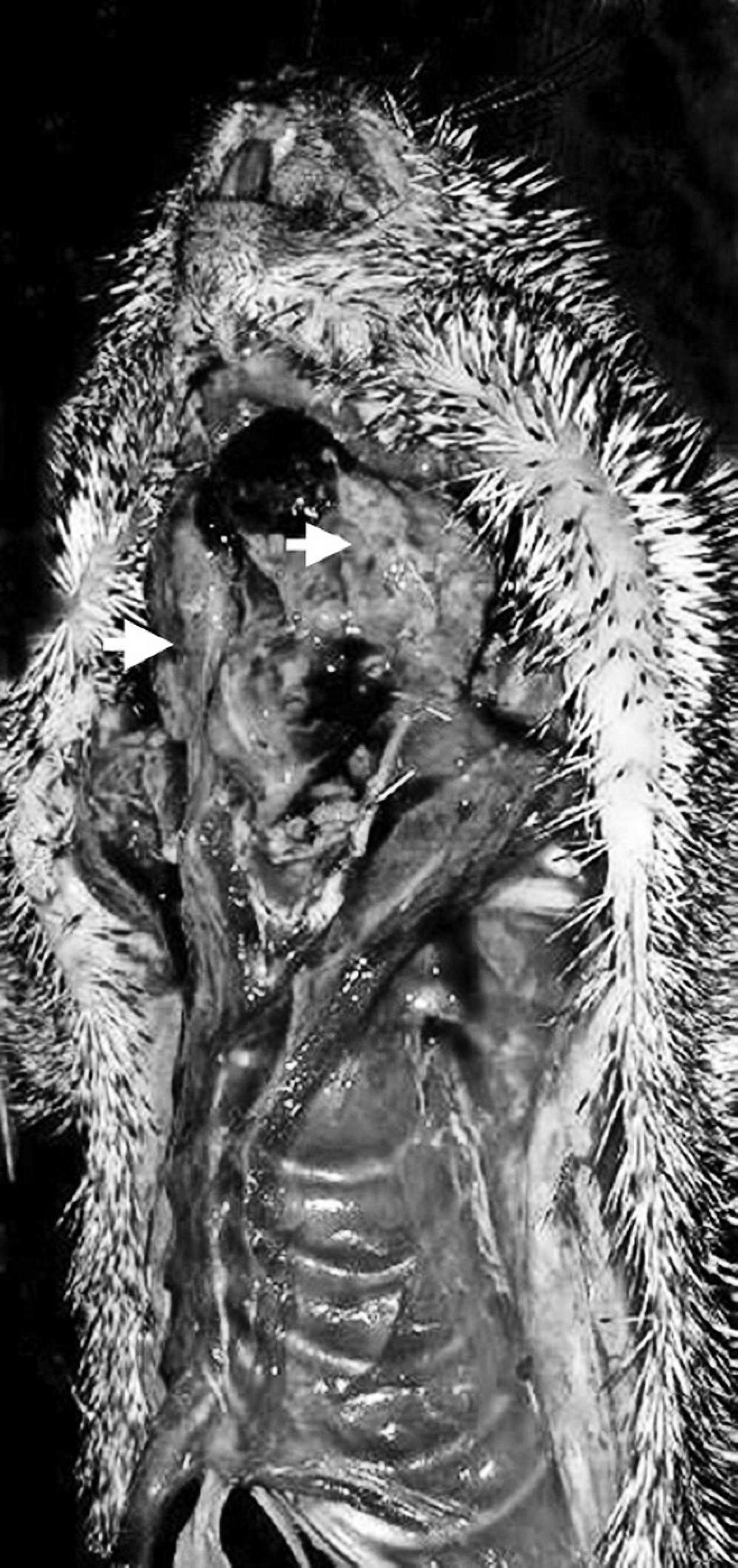
Brazilian porcupine (Coendou prehensilis) with T-cell lymphoma. A tumoral mass replaces the salivary glands and lymph nodes in the mandibular and cervical regions (arrows).
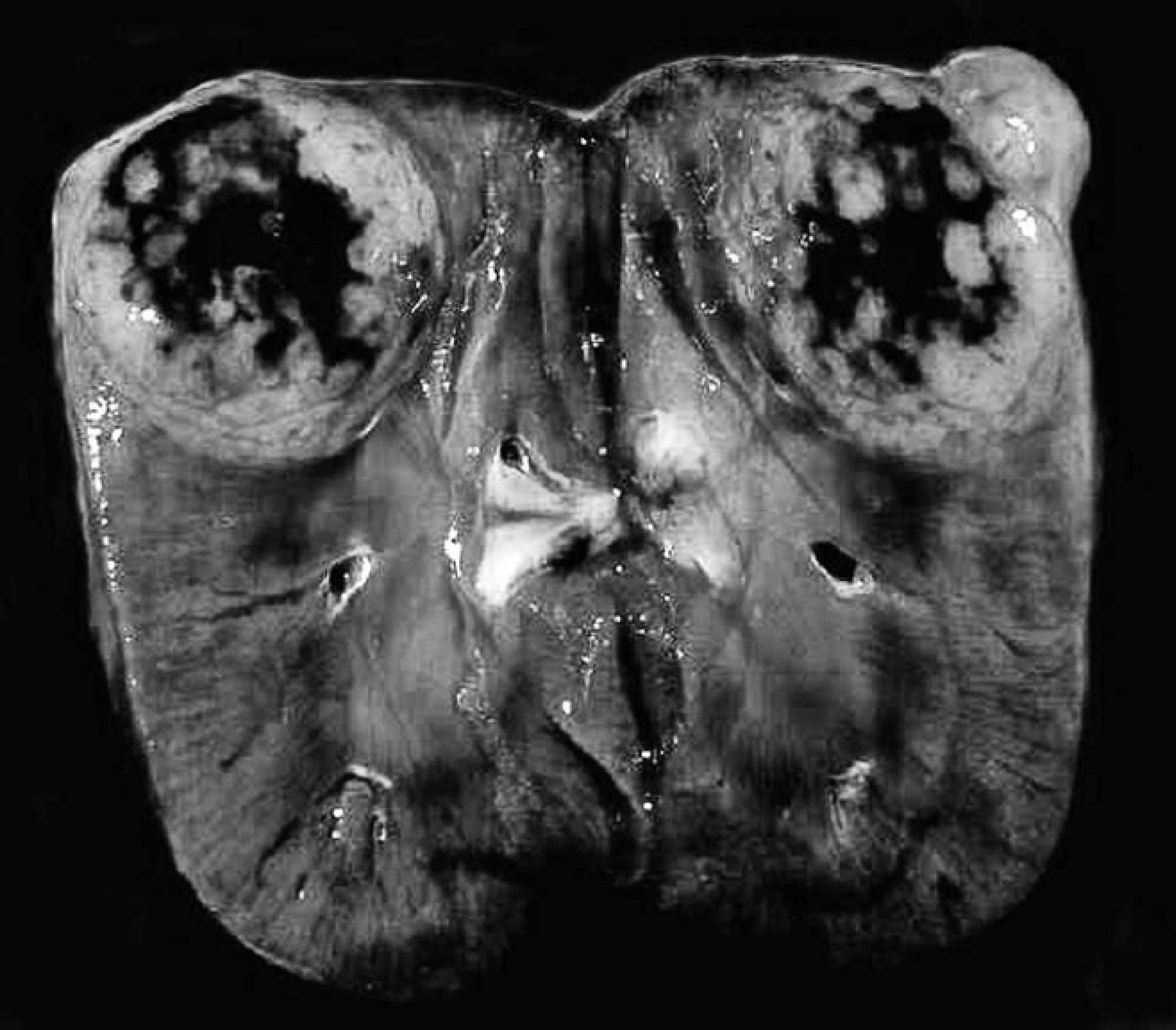
Brazilian porcupine (Coendou prehensilis) with T-cell lymphoma. A large white nodule with central necrosis and hemorrhage is embedded in the renal parenchyma and bulges slightly from the capsular surface.
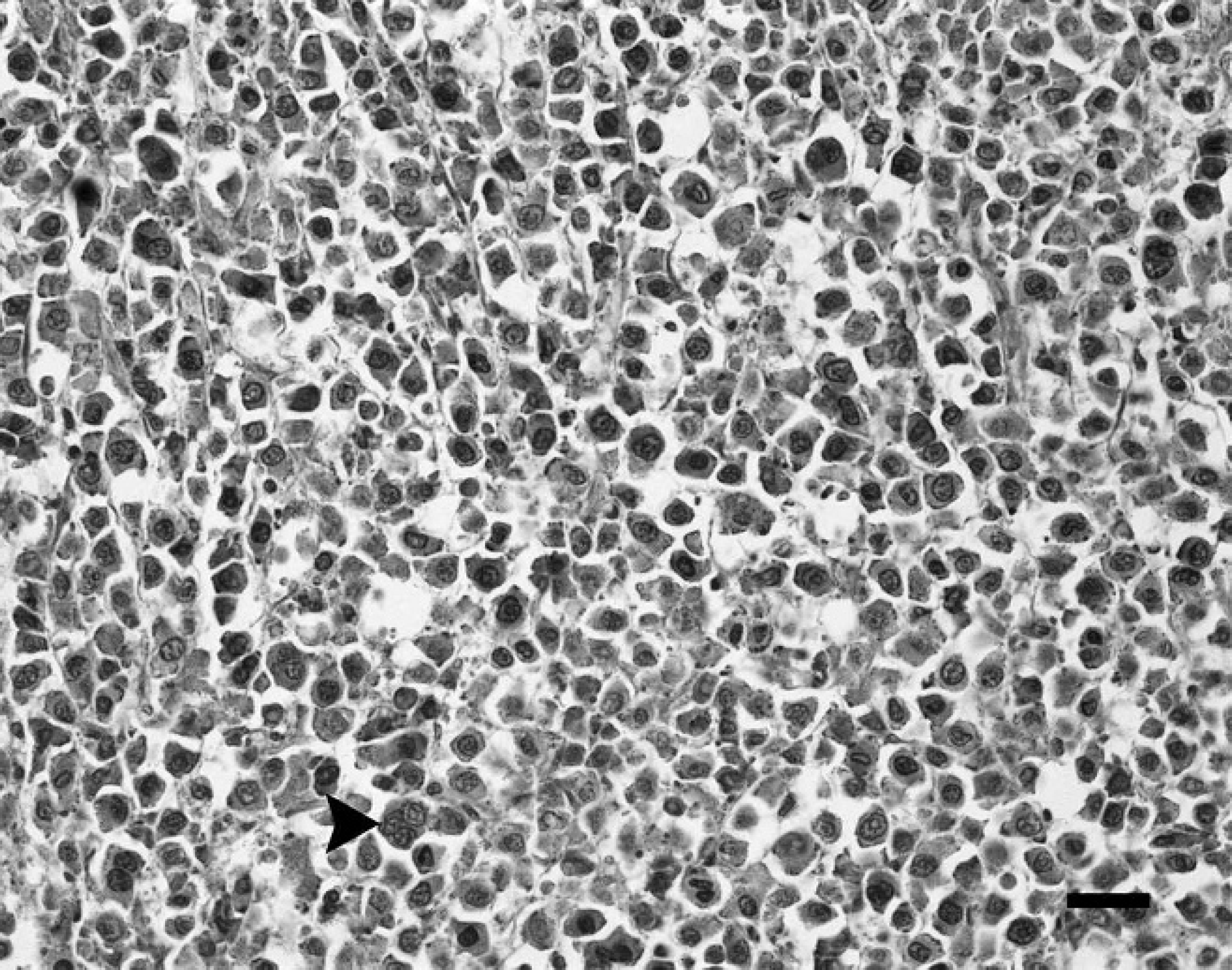
Brazilian porcupine (Coendou prehensilis) with T-cell lymphoma. The cervical lymph node is infiltrated by markedly pleomorphic, neoplastic lymphocytes. Cells with multiple nuclei (arrowhead) are commonly observed. Hematoxylin and eosin. Bar = 25 μm.
Histologically, the cervical mass consisted of cervical lymph nodes that were extensively effaced by sheets of large, markedly pleomorphic, neoplastic lymphocytes (Fig. 3), with scant amount of extracellular stroma and cellular infiltration of the nodal capsule. Individual neoplastic cells had moderate amount of lightly eosinophilic cytoplasm. Nuclei were round or cleaved with vesicular chromatin and 1–3 large nucleoli. Scattered binucleate and multinucleate lymphocytes (3–4 nuclei) were present, which had abundant cytoplasm. Three or 4 mitotic figures, some of which were atypical, were noted per high power field of view (400×). A few salivary gland acini that were extensively infiltrated by the tumor cells were observed in one of the tissue sections. Neoplastic lymphocytes had also extensively invaded the renal parenchyma, replacing tubules and glomeruli. Dense nests and cords of neoplastic cells, identical to those observed in the cervical lymph nodes and salivary gland, were also observed in the liver and in the lung. Additionally, neoplastic cells were seen in blood vessels of the cervical lymph nodes, kidney, and liver. Aggregated neoplastic lymphocytes were also observed in the lymphoid follicles of the spleen. Moderate lymphoid hyperplasia was observed in the mesenteric lymph nodes. Immunohistochemistry was performed on sections of cervical and mesenteric lymph node, liver, kidney, and spleen, as previously described. 6 The neoplastic cells in the cervical lymph node, lung, liver, and kidney were positive to cluster of differentiation (CD)3 (clone A0452 a ) and negative to CD79a (clone M7051 a ), indicating that they were T lymphocytes. Sections of the histologically normal mesenteric lymph node of this animal were used as a positive control for immunohistochemical staining.
Gross and histologic changes in the porcupine in the present study were consistent with disseminated malignant round cell neoplasm of T-lymphocyte origin that was based on immunohistochemistry. The dyspnea reported in the porcupine most likely was caused by the tumor compressing the trachea. The subcutaneous edema in the mandibular and cervical region of this animal likely occurred because of inefficient lymphatic drainage as a result of the extensive neoplastic cell proliferation within the cervical lymph nodes.
Lymphoid tumors commonly involve several organs, including lymph nodes, spleen, liver, kidney, heart, gastrointestinal tract, and bones. 1 Animals with advanced T-cell lymphoma can develop a leukemia-like blood picture as a result of metastases to the blood and bone marrow. This process is sometimes called leukemic lymphoma. 8 In the present case, neoplastic T cells were observed within blood vessels in sections of multiple tissues, and a complete blood cell count revealed mild leukocytosis characterized by mild lymphocytosis. Lymphocytosis may occur with lymphoid neoplasia, but the complete blood cell count was not suggestive of overt lymphocytic leukemia. The increase in BUN and creatinine concentrations is likely the result of extensive neoplastic involvement of the renal parenchyma with decreased filtration of nitrogenous wastes. Lymphoma has been reported in several different species. For example, lymphoid proliferations were identified as T immunophenotype in a guinea pig (Cavia porcellus) 7 and in a white-tailed deer (Odocoileus virginianus). 3 A multicentric B-cell lymphoma was reported in captive greater hedgehog tenrecs (Setifer setosus). 2 A T-cell–rich B-cell lymphoma also was identified in a ring-tailed lemur (Lemur catta). 5 The round cell neoplasm of the porcupine in the present case represents a malignant lymphoma of T-cell origin; however, no information is currently available about the biologic behavior of this neoplasm in porcupines.
Footnotes
a.
Dako North America Inc., Carpinteria, CA.