
Abstract
Although the novel Brazilian porcupinepox virus (BPoPV) can infect wild porcupines, its lethality and zoonotic potential are not well-established. In May 2021, a free-ranging neotropical porcupine (Coendou longicaudatus boliviensis) rescued from the natural savanna in the Brazilian Cerrado, Mato Grosso State, was presented with a lethal poxviral infection. Clinical signs and PCR detection of BPoPV supported the diagnosis. Poxviral lesions included erythema, exfoliative dermatitis, and erosions involving mainly the face, hindlimb, and vulva. Histologically, the lesions consisted of proliferative and necrotic dermatitis, intraepidermal and follicular pustules, and intracytoplasmic inclusion bodies in keratinocytes. Phylogenetic analysis revealed BPoPV strains closely related to other rodent-infecting poxviruses. This poxviral infection resulted in the death of a specimen of C. l. boliviensis; the effect on species conservation, and the potential of spillover into humans and other vertebrates remain unknown.
Poxviruses have historically posed challenges to the health of humans, domestic animals, and wildlife. Aside from the notorious variola virus, the causative agent of one of the deadliest infectious diseases in history,2,8,3 various poxviruses infect vertebrates, including humans. 17 The family Poxviridae comprises 22 genera and 83 species,14,17 of which at least 3 genera can cause zoonotic infections—Parapoxvirus, Yatapoxvirus, and Orthopoxvirus. Despite the availability of epidemiologic information about the host range for most orthopoxviruses, such as cowpox, mpox (monkeypox), vaccinia, and variola viruses, the emergence of generalist poxviruses reveals their high potential for spillover into novel hosts, as evidenced by the reported case of human-to-dog transmission during the current human mpox outbreak. 11
The host assemblage of poxviruses has been proven to be more broad-ranging than formerly established, and rodents can serve as virus reservoirs.4,12 A new poxvirus species has been detected in the Brazilian porcupine (Coendou prehensilis) in Uberlândia, Minas Gerais (MG), Brazil. 7 This poxvirus was tentatively named Brazilian porcupinepox virus (BPoPV; Poxviridae). Brazilian porcupines are listed as Low Concerning in the IUCN Red List (https://www.iucnredlist.org/species/101228458/22214580); however, the reported poxvirus-related death of C. prehensilis warns conservationists of a threat to the species. 7
In May 2021, the Department of Mato Grosso State for the Environment (Secretaria de Estado de Meio Ambiente, Mato Grosso; SEMA/MT) encountered a Cerrado porcupine (Rodentia, Erethizontidae, C. longicaudatus boliviensis). The individual was severely dehydrated and was immediately referred to the Universidade Federal de Mato Grosso Veterinary Hospital (HOVET-UFMT), Cuiabá, Mato Grosso. After a thorough physical examination, the rodent was admitted to the Wildlife Unit of the HOVET-UFMT. Clinical signs included wrinkled and scaly erythema (mainly of the head, hindlimb, and vulva) and lethargy; hematologic findings included metabolic acidosis and hypergammaglobulinemia. Unfortunately, the patient died the next day despite supportive care and was submitted immediately for autopsy.
Gross findings in this Cerrado porcupine comprised typical poxviral skin lesions, consistent with reported infection by BPoPV. 7 Erythematous papules were in clusters on several regions (auricular, nasal, perioral, periocular, chin, perianal, genital, and thigh) and tended to coalesce into plaques with an irregular surface, separated by superficial-to-deep and moderately erythematous striae. The marked epidermal thickening of the eyelids resulted in bilateral occlusion of the palpebral fissures. In addition, the nose, lips, and muzzle had superficial erosions and ochre crusting. Finally, the left cranial lung lobe was markedly enlarged with a hemorrhagic surface. Lesions were not observed in other organs.
Histologic features included moderate diffuse parakeratotic hyperkeratosis with neutrophil debris and degenerate keratinocytes with gram-positive cocci within crusts in the superficial stratum corneum. In addition, there was moderate-to-marked neutrophilic folliculitis. The epidermis had extensive necrotic areas; intercellular edema; mild-to-moderate, random, multifocal acantholysis; and psoriasiform hyperplasia; keratinocytes often had eosinophilic cytoplasmic B-type inclusion bodies, and ballooning degeneration (Fig. 1). In the dermis was a multifocal-to-coalescent lymphoplasmacytic infiltrate and edema, with myxoid material in the papillary layer. In addition to focal hemorrhage, the lungs had diffuse thickening of alveolar septa, a proliferation of type II pneumocytes, and a mononuclear interstitial infiltrate. Bronchi and bronchioles had moderate epithelium hyperplasia and a submucosal lymphoplasmacytic infiltrate. Structural abnormalities were not seen in other tissues.
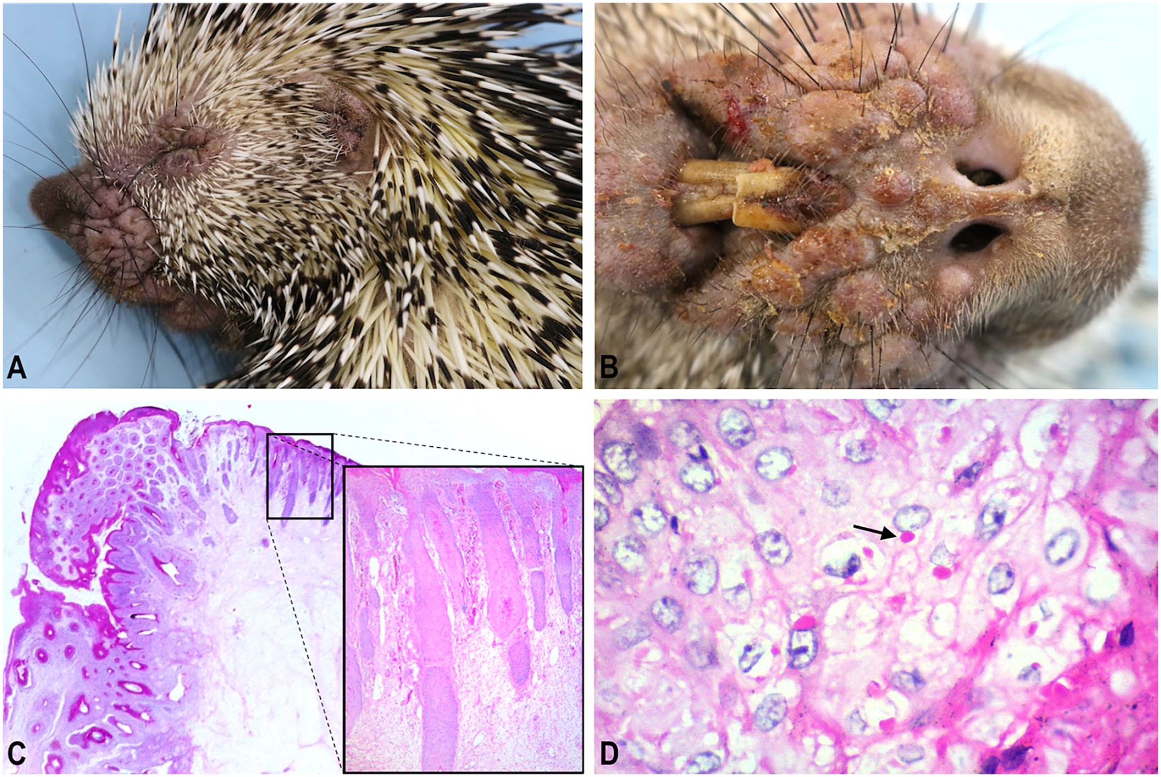
Brazilian porcupinepox virus infection in a free-ranging neotropical porcupine.
DNA was extracted from the skin with phenol–chloroform. 10 We utilized a low-GC pan-chordopoxvirus PCR assay to amplify a 231-bp fragment, as described elsewhere. 9 The acquired amplicons were purified (Agencourt AMPure XP; Beckman Coulter), bidirectionally sequenced (ABI Prism 3500 genetic analyzer; Life Technologies), and further confirmed by BLAST (https://blast.ncbi.nlm.nih.gov/Blast.cgi). We deposited the obtained sequence in GenBank (MZ709431). The evolutionary history was inferred using the maximum-likelihood method and Tamura 3-parameter model on MEGA11. 13 Phylogenetic analysis revealed that the BPoPV strains, including the one detected in our study and another reported elsewhere (MN692191), formed a well-supported monophyletic clade (Suppl. Fig. 1). We also found this clade to be closely related to Cotia virus, another rodent-infecting Brazilian poxvirus (KM595078, HQ647181), which comprises the putative genus Oryzopoxvirus. 15
Rodents other than porcupines can act as poxvirus natural hosts. Myxoma and fibroma viruses (Poxviridae, Chordopoxvirinae, Leporipoxvirus) cause localized benign tumor-like lesions in rabbits and squirrels. 16 Squirrel fibroma virus reportedly causes squirrel fibromatosis, which leads to alopecic dermal nodules with typical poxviral lesions in red (Tamiasciurus hudsonicus), gray (Sciurus carolinensis), and fox (S. niger) squirrels.1,16 Given the non-lethal occurrence of poxviral infections, rabbits and squirrels have been strongly suggested as potential reservoirs. 4 Northern pygmy mice (Baiomys taylori) have been reported with epidermal proliferative lesions on the tail and paws caused by a poxvirus; this new species was tentatively named Brazospox virus. 6
BPoPV was reported in Uberlândia, MG, 7 ~1,000 km from Cuiabá, MT. Our finding raises concerns about the impact of BPoPV infection in Coendou spp. in a variety of distinct South American biomes. Our case and the case reported in Uberlândia had similar clinical findings, macroscopic and microscopic lesions, and fatal outcomes. The intensity and distribution of these lesions are critical; they help explain the severity of the clinical picture and are related to the cause of death of our case. Wildlife veterinarians observed similar clinical signs in free-ranging porcupines, with a case fatality rate of >75%. 7 BPoPV infection in a hairy dwarf porcupine (Coendou spinosus) from São Paulo was treated successfully. 5 The susceptibility spectrum, pathogenicity, and disease progression of BPoPV infections in rodents or other taxa remain to be resolved.
Supplemental Material
sj-pdf-1-vdi-10.1177_10406387231161340 – Supplemental material for Brazilian porcupinepox virus infection in a free-ranging neotropical porcupine in Mato Grosso, Brazil
Supplemental material, sj-pdf-1-vdi-10.1177_10406387231161340 for Brazilian porcupinepox virus infection in a free-ranging neotropical porcupine in Mato Grosso, Brazil by David J. F. Silva, Janaina M. A. R. Moreira, Marlon Ribeiro, Fernanda H. Maruyama, Thaís O. Morgado, Luciano Nakazato, Valeria Dutra, Marcos A. Souza and Edson M. Colodel in Journal of Veterinary Diagnostic Investigation
Footnotes
Acknowledgements
We thank Raphael Neres, Morgana Hennig, and Stephanni Pimentel, staff at the Wildlife Section of HOVET-UFMT. We also thank the staff of the Department of Mato Grosso State for the Environment.
Declaration of conflicting interests
The authors declared no potential conflicts of interest with respect to the research, authorship, and/or publication of this article.
Funding
David J.F. Silva receives a scholarship from the Ministry of Education, Marlon Ribeiro receives a fellowship from CNPq, and Fernanda H. Maruyama receives a fellowship grant awarded by CAPES Foundation.
Supplemental material
Supplemental material for this article is available online.
References
Supplementary Material
Please find the following supplemental material available below.
For Open Access articles published under a Creative Commons License, all supplemental material carries the same license as the article it is associated with.
For non-Open Access articles published, all supplemental material carries a non-exclusive license, and permission requests for re-use of supplemental material or any part of supplemental material shall be sent directly to the copyright owner as specified in the copyright notice associated with the article.