
Abstract
Avian influenza virus (AIV) is one of the most important zoonotic pathogens because of its potential to cause severe disease outbreaks in avian and human hosts. Virus isolation in embryonated chicken eggs (ECEs) remains a gold standard technique for AIV detection. However, some laboratories prefer molecular methods, such as real-time quantitative reverse transcription polymerase chain reaction (qRT-PCR), for initial sample screening because of their high throughput sample processing and rapid results. Samples found positive on real-time qRT-PCR are then inoculated in ECEs for virus isolation and characterization. This approach is based on the premise that real-time qRT-PCR will detect all AIV-positive samples. The current study aimed to determine if AIV can be isolated from cloacal samples of waterfowl that were initially found to be negative by real-time qRT-PCR screening. Quantitative RT-PCR–negative cloacal samples (1,369) were inoculated for virus isolation in commercial nonspecific pathogen–free ECEs. After 4 days of incubation, the allantoic fluids were harvested and inoculated in fresh ECEs for a second passage. Allantoic fluids from 147 samples were positive for hemagglutination with chicken erythrocytes. Of the 147 hemagglutination-positive allantoic fluids, 82 were AIV positive when confirmed with real-time qRT-PCR. Ten isolates were subtyped as H7N2 (n = 7), H7N1, H1N2, and H2N2. In addition, N subtype could be determined for isolates from an additional 25 samples. These results highlight the fact that screening by realtime qRT-PCR may result in some false-negative cloacal samples for AIV.
Influenza A virus (family Orthomyxoviridae, genus Influenzavirus A) is a common cause of respiratory tract infections in a variety of species, including human, avian, swine, canine, and equine. 23 The influenza viruses are classified into subtypes on the basis of antigenic differences in their surface glycoproteins: hemagglutinin (HA) and neuraminidase (NA). 23 On the basis of the severity of clinical signs and mortality rate in experimentally inoculated chickens, Avian influenza virus (AIV) can be categorized into 2 pathotypes: high-pathogenicity avian influenza (HPAI) and low-pathogenicity avian influenza (LPAI). Highly pathogenic AIV is a “list A” disease of the World Organization for Animal Health (OIE). 24
To date, 16 HA and 9 NA subtypes have been detected, and any combination of them can be found in nature. Of the possible 144 such combinations, almost all have been detected in wild birds and waterfowl, 9 but only H5 and H7 subtypes represent a potential to be HPAI. The HPAI H5 and H7 subtypes can arise from avirulent or LPAI subtypes by mutation or acquisition of specific sequence changes, such as losing potential glycosylation sites or gaining multiple basic amino acid sequences in the connecting peptide of HA. 13 Therefore, all H5 and H7 AIV strains must be reported to the OIE 24 due to their potential risk of being HPAI. In addition, many such strains are now known to be zoonotic in nature.
Recent outbreaks of avian influenza H5N1 in poultry and human beings have highlighted the important role of routine surveillance and early detection of AIV in waterfowl, which are the natural reservoirs of AIV. 10,23 The aquatic birds of the orders Anseriformes (ducks, geese) and Charadriiformes (shorebirds) can be infected with AIV but do not normally show clinical signs. 19 Also, the virus has a predilection for the gastrointestinal tract in these birds and is shed primarily in their feces, 17 except that H5N1 lineage virus is shed primarily from the upper respiratory tract. The routine surveillance of AIV strains from these reservoirs is helpful in epidemiologic studies and in selection of up-to-date strains for developing appropriate vaccines. 15
Historically, virus isolation in embryonated chicken eggs (ECEs) is the standard technique of virus detection followed by antigenic characterization of the circulating strains using classical methods of hemagglutination inhibition (HI) and neuraminidase inhibition (NI). 14 Because these standard methods are laborious, expensive, and time consuming, there has been an increase in the use of molecular methods of AIV detection and characterization in many laboratories. 7 Molecular methods, such as reverse transcription polymerase chain reaction (RT-PCR), have enormously simplified virologic surveillance in recent years because of their high sensitivity and speed. 3 Rapid molecular tools are especially useful during epidemics, because virus isolation in ECEs may take 1–2 weeks. For routine surveillance, several laboratories use real-time quantitative RT-PCR (qRT-PCR) as a screening tool followed by virus isolation and subtyping. 11
One noticeable concern with any diagnostic method is the potential of obtaining false-negative results. 4 In RT-PCR, this may occur due to the presence of RT-PCR inhibitors, low virus titers, poor RNA extraction, mismatched primers, degradation of target RNA before amplification, errors in setting up a reaction, or a degraded reagent. 21 Another limitation is that nucleic acid–based testing requires routine updating of the primers because of frequent antigenic drift and mutations in influenza A viruses. Thus, isolation of AIV from RT-PCR–negative samples has been previously reported. 18 The present study was conducted to expand on these results and to determine if AIV can be isolated from real-time qRT-PCR–negative cloacal samples by inoculation in commercial nonspecific pathogen–free (nSPF) ECEs.
From June to September 2008, cloacal swabs from 7,260 migrating waterfowl were collected. The birds were trapped and sampled in the states of Minnesota and North Dakota from 29 different locations using night lighting, rocket netting, and hunter harvesting. The cloacal swabs were placed in brain heart infusion broth containing penicillin (10,000 units/ml), streptomycin (10,000 μg/ml), gentamicin (5,000 μg/ml), and amphotericin B (50 μg/ml). The samples were transported to the laboratory in liquid nitrogen and stored at −80°C until processed. All samples were screened by real-time qRT-PCR targeting a conserved region of the matrix gene within influenza A viruses. 18 Samples with threshold cycle values of less than 36 were considered positive for AIV. 1 Samples (1,369) were randomly selected that were negative for AIV by real-time qRT-PCR and tested for virus isolation in nSPF ECEs. After 2 blind passages in ECEs, the allantoic fluids were collected and tested for HA. All HA-positive allantoic fluids were tested by real-time qRT-PCR 18 followed by subtyping of real-time qRT-PCR–positive samples by RT-PCR using previously described methods for HA 11 and NA 2 typing.
In total, 1,369 cloacal samples, which were earlier found to be negative for AIV by real-time qRT-PCR, were used in the present study. The samples were collected from different species of waterfowl (Table 1), with the maximum number coming from mallards (n = 707). After 2 passages in ECEs, the allantoic fluids were harvested and tested by HA; 147 samples were found to be HA positive. On testing of these 147 HA-positive samples by real-time qRT-PCR, 82 were confirmed as AIV. Of these, 10 isolates could be subtyped completely (both HA and NA), while only NA subtype could be determined for isolates from an additional 25 samples. Five samples showed mixed infection with 2 different N subtypes (N1 and N2; Table 1).
Eight of the 10 completely subtyped isolates were of the H7 subtype. To determine their pathogenic potential (HPAI or LPAI), the HA cleavage site of these 8 H7 isolates was amplified, 20 and DNA bands of 241 base pairs corresponding to the cleavage site of H7 subtype were excised, purified, and sequenced. Deduced amino acid sequences were analyzed to determine the potential pathogenicity (HPAI or LPAI) of the isolates. The amino acid analysis of sequences thus obtained confirmed them to be LPAI.
Several techniques are available for the detection of AIV in waterfowl and other hosts. The specificity, sensitivity, and rapidity of these techniques are important factors when considering the most appropriate technique to be used in a particular situation. Virus isolation remains the standard technique for the detection and propagation of viruses so that live viruses can be used for further characterization and in up-to-date vaccine production. 23 Real-time qRT-PCR has been extensively used for the detection of AIV from waterfowl because of its low cost and high through-put. 1 However, due to frequent mutations in influenza viruses, the sequence of primer sets used in PCR-based detection must be appropriate for the detection of currently circulating strains. 8 Also, it is epidemiologically more important to determine the circulating subtypes of AIV rather than to determine only if a sample is positive or negative for AIV.
In the present study, a large number of real-time qRT-PCR–negative cloacal samples (n = 1,369) were examined for the presence of AIV by virus isolation. Reports on the isolation of AIV from real-time qRT-PCR–negative samples are scattered throughout the veterinary literature, but no comprehensive study is available on this subject. Results from the current study reinforce the observation that some real-time qRT-PCR–negative samples can yield AIV by virus isolation. Out of 147 HA-positive samples, only 82 were confirmed as AIV by real-time qRT-PCR. Although molecular methods are considered more sensitive than virus isolation, 8 the failure of real-time qRT-PCR to detect all AIV in the present study can partially be explained by the presence of non-AIV hemagglutinating viruses (e.g., Newcastle disease virus [NDV] and other paramyxoviruses). In fact, 132 of the 147 HA-positive samples were found to be positive for NDV by RT-PCR. 6,13 Also, the presence of PCR inhibitors in cloacal samples 1,4,5 is known to yield false-negative results in PCR. To rule this out, approximately 10% of the cloacal samples were tested by real-time qRT-PCR in which an internal control was included. No PCR inhibitors were encountered in those samples.
Only 10 of the 82 isolates could be fully subtyped, and isolates from an additional 25 samples could be subtyped only for NA using NA-specific primers. It is possible that the amount of virus in untypable isolates was not sufficient for subtyping, because all AIV strains may not readily adapt to grow to detectable titers within 2 ECE passages. 18 Additional blind passages of such samples in ECEs may improve the rate of virus isolation and identification. In a related study, it was observed that the presence of mixed infection (e.g., AIV, NDV) in a single sample may allow overgrowth of NDV resulting in suppression of AIV. 6 For such samples, a method to inhibit growth of NDV was developed by treating samples with anti-NDV antibody before egg inoculation to promote growth of AIV. 6 The detection of mixed infection with 2 different AIV subtypes in a single sample is not surprising. Mixed infections of waterfowl with more than one HA and/or NA subtype have been reported. 10,16,22
Detection of Avian influenza virus in real-time quantitative reverse transcription polymerase chain reaction (qRT-PCR)–negative cloacal samples of waterfowl. *
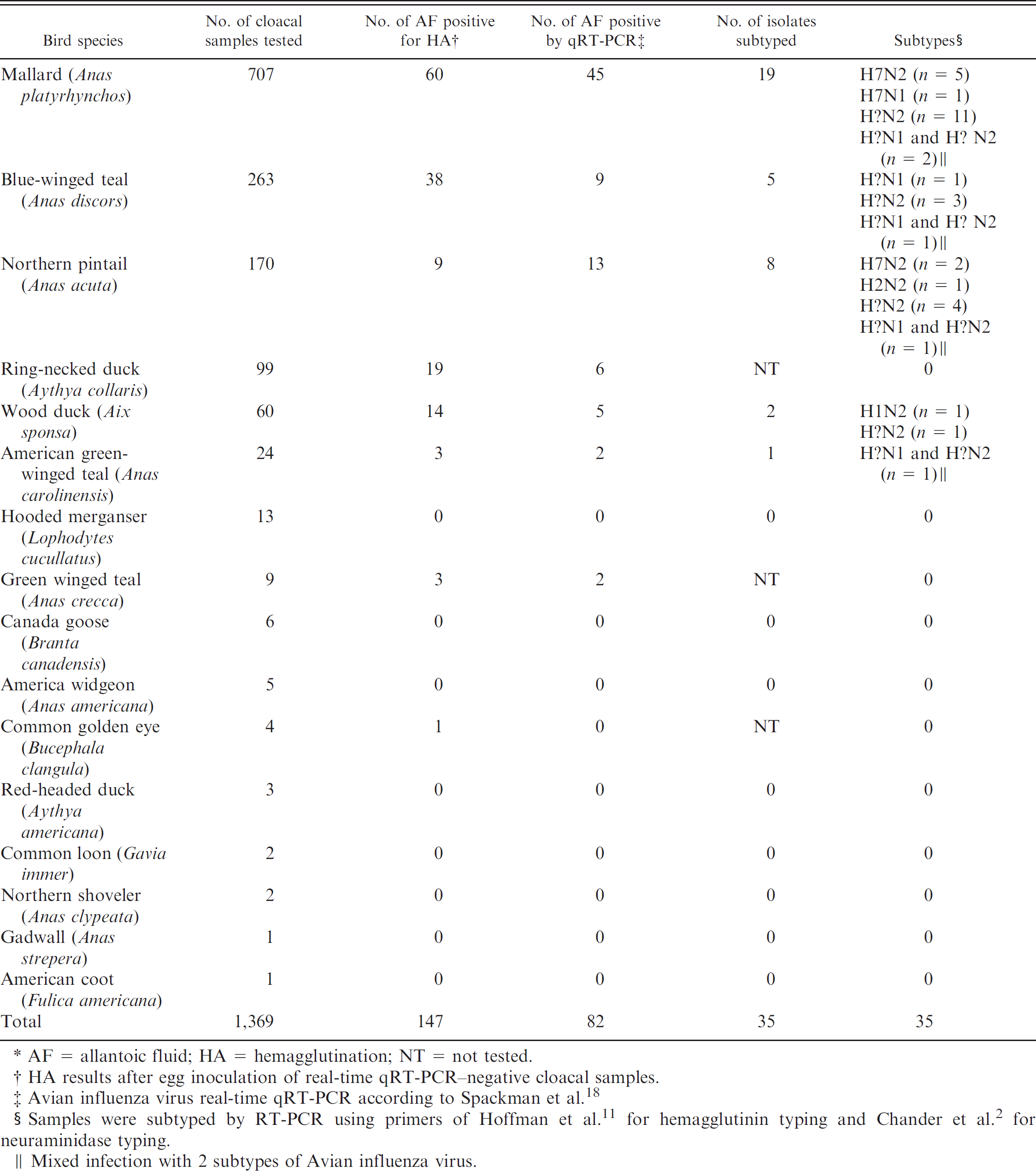
AF = allantoic fluid; HA = hemagglutination; NT = not tested.
HA results after egg inoculation of real-time qRT-PCR–negative cloacal samples.
Avian influenza virus real-time qRT-PCR according to Spackman et al. 18
Samples were subtyped by RT-PCR using primers of Hoffman et al. 11 for hemagglutinin typing and Chander et al. 2 for neuraminidase typing.
Mixed infection with 2 subtypes of Avian influenza virus.
Another objective of the present study was to determine if commercial nSPF can be used for growing AIV. Essentially all laboratories currently use SPF eggs for isolation of AIV. Results of the current study indicate that it may not be necessary to use costly SPF eggs unless an AIV vaccine is used in the commercial flocks. Because no AIV vaccine is currently used for poultry in the United States, an inexpensive source of ECE was considered safe to use in the present study. However, before using commercial eggs for AIV isolation, a representative number should be examined and confirmed to be negative for anti-AIV antibodies. In AIV surveillance, it is important to perform virus isolation to obtain a clear picture of circulating AIV and to understand that relying solely on real-time qRT-PCR may yield spurious results.
Acknowledgements. This work was funded in part with federal funds from the National Institute of Allergy and Infectious Diseases, National Institutes of Health, Department of Health and Human Services, under contract no. HHSN266200700007C. Its contents are solely the responsibility of the authors and do not necessarily represent the official views of the National Institutes of Health. The authors thank the Egyptian Cultural and Educational Bureau in Washington, DC, Zagazig University in Egypt, and the Egyptian Ministry of Higher Education and State for Scientific Research for their financial support and scientific supervision of the PhD research scholar M. Ezzat El Zowalaty.