
Abstract
The present work aimed to study the behavior of acute phase proteins (haptoglobin, serum amyloid A, acid soluble glycoprotein, fibrinogen, and albumin) in fasting-induced pregnancy toxemia in goats and their relationship with classical indicators of this disorder such as beta-hydroxybutyrate and nonesterified fatty acids in the blood and decreased urine pH and ketonuria. Twelve adult Murciano–Granadina goats at the final stage of gestation were used in this experiment. Pregnancy toxemia was induced in 6 goats by fasting for 72 hr. The other 6 animals were used as control group. Ketonuria was present in 4 out of 5 fasting animals at 24 hr and in all fasting animals at 48 hr of fasting. Serum nonesterified fatty acids were significantly increased at 24, 48, and 72 hr of fasting. Beta-hydroxybutyrate and haptoglobin achieved significantly increased concentrations at 48 hr and 72 hr, respectively, remaining increased during the entire study. Serum amyloid A, acid soluble glycoprotein, fibrinogen, and albumin were not affected by fasting. In conclusion, acute phase proteins (including haptoglobin) seemed not to have an advantage over traditional markers in diagnosis of fasting-induced pregnancy toxemia in goats.
Introduction
Pregnancy toxemia is a metabolic disorder that affects goats during the last period of gestation, occurring most frequently in animals with high body condition with 2 or more fetuses. 21 Undernutrition, due to great energy demand by fetuses at the final stage of pregnancy, and obese body condition are the main factors that result in decreased glucose and excessive lipid mobilization, and consequently, ketonemia. 2 It has been demonstrated that hypoglycemia leads to production of ketone bodies in a manner inversely proportional to the plasma glucose concentrations. 26 The diagnosis of this disease is based on history, clinical signs, and detection of ketone bodies such as beta-hydroxybutyrate (BHB) in serum or urine. 14 Morbidity may approach 5–20% in farm goats, and mortality in nontreated animals may be as high as 80%. 21
Acute phase proteins (APPs) have been proposed as sensitive and rapid indicators of inflammatory disturbances in ruminants. 5 The major APPs in ruminants that have been described in the literature are haptoglobin (Hp) and serum amyloid A (SAA). 16 In goats, Hp and SAA could be considered as major APPs, while acid-soluble glycoprotein (ASG) and fibrinogen may be considered as moderate. 10 Previous studies in ruminants suggested a relationship between selected APPs and lipid mobilization. One study 18 reported increased Hp concentration in cows with fatty liver, compared with the values in healthy cows (466 mg/l vs. 0.01 mg/l). In addition, increased Hp levels in cows around parturition have been described. It has been postulated to be related to negative energy balance, since cows with high milk Hp also showed high non-esterified fatty acid (NEFA) concentration in serum. 11 A significant correlation has been also reported between Hp and BHB in lactating goats (Trevisi E, D'Angelo A, Gaviraghi A, et al.: 2005, Blood inflammatory indices in goats around kidding. Ital J Anim Sci 4:404. Abstract).
The previous findings reveal a possible relationship between Hp and markers of negative energy status in ruminants and led the current authors to study the APP response in pregnancy toxemia in goats. The present work was aimed at determining the concentration and evolution of different APPs, such as Hp, SAA, ASG, fibrinogen, and albumin, in fasting-induced pregnancy toxemia in goats and comparing them with other traditional biochemical markers such as BHB, NEFA, glucose, triglycerides, and cholesterol. The current study presents an evaluation of these proteins in the disease and possible practical applications of APPs in the diagnosis and management of pregnancy toxemia in goats.
Materials and methods
Animals and diet
Twelve healthy Murciano–Granadina multiparous goats of similar age (mean: 4.3 years, range: 3–5 years) from a farm in the Murcia Region of Spain were used in the present experiment. This breed is characterized by milk production of more than 600 kg of milk per lactation. Weight ranged between 45 and 55 kg (mean: 48 kg), which was within breed parameters for female individuals (40–55 kg). The mean body condition score of the goats was 3.5–4.0 on a scale of 1–5 (Fredricks G: 1993, Using body condition score to evaluate feeding management. In: Proceedings of the American Dairy Goat Association National Convention, p. 78. Portland, OR), indicating that the goats had adequate fat reserves.
All goats were inseminated after synchronization with 3-mg norgestimate ear implants and a 7.5-mg injection of dinoprost a 13 days after the implant. Forty-five days after insemination, pregnancy was diagnosed by transabdominal ultrasonography. Approximately 6 weeks prior to the scheduled parturition, a new ultrasonography was completed to check that all goats had a minimum of 2 fetuses.
Goats were fed ad libitum on alfalfa hay of 0.6 milk forage unit (UFL)/kg dry matter (DM), 156 g crude protein (CP)/kg DM, and 463 g neutral detergent fiber (NDF)/kg DM plus 250 g of concentrate/day at 2 months gestation, increasing gradually to 500 g/day per head for the last 4 weeks of pregnancy. The concentrate used (1.05 UFL/kg DM, 160 g CP/kg DM, and 246 g NDF/kg DM) was a mixture of corn, barley, oat, cotton, and lupine grains; sunflower meal; citrus pulp; and dried distillers grains pellets; and a pellet concentrate, where vitamin and mineral supplements were included. The concentrate was offered in 2 equal portions at 8:00 AM and 4 PM. The animals had free access to water.
Approximately 4 weeks prior to the scheduled parturition (at 120 days from insemination), goats were randomly divided into 2 groups—control group (n = 6) and fasting group (n = 6)—and housed in 2 separated pens with straw bedding. The control group was fed normally during the study. After a period of 2 weeks for adaptation to housing in separated pens, pregnancy toxemia was induced in the fasting group by allowing access only to water for 72 hr. 26 Animals were examined daily to detect clinical signs of pregnancy toxemia. After 72 hr of starvation, the first clinical signs were observed; at this point, feed was reintroduced ad libitum to all animals of the fasting group using the same diet that was used for the control group. All procedures were done with the approval of the Ethical Committee of the University of Murcia (Spain).
Sampling
Blood and urine samples were collected every morning at 10:00 AM in both groups of animals, 1 day before the induction of fasting, and daily during the following 5 days. Blood samples were collected in vacuum tubes b with heparin (5 ml) for fibrinogen analysis and in plain tubes (10 ml) for the rest of the biochemical analyses. Urine samples were obtained by voluntary miction or induced by covering the nose and the mouth of the goat for few seconds (Ortolani E: 2003, Diagnóstico de doenças nutricionais e metabólicas por meio de exame de urina em ruminantes [Diagnostic of nutritional and metabolic diseases using urine examination in ruminants]. In: Anais do I Simpósio de Patologia Clínica Veterinária da Região Sul do Brasil, ed. Gonzalez FHD, Campos R, pp. 91–102. Universidade Federal do Rio Grande do Sul, Porto Alegre, Brazil). In fasted goats, sampling at 72 hr was performed prior to feed reintroduction to avoid any influence in the results. Samples were kept in a thermal box for transport to the laboratory. Blood in plain tubes was centrifuged (2,000 × g for 10 min), and serum was stored at −20°C until biochemical analysis.
Urine and serum metabolites analysis
Determination of the urine pH was made with a digital pH meter. c Chemical analysis of urine was conducted using reagent strips. d The following serum metabolites were determined by commercially available colorimetric methods: glucose, e triglycerides, e total cholesterol, e BHB, f and NEFA. f
Acute phase proteins analysis
Albumin was measured using a colorimetric technique. e Serum concentrations of Hp were quantified by a spectrophotometric method using a commercial kit. g The method of ASG determination was based on a technique described in 1989 17 and modified in 1996. 6 Serum SAA concentration was measured using a commercially available enzyme-linked immunosorbent assay (ELISA) kit g according to the manufacturer's instructions. Fibrinogen was measured in plasma samples by the heating precipitation method at 56°C. 25 All methods for APP determination have been previously validated at the authors' laboratory in goats, giving intra-assay and interassay coefficients of variation within 1.36–4.47% and 3.69–12.68%, respectively. Limits of detection were 0.02 g/l for Hp, 0.20 g/l for ASG, and 3.06 mg/l for SAA. 10 All colorimetric methods were performed in an automatic analyzer. h The final absorbance in ELISA methods was measured in a microtiter plate reader i at 450 nm wavelength.
Statistical analysis
Arithmetic means and standard deviations were calculated using routine descriptive statistical procedures. Kolmogorov–Smirnov test was used to assess normality of data, giving a nonparametric distribution. Therefore, all data were normalized by logarithmic transformation before analysis. To assess differences between control and fasting groups, 2-way analysis of variance of repeated measures with Bonferroni posttest was performed. j A P value < 0.05 was considered statistically significant. Correlation between APPs and other biochemical parameters were analyzed using Pearson correlation analysis; data obtained from all animals during the study period were included in this analysis.
Mean values of urinary pH and units of ketonuria (acetoacetic acid) after the induction of pregnancy toxemia in goats by fasting for 72 hr (n control = 6, n fasting = 5). *
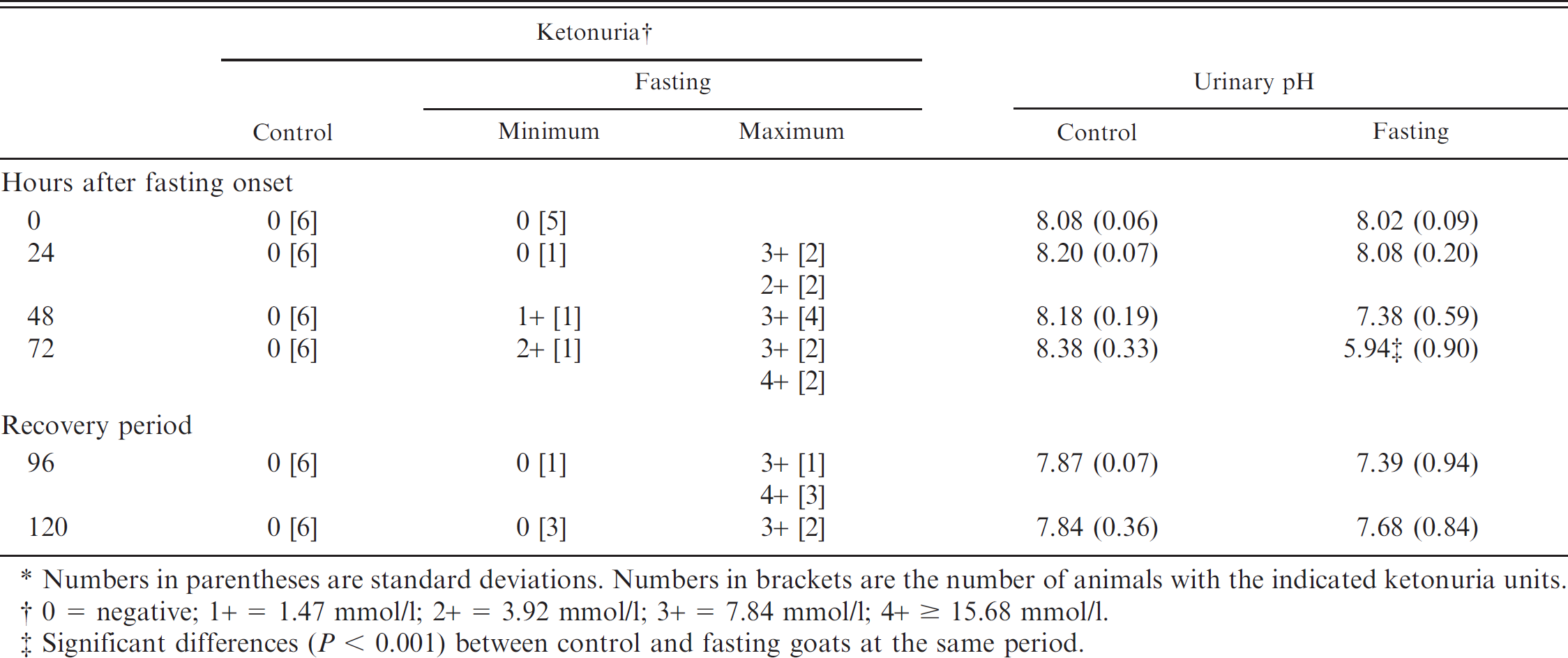
Numbers in parentheses are standard deviations. Numbers in brackets are the number of animals with the indicated ketonuria units.
0 = negative; 1+ = 1.47 mmol/l; 2+ = 3.92 mmol/l; 3+ = 7.84 mmol/l; 4+ ≥ 15.68 mmol/l.
Significant differences (P < 0.001) between control and fasting goats at the same period.
Results
Clinical signs of early pregnancy toxemia started to appear after 72 hr of fasting, when 5 out of 6 goats (83.3%) showed depression, muscle tremors, and staggering gait. At this moment, feed was reintroduced for all the animals. The clinical signs disappeared in all animals within a few hours after feed was reintroduced. One of the fasted goats kidded the last day of the experiment, so the values of this goat were excluded from statistical analysis.
Mean values of glucose, non-esterified fatty acid (NEFA), beta-hydroxybutyrate (BHB), triglycerides, and cholesterol after the induction of pregnancy toxemia in goats by fasting for 72 hr (n control = 6, n fasting = 5). *
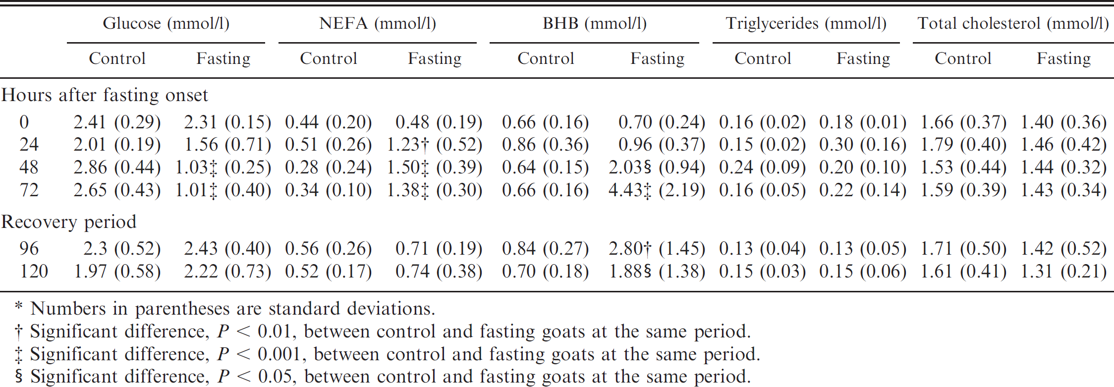
Numbers in parentheses are standard deviations.
Significant difference, P < 0.01, between control and fasting goats at the same period.
Significant difference, P < 0.001, between control and fasting goats at the same period.
Significant difference, P < 0.05, between control and fasting goats at the same period.
Moderate to severe ketonuria was evident at 24 hr after feed withdrawal, and the urinary pH decreased significantly at 72 hr of fasting. During the recovery period, urinary pH returned to normal in all animals, whereas ketonuria was present in 2 goats of the fasting group at the end of the study (Table 1).
Compared with the control group, glucose values significantly (P < 0.001) decreased in the fasting group after 48 hr of fasting. NEFA were significantly (P < 0.01) increased in the fasting goats after 24 hr and peaked at 48 hr of fasting. BHB was significantly (P < 0.05) higher in the fasting group than the control after 48 hr of fasting and peaked at 72 hr. Glucose and NEFA normalized as soon as feed was reintroduced, whereas BHB was significantly higher in the fasting group throughout the study (Table 2). Triglycerides and total cholesterol values did not show any significant difference between the control and fasting group (Table 2).
Mean values of haptoglobin (Hp), serum amyloid A (SAA), acid-soluble glycoprotein (ASG), fibrinogen, and albumin after the induction of pregnancy toxemia in goats by fasting for 72 hr (n control = 6, n fasting = 5). *
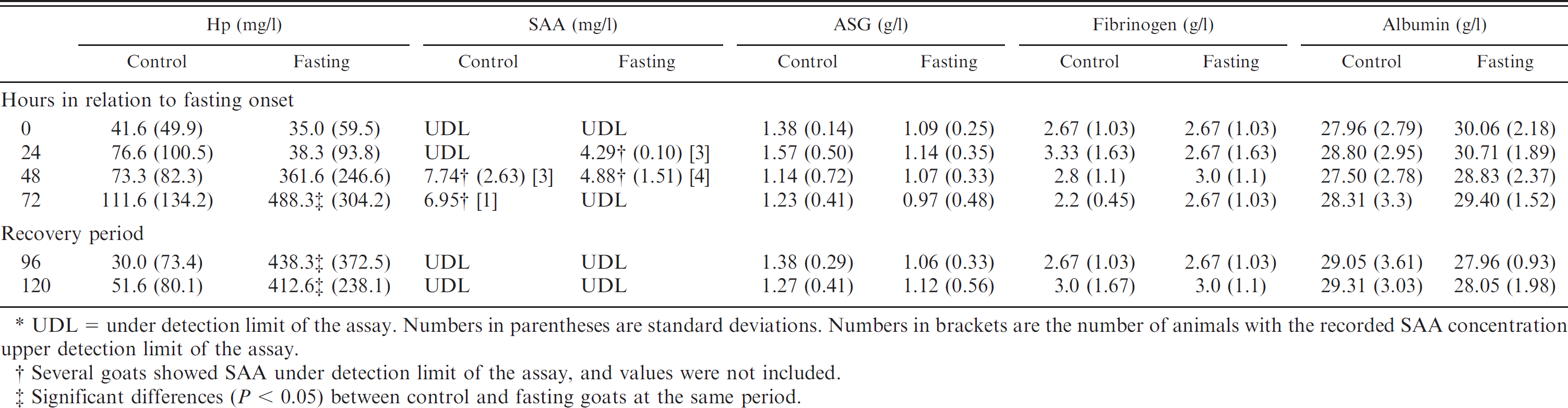
UDL = under detection limit of the assay. Numbers in parentheses are standard deviations. Numbers in brackets are the number of animals with the recorded SAA concentration upper detection limit of the assay.
Several goats showed SAA under detection limit of the assay, and values were not included.
Significant differences (P < 0.05) between control and fasting goats at the same period.
Haptoglobin values tended to be increased in the fasting group at 48 hr and were significantly (<0.05) increased at 72 hr, remaining elevated during the recovery period (Table 3). The values of ASG, fibrinogen, SAA, and albumin were not affected by fasting.
The Pearson coefficient of correlation between Hp and BHB during the fasting period was 0.84 (P < 0.05). Correlation between other APPs and metabolites did not show any significance.
Discussion
The first clinical signs of pregnancy toxemia in sheep usually appear when the blood concentration of ketone bodies reaches 4 mmol/l. 7 In the present experiment, clinical signs of pregnancy toxemia, characterized by staggering gait, muscle tremors, and depression, were evident in the goats after 72 hr of fasting when the mean concentration of serum BHB was 4.43 mmol/l.
Ewes and goats with BHB concentrations of 0.86–1.6 mmol/l are classified as having mild or subclinical pregnancy toxemia. 3,19 According to these criteria, fasted goats in the current study had subclinical pregnancy toxemia 24 hr after fasting when BHB values were 0.96 mmol/l. Also, NEFA were increased, and ketonuria appeared after 24 hr of fasting, but clinical signs were not evident at this time.
In the present study, Hp concentrations tended to be increased at 48 hr of fasting in goats and were significantly increased at 72 hr. Concentrations of the other APPs, such as ASG, fibrinogen, SAA, and albumin, were not affected by the induction of pregnancy toxemia. SAA showed great variability with undetectable concentrations at multiple time points. A few goats (3 goats at 24 hr and 4 goats at 48 hr in the fasting group, and 3 goats at 48 hr and 1 goat at 72 hr in the control group) had a measurable concentration of SAA but a consistent change associated with pregnancy toxemia was not observed. In all instances, SAA concentrations were lower than 10 mg/l, which could be a normal variation for this species. 10 Other authors have reported increases in Hp without increases in other APP such as alpha 1–acid glycoprotein after starvation of cows. 27 Also, ketosis did not produce any change in serum transferrin (an APP) in cows. 15 Moreover fasting has been related to a decrease of inflammation status by decreasing interleukin-6 in humans. 8 These observations and the lack of increase in other APPs evaluated in the current study suggest that the increase in Hp observed in the present study was not part of an inflammatory or acute phase response.
Increases in Hp found in goats with pregnancy toxemia could be related to the changes in lipid metabolism that occurs in this process. In cows, nonfeeding induces NEFA mobilization. 1,22 When a fasted period of 3 days was applied to a group of calves, NEFA mobilization was observed on the first fasting day. 13 In the same study, Hp was also observed to be increased but lagged behind the rise in NEFA and was not increased in most calves after 2–3 days of refeeding. 13 There are some evidences of a relationship between Hp and lipids. In humans, Hp is synthesized by adipocytes as well as by hepatocytes, 9 and Hp locus is close to the loci of lipid-related enzymes such as lecithin:cholesterol acyltransferase and cholesteryl ester transfer protein. 20 Moreover, Hp is attached to high-density lipoproteins (HDL) and apolipoproteins (apo) such as apoA-I 24 and apoE 4 in humans, and to HDL and very high-density lipoproteins in cows. 12
Significant increases in Hp appeared in fasted goats at the same time as the clinical signs in the present study. However, it seems that there is no correlation between severity of clinical signs and Hp concentration because the only goat that did not show clinical signs had high Hp values (570 mg/l). In addition, Hp does not seem to be a good marker of subclinical pregnancy toxemia because other markers such as plasma NEFA and ketone bodies in urine showed significant changes at 24 hr. Similar results have been reported in cows with subclinical ketosis showing increased concentrations of BHB and normal Hp concentrations. 23
In conclusion, fasting in goats causes a significant increase in Hp values but not in other APPs. However, a significant increase in Hp was not observed until the appearance of clinical signs, so it should not be used for the diagnosis of subclinical pregnancy toxemia and seemed not to have clear advantages over the traditional markers of pregnancy toxemia such as BHB or ketonuria.
Acknowledgements
This study was supported by the Seneca Foundation from the Regional Government of Murcia (Spain) and by the National Council for Scientific and Technological Development (CNPq) in Brazil.
Footnotes
a.
Lutalyse, Pharmacia and Upjohn Co., Kalamazoo, MI.
b.
BD, Franklin Lakes, NJ.
c.
Basic 20, Crison Instruments SA, Barcelona, Spain.
d.
Multistix® 10SG, Siemens Healthcare Diagnostics, Deerfield, IL.
e.
Spinreact SA, Girona, Spain.
f.
Randox Laboratories Ltd., Crumlin, United Kingdom.
g.
Tridelta Development Ltd., Greystones, County Wicklow, Ireland.
h.
Cobas Mira Plus, ABX Diagnostica, Montpellier, France.
i.
PowerWave XS, BioTek Instruments Inc., Winooski, VT.
j.
Version 5.0, GraphPad Software Inc., La Jolla, CA.