
Abstract
A disease outbreak of high morbidity and high mortality in bison (Bison bison) was investigated. Clinical signs included lameness, swollen joints, respiratory distress, and lethargy. Fifty-three of 194 animals died. Cows between 5 and 10 years of age were the most affected group, in which 40 of 88 animals died. Necropsies were performed on several animals. There were abscesses in the lung and liver, as well as fibrinosuppurative pleuritis, polyarthritis, and disseminated microabscesses in various organs. No significant bacteria were isolated by routine aerobic cultures of lung and liver from 2 representative cases. However, Mycoplasma cultures were positive. Polymerase chain reaction tests on the isolated bacteria were positive for Mycoplasma bovis. Histologically, the abscesses were characterized by areas of necrosis with variable mineralization rimmed by granulomatous inflammation and fibrous tissue. No new animals had been introduced into the herd, but a cattle herd was present adjacent to the affected bison herd. Two restriction fragment length polymorphism techniques were used to compare the bison isolate and another bison isolate from an outbreak in North Dakota with a field isolate of M. bovis from cattle and with a laboratory control strain of M. bovis; the isolates and control strain were found to be similar. The isolates and the control were sequenced and compared with sequences in GenBank. Bison isolates were more than 99% homologous to M. bovis sequences in GenBank. It was concluded that M. bovis in bison can cause disseminated infection with a high morbidity and mortality and that bison isolates are similar to bovine M. bovis isolates.
Mycoplasma bovis in bovines is known to cause pneumonia, mastitis, arthritis, abortion, and keratoconjunctivitis. 7 Although there are several reports on Mycoplasma-induced diseases in bovines, 3,10 only 1 report was found on Mycoplasma infection in bison. 2 Mycoplasma bovis infection causes significant economic loss to the cattle industry. 6,7 Once on the verge of extinction, bison numbers have substantially increased from approximately 1,000 in the late 1800s to approximately 500,000 in North America. 4 Increasing demand for bison meat, primarily because of its nutritional value, has led to the growth of the commercial bison industry (Firmage-O'Brien K: 2008, Bison on the comeback trail. Canadian Agriculture at a Glance. Statistics Canada Catalogue no. 96-325-XIE2007000. Available at http://www.statcan.gc.ca/pub/96-325-x/2007000/article/10504-eng.pdf. Accessed January 10, 2010). Because of such growth, it has become important to understand the diseases that affect bison. Although there are publications describing the importance of such diseases as malignant catarrhal fever, 8 anthrax, 11 and brucellosis 9 in bison, there is only a single publication reporting mycoplasmosis in bison. 2 The current report investigates an outbreak of disease in bison characterized by high morbidity and mortality, in which M. bovis was identified as the cause.
The disease outbreak occurred from mid-September 2006 to January 2007 in Kansas. The herd contained a total of 194 bison, along with a few elk. Among the bison, 97 were adults (5–10 years old), 50 were in the range of 1–2 years, and 47 were calves. There had been no introduction of new animals in the 4 years before the outbreak. A cattle herd was present adjacent to the bison herd. The affected bison were weak, with swollen joints, lameness, respiratory distress, and mastitis. Most of the animals died naturally, and a few were euthanized because of the poor clinical condition. Fifty-three bison died during the disease outbreak. The percentage of mortality in different age groups is shown in Table 1. Necropsies were performed on several animals by the submitting veterinarian, with samples from 2 bison submitted to Kansas State Veterinary Diagnostic Laboratory (KSVDL case nos. 06-57154 and 07-03804). The gross lesions included mucopurulent effusion in multiple joints; disseminated microabscesses in many organs, including spleen, mesentery, lungs (Fig. 1A), liver (Fig. 1B), uterus, and intestine; pericardial effusion; fibrinopurulent pleuritis; and mastitis. The abscesses in the lung (present in all lobes and up to 40 cm in diameter) and liver were very large.
Microscopically, there were multifocal areas of necrogranulomatous inflammation in the lung. These were characterized by central areas of necrosis, which were occasionally mineralized, surrounded by a rim of degenerate neutrophils, lymphocytes, plasma cells, and macrophages, which were further surrounded by layers of fibrous connective tissue (Fig. 1C). Similar lesions were present in the liver (Fig. 1D). The microscopic changes are similar to M. bovis–induced microscopic lesions in cattle. 3,10
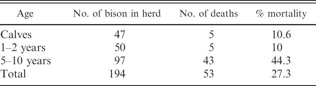
Details of the bison herd affected with Mycoplasma bovis during the outbreak.
In 1 bison (KSVDL 06-57154), Mycoplasma culture was performed on synovial fluid, and aerobic bacterial culture was performed on liver, lung, and spleen. In the other bison (KSVDL 07-03804), aerobic and Mycoplasma cultures were performed using lung and liver. No significant bacteria were isolated by routine aerobic cultures. Mycoplasma sp. was isolated in Mycoplasma-specific cultures. Mycoplasma isolated from the lung and liver of KSVDL 07-03804 was used for further tests (hereafter, Kansas [KS] isolate). In addition to these specimens, an isolate of M. bovis from another outbreak of mycoplasmosis in bison (hereafter, North Dakota [ND] isolate) was compared with the KS isolate. 2
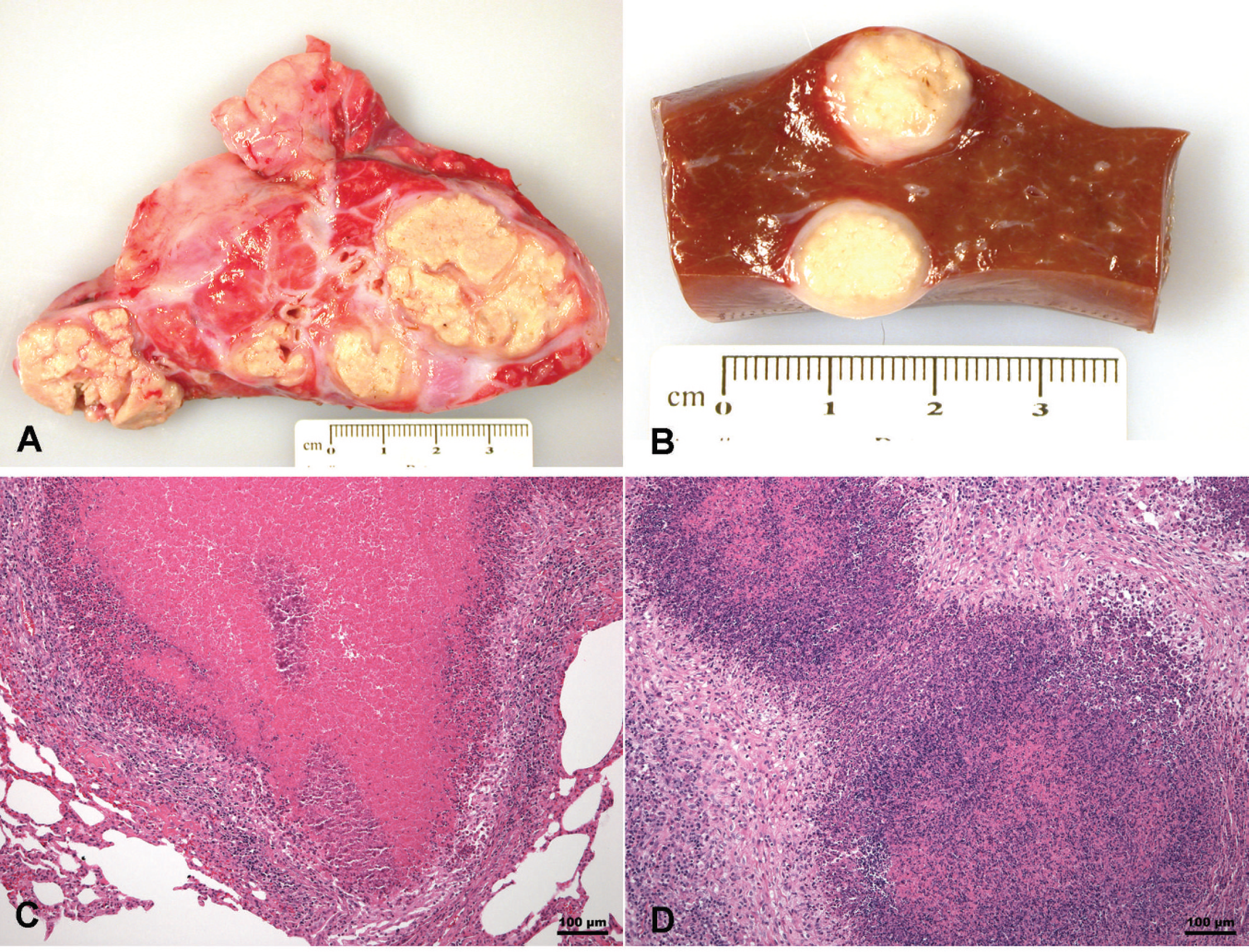
Gross and microscopic changes in Mycoplasma bovis–infected bison (Bison bison). Lung (
To identify the species of Mycoplasma, the isolates (ND and KS) were compared with known M. bovis (KSVDL 06-11241) and laboratory control (KSU-TY-1) using M. bovis–specific polymerase chain reaction (PCR) and 2 different restriction fragment length polymorphism (RFLP) techniques. The ND and KS isolates streaked on Mycoplasma agar plates a were inoculated into Mycoplasma broth. One milliliter of culture fluid was centrifuged at 14,000 × g, and the cell pellet was used to extract nucleic acid using a commercial kit. b The nucleic acid was used for further characterization of the bacteria.
The PCR was performed using M. bovis–specific (MBV-F 5′-TGATAGCAATATCATAGCGGC-3′; MBV-R 5′-GTAGCATCATTTCCTATGCTAC-3′) primers at a 250 nM concentration in a 25-μl reaction mixture containing 12.5 μl of 2× SYBR Green supermix, c sterile water to a volume of 22.5, and 2.5 μl of sample nucleic acid. Amplification was performed by an initial denaturing/activation step of 95°C for 5 min, followed by 45 cycles of 95°C for 10 sec, 55°C for 15 sec, and 72°C for 30 sec with the SYBR Green fluorescence recorded at the 72°C extension step of each cycle. The bacteria were positive for M. bovis.
For RFLP, 2 different methods were used. First, a method described previously (Lauerman L: 1998, Nucleic acid amplification assays for diagnosis of animal diseases. American Association of Veterinary Laboratory Diagnosticians) was used. A Mycoplasma spp. forward primer (CGP1-F 5′-ACACCATGGGAGCTGGTAAT-3′) and reverse primer (CGP1-R 5′-CTTCATCGACTTTCAGACCCAAGGCAT-3′) were used at a 250 nM concentration in a 100-μl reaction mixture containing 12.5 μl of 2× iQ Supermix, d sterile water to a volume of 90, and 10 μl of sample nucleic acid. Ten microliters of each reaction was utilized to demonstrate the presence of an approximately 490-bp fragment on electrophoresis in a 2% agarose gel (data not shown). Fifteen microliters of each (KS, ND, and bovine isolates and laboratory control) amplicon from Mycoplasma spp. The PCR fragments were digested separately in a 30-μl final reaction volume using restriction enzymes (Mse I, e TSP509 I, f Dra I, g and Rsa Ih) following manufacturer-recommended reaction conditions and restriction buffers. After digestion, 6× blue orange loading dye was added to the restriction reaction tube, mixed well, and loaded onto a 5% Tris–borate–ethylenediamine tetra-acetic acid (EDTA) pre-made polyacrylamide gel i for electrophoresis.
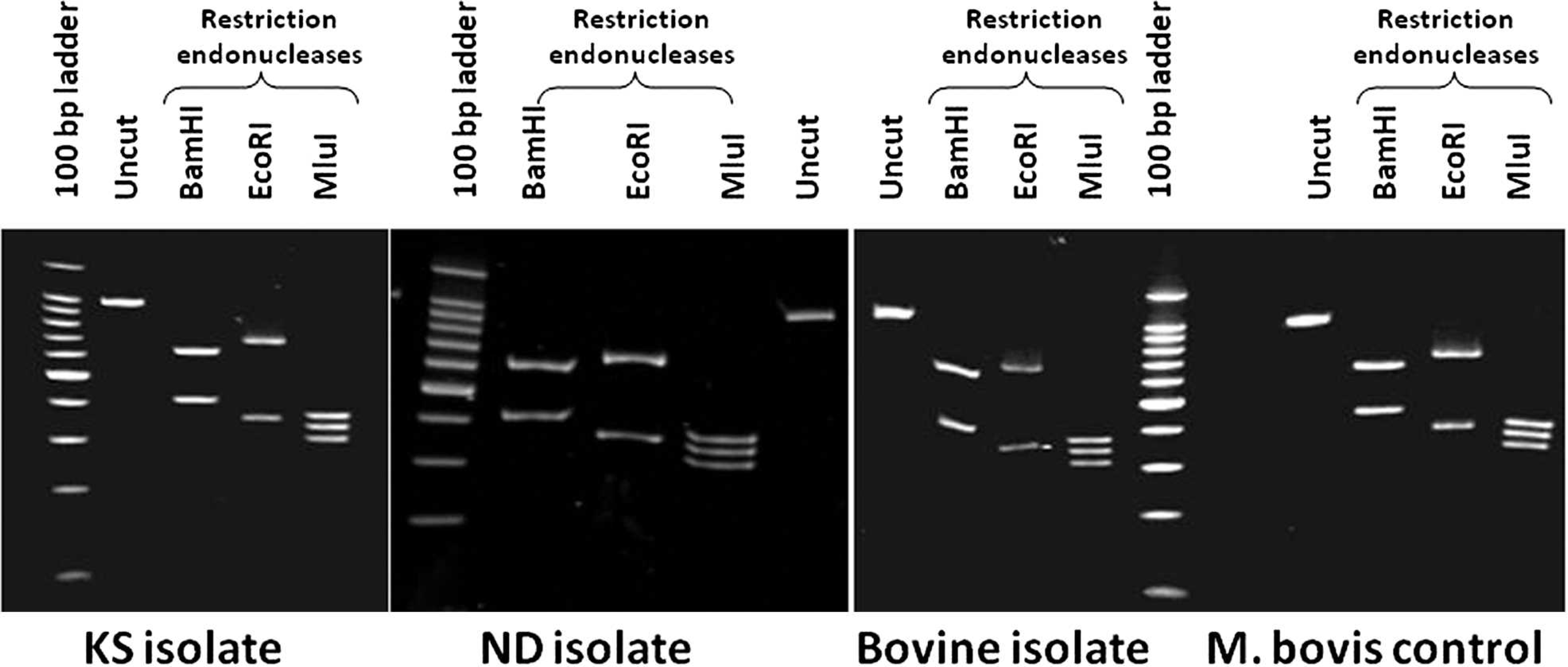
Restriction fragment length polymorphism analysis of Kansas (KS) and North Dakota (ND) isolates and their comparison with a bovine Mycoplasma bovis isolate (KSVDL 06-11241) and a M. bovis laboratory control (KSU-TY-1).
A second 16S RFLP was performed according to the method describe previously 1 for Mycoplasma spp. Fifteen microliters of each amplicon generated by the 16S PCR were digested in separate reactions with restriction enzymes (EcoR I, j Mlu I, k and BamH Il) following manufacturer-recommended reaction conditions and restriction buffers. After digestion, 6× blue orange loading dye was added to the restriction reaction tube, mixed well, and loaded onto a 5% Tris–borate–EDTA precast polyacrylamide gel i for electrophoresis.
The RFLP patterns were the same between M. bovis isolates from bison (KS and ND isolates) and bovine (KSVDL 06-11241) and laboratory control (KSU-TY-1). The RFLP performed according to the method previously cited (Lauerman L: 1998, Nucleic acid amplification assays for diagnosis of animal diseases) yielded a pattern (data not shown) consistent with the expected pattern for M. bovis. The fragment sizes for the 16S RFLP patterns were all consistent with the sizes suggested previously 1 for M. bovis, with EcoR I digestion resulting in bands of 340 and 661 bp; Mlu I digestion resulting in bands of 297, 332, and 372 bp; and BamH I digestion resulting in bands of 414 and 587 bp (Fig. 2).
Further, the sequences of bison isolates were compared with the bovine isolate and the laboratory control. The 16S ribosomal DNA gene of each M. bovis isolate from either a bovine or a bison source was amplified using previously described primers. 1 Presence of an approximate 1,000-bp fragment was verified by 1.5% agarose electrophoresis and then cloned into the pCR8/GW/TOPO TA plasmid vector m according to the manufacturer's instructions. The 16S ribosomal DNA gene plasmid clones were sequenced at the University of Arkansas Medical Sciences DNA Sequencing Laboratory (Little Rock, Arkansas). Sequences were analyzed and aligned using the MEGA 4.0 software package. 13
The 16S sequences of KS (GenBank accession no. GU993858) and ND (GenBank accession no. GU993861) M. bovis isolates were more than 99% homologous to the sequence of M. bovis bovine isolate (KSVDL 06-11241, GenBank accession no. GU993859) and M. bovis control (KSU-TY-1, GenBank accession no. GU993860; Fig. 3). All of these isolates are 99% homologous to M. bovis sequence in GenBank (GenBank accession no. U02968).
The present investigation implicates M. bovis as an agent causing high morbidity and mortality in bison. Mycoplasma bovis infection in cattle results in pneumonia, arthritis, otitis media, keratoconjunctivitis, and mastitis. Calves develop pneumonia, arthritis, and otitis media. Reports in adult dairy cattle describe mastitis, with feeder and stocker cattle experiencing pneumonia and arthritis. 6 In comparison, the disease occurrence in bison appears to vary slightly. In the KS outbreak, the majority of affected bison were in the 5–10-year age group, whereas calves and 1–2-year-old bison were affected to a lesser degree. Mycoplasma bovis induced a disseminated disease resulting in abscesses or infection of multiple organs, including lung, liver, mesentery, intestine, uterus, mammary gland, and joints. The ND outbreak 2 reported primary involvement of lungs and joints. It is difficult to speculate on the variation in lesion patterns between the KS and ND outbreaks. In calves experimentally infected with M. bovis, bacteria was isolated from liver, spleen, and kidneys at 21 days postinfection. 12 Mycoplasma bovis was immunohistochemically detected in liver and kidney in an additional report of naturally infected calves. 5 These findings highlight the ability of M. bovis to disseminate hematogenously to various organs. In the KS outbreak, the disease process was very likely chronic, leading to septicemia and seeding of organisms into different tissues.
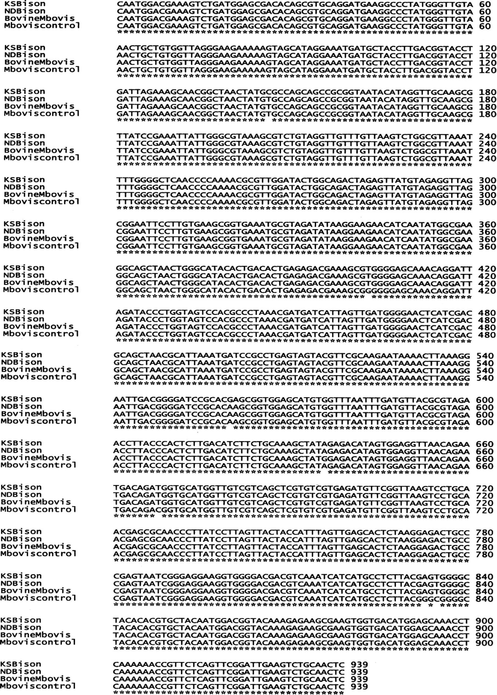
Sequence data for Kansas isolate (KSBison), North Dakota isolate (NDBison), Mycoplasma bovis isolate from cattle (BovineMbovis), and laboratory control (Mboviscontrol). The KS and ND isolates are more than 99% homologous to the M. bovis isolate from cattle (KSVDL 06-11241) and the laboratory control (KSU-TY-1).
To identify the pathogen, bacterial isolates from KS and ND were sequenced and compared using BLAST (http://www.ncbi.nlm.nih.gov/blast/Blast.cgi). Both the isolates were 99% homologous to M. bovis sequences in GenBank and were also 99% homologous to sequences of Mycoplasma agalactiae. However, the RFLP pattern was consistent with a previously described M. bovis pattern (Lauerman L: 1998, Nucleic acid amplification assays for diagnosis of animal diseases). Also, bison samples were positive by PCR for M. bovis when M. bovis–specific primers were used. Based on the findings of the current study, routine diagnostic tests, such as culture, PCR, and RFLP, used for diagnosis of M. bovis in bovine samples will be useful in diagnosing M. bovis infection in bison.
The present investigation highlights the importance of M. bovis infection in bison. The source of pathogen remains unknown; however, it is reasonable to speculate that the cattle adjacent to the affected bison herd are the probable candidates. In view of recent reports describing the severity of mycoplasmosis in bison, it is important for producers to adopt preventive measures avoiding M. bovis exposure and infection.
Acknowledgements
The authors thank the Director of Kansas State Veterinary Diagnostic Laboratory, Dr. Gary Anderson, for the financial support for this project. The authors also thank Dr. Byron Bachman, Lindsborg Veterinary Hospital, Lindsborg, Kansas, for performing the necropsies, submitting the appropriate samples, and providing a detailed history; and Mr. Cliff Peterson, Maxwell Refuge manager, for providing details on the bison herd.
Footnotes
a.
Catalog no. G102, Hardy Diagnostics, Santa Maria, CA.
b.
Qiagen Viral RNA Kit (catalog no. 52906), Qiagen Corp., Valencia, CA.
c.
Catalog no. 170-8880, Bio-Rad Laboratories, Hercules, CA.
d.
Catalog no. 170-8860, Bio-Rad Laboratories, Hercules, CA.
e.
Catalog no. R0525S, New England Biolabs Inc., Ipswich, MA.
f.
Catalog no. R0576S, New England Biolabs Inc., Ipswich, MA.
g.
Catalog no. R0129S, New England Biolabs Inc., Ipswich, MA.
h.
Catalog no. R0167S, New England Biolabs Inc., Ipswich, MA.
i.
Catalog no. 1161-1127, Bio-Rad Laboratories, Hercules, CA.
j.
Catalog no. R6011, Promega Corp., Madison, WI.
k.
Catalog no. R6381, Promega Corp., Madison, WI.
i.
Catalog no. R6021, Promega Corp., Madison, WI.
m.
Catalog no. K2500-20, Invitrogen Corp., Carlsbad, CA.