
Abstract
Atherosclerosis is a common disease in pet birds, particularly in psittacines, and is frequently found when performing postmortem examinations on adult and old dogs, in which it is mainly associated with endocrine diseases, such as hypothyroidism and diabetes mellitus. However, atherosclerosis is poorly documented in reptiles and consequently poorly understood. In the current case report, atherosclerosis and pericardial effusion were diagnosed in a 2-year-old male central bearded dragon (Pogona vitticeps) based on ultrasound visualization, necropsy, and histologic examination.
A 0.4 kg, 2-year-old, male, captive-born central bearded dragon (Pogona vitticeps) was presented with a 3-week history of anorexia, extreme lethargy, and postural abnormality. The animal was purchased in a pet store and lived in isolation in a terrarium. Upon questioning, the owner described the general husbandry as adequate and appropriate in terms of ultraviolet B exposure, photoperiod, humidity, substrate, and temperature gradients. However, the animal's diet was unbalanced, consisting essentially of house crickets (Acheta domesticus), yellow mealworms (Tenebrio molitor), kingworm beetles (Zophobas morio), and greater wax moth larvae (Galleria mellonella). Vegetables, necessary for the omnivorous requirements of this species, were rarely if ever given.
Physical examination revealed scoliosis, poor body condition, and lethargy. No additional abnormal findings were observed on physical examination. Auscultation revealed a normal heart rate (60 beats/min) and a regular cardiac rhythm. 25
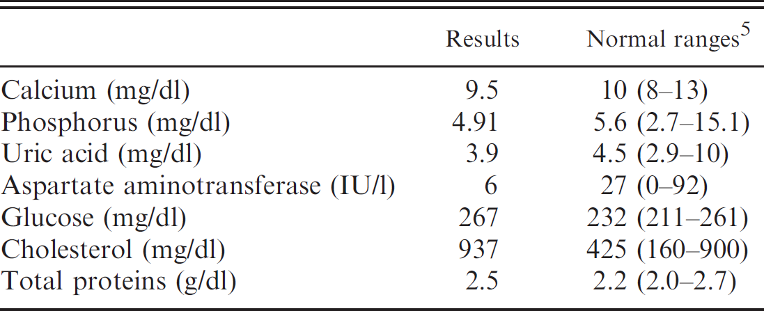
No biochemical abnormalities were noted except mild hyperglycemia and hypercholesterolemia (Table 1). 5 Cardiomegaly was observed on survey thoracic radiographs (Fig. 1). 21 No lytic or proliferative osseous lesions were observed in association with the scoliosis. A 2-dimensional echocardiographic examination was performed in ventrodorsal recumbency using a ventral approach (Hochleitner C, Hochleitner M: 2004, Ultrasound in reptiles. Proceedings of the Association of Reptilian Amphibian Veterinarians, pp. 41–44. Naples, FL). a , 3,8,15,17,19,20 The apical, transventricular, and subarterial short-axis sections of the heart revealed pericardial effusion, normal myocardium, and a 3 mm × 4 mm mass located close to the aortic arches and the pulmonary trunk, surrounded by anechoic liquid (Fig. 2). A long-axis transarterial section revealed normal atrioventricular junctions. 3,19 Echo-guided fine-needle aspiration of the pericardial space was performed. 18 Cytology revealed large numbers of erythrocytes without associated inflammatory cells, compatible either with blood contamination of the effusion during pericardiocentesis or with hemopericardium. The owner elected euthanasia.
Upon postmortem examination, a firm plaque-like mass of yellow tissue was found close to the base of the major arteries (Fig. 3). Pericardial effusion was confirmed. The liver was enlarged, friable, and pale yellow. Tissues submitted for histopathology included the base of the large arteries with the mass, as well as thoracic vertebrae and liver. Samples were routinely sectioned at 5-μm thickness and stained with hematoxylin and eosin. All vessels examined were similarly affected, with variable severity. Alterations included segmental to circumferential plaquelike thickening of the intima. Plaques were composed of mature, well-vascularized fibrous connective tissue, frequently overlain by a thick layer of fibrin and few erythrocytes. Collagen fibers within the plaque were often separated by aggregates of lipid-laden macrophages, erythrocytes, and/or hemosiderin-laden macrophages. These histologic changes were consistent with atherosclerotic deposits, early aneurysm formation, and chronic and acute hemorrhage. The lumen of the affected vessels had partial to almost complete obstruction by combined atheromatous plaques and thrombi (Fig. 4). Histologic examination of the liver was consistent with severe hepatic lipidosis. Microscopic examination of the thoracic vertebra did not show any histopathologic lesions associated with the abnormal posture in this animal.
Atherosclerosis is a disease of large- and medium-sized muscular arteries, characterized by inflammation of smooth muscle cells and formation of atherosclerotic plaques composed of necrotic cores; calcium deposits; and an accumulation of modified lipids, endothelial cells, leukocytes, and foam cells. 1,4,6,7,11,22,24 Many components of the vascular, metabolic, and immune systems are involved in this process. 2,6,9–12,16,22–24 Buildup of material and infiltrates leads to vascular remodeling, acute and chronic luminal obstruction, abnormalities of blood flow, and diminished oxygen supply to target organs. 11,22 In human beings, atherosclerosis is the most common pathologic process leading to cardiovascular disease. 9,22
Blood biochemical evaluation.
Various cardiovascular lesions have been described in reptiles. Most commonly, these lesions include endocarditis, myocarditis, and pericarditis, but infarcts, cardiomyopathy, parasitic infestation, and even tumors have been sporadically reported. 3,7,18,19 Reports of atherosclerosis in reptiles are rare. 4,7,24 Aortic atherosclerosis is found less frequently in reptiles when compared with captive wild birds and to a lesser extent in domestic and wild mammals. 13,22 Little is known about the risk factors predisposing animals to this disease, but in birds, development of this lesion may be the result of a high concentration of plasma cholesterol, an unbalanced diet, stress, hepatic lipidosis, and/or lack of exercise. 12 Given the dietary imbalances and elevation of plasma cholesterol in the individual described in the present case report, it could be speculated that similar risk factors exist in reptiles. A genetic predisposition to atherosclerosis has also been demonstrated in pigeons. 23 Strains of rabbits, in which diet has been shown to play a role in the development of atherosclerosis, have been used as an animal model of disease due to their propensity to develop vascular lesions. 2 Low-density lipoprotein remains the most important risk factor for development of atherosclerosis in humans and has been also associated with atherosclerosis in animals, including reptiles. 4,9,14,22 However, immune and inflammatory mechanisms of atherosclerosis have gained tremendous interest in the past 20 years. 9,10,14,16 New data suggest an important role for chemokines and chemokine receptors in atherosclerosis and highlight a network of cytokines that modulate the immune response and inflammation of the arterial wall. 9 Because it has been shown in humans that all phases of atherosclerosis are regulated by inflammatory mechanisms, the possible impact of chronic inflammation in the development of atherosclerosis in animals and the importance of preventive diagnosis should be considered. 9
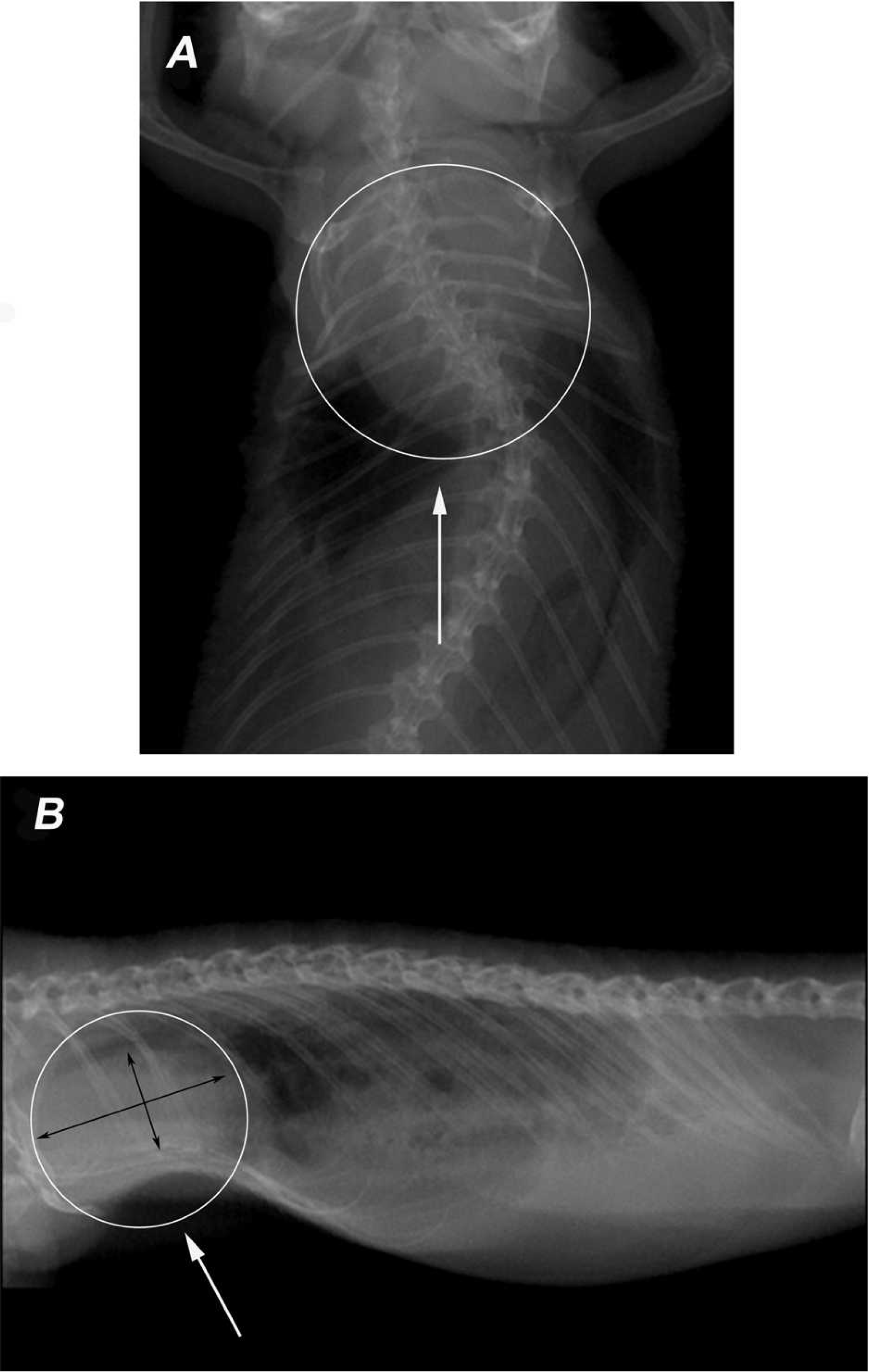
Survey thoracic radiographs. Cardiomegaly is present (arrows):
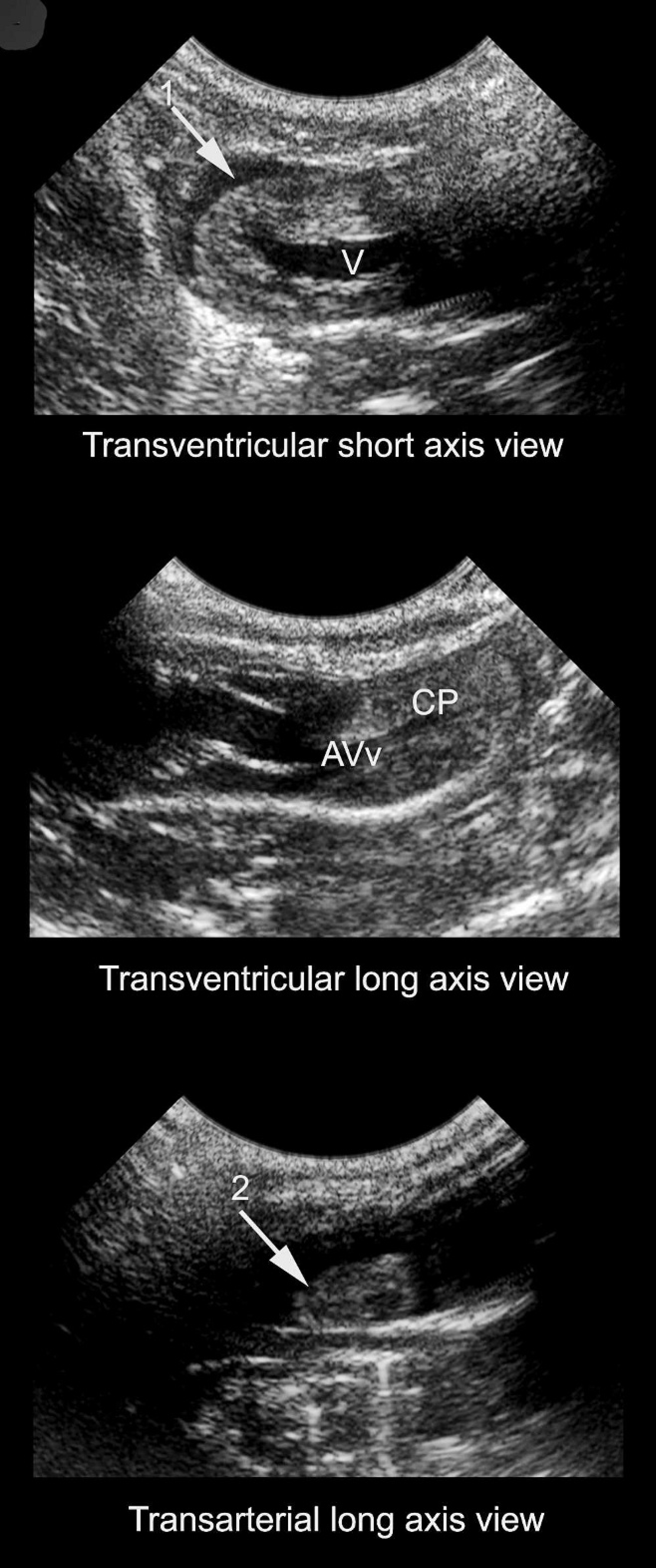
Two-dimensional echocardiogram. This transventricular short-axis view shows a transversal section of the ventricle (V) surrounded by pericardial fluid (arrow 1). The ventral cavum pulmonale (CP) and the atrioventricular valve (AVv) are partially visualized. The transarterial long-axis section shows a mass close to the 2 aortic arches (arrow 2).
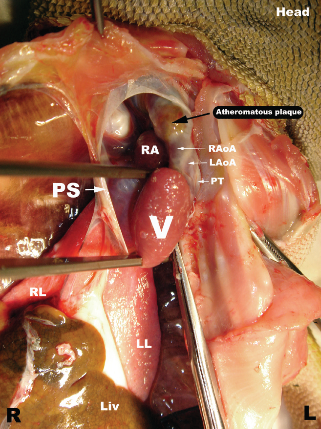
Necropsy. The opening through the pericardial sac shows the previously visualized yellow plaque-like mass located near the heart base and attached to the arterial efferent trunks. R = right; L = left; PS = pericardial sac; RA = right atrium; RAoA = right aortic arch; LAoA = left aortic arch; PT = pulmonary trunk; V = ventricle; RL = right lung; LL = left lung; Liv = liver.
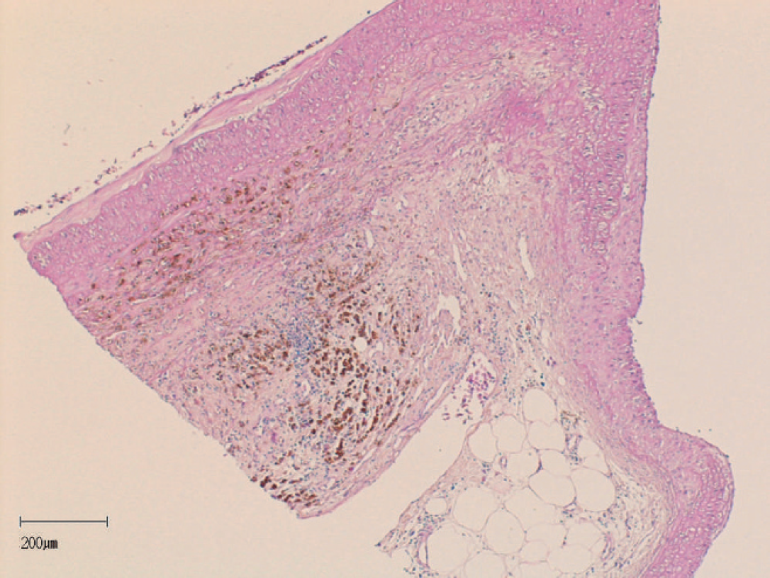
Histology. Microscopic section of the aortic arches shows thickening of the vascular wall by atherosclerotic deposits and chronic hemorrhage. Hematoxylin and eosin. Bar = 200 μm.
As in human and small animal medicine, ultrasound examination is probably the technique of choice for antemortem diagnosis of atherosclerosis (Hochleitner C, Hochleitner M: 2004, Ultrasound in reptiles). 3,8,17,19,20 The current case is another illustration of potential consequences of dietary imbalances in reptiles. Nutritional diseases, such as metabolic bone disease, vitamin A deficiency, and hepatic lipidosis, are rare in wild reptiles but fairly common in captive animals. The increased incidence of atherosclerosis in captive reptiles is often related to a lack of knowledge of the natural diet of these species by their keepers.
Footnotes
a.
MyLab®, Biosound Esaote, Genova, Italy.