
Abstract
The purpose of the current study was to compare the molecular detection rate of Lawsonia intracellularis between feces and rectal swabs collected from 42 foals with suspected equine proliferative enteropathy (EPE). Fecal samples and rectal swabs were processed for DNA purification by using an automated extraction system. The purified DNA was then analyzed by real-time polymerase chain reaction (PCR) for the presence of the aspartate ammonia lyase (aspA) gene of L. intracellularis. Absolute quantitation was calculated by using a standard curve for L. intracellularis and expressed as copy numbers of the aspA gene of L. intracellularis per microliter of purified DNA. The combined PCR detection rate for L. intracellularis was 90%, with 38 foals testing PCR positive in feces (33 samples), rectal swabs (32), or both (27). Six foals tested PCR positive only in feces, whereas 5 tested positive only in rectal swabs. Feces yielded a significantly higher aspA gene copy number of L. intracellularis than rectal swabs. Feces and rectal swabs tested PCR negative from 4 foals. In conclusion, the results showed that feces yielded similar numbers of PCR-positive results, with a higher L. intracellularis aspA gene load than rectal swabs. By analyzing dual samples, the PCR detection rate for L. intracellularis increased from 76% and 79% for rectal swabs and feces, respectively, to 90%. Rectal swabs should be considered as an alternative sample type for EPE-suspected patients with decreased or no fecal output.
Equine proliferative enteropathy (EPE) is a disease of foals caused by the obligate intracellular organism Lawsonia intracellularis. This emerging disease affects weanling foals and causes fever, lethargy, peripheral edema, diarrhea, colic, and weight loss. 6 The antemortem diagnosis of EPE may be challenging and relies on the presence of hypoproteinemia, exclusion of common enteric diseases, thickening of segments of the small intestinal wall observed on abdominal ultrasonography, positive serology, and molecular detection of L. intracellularis in feces. 7
Current methods used for serologic diagnosis of proliferative enteropathy (PE) use L. intracellularis cultured in enterocytes or a preparation of L. intracellularis on slides as the antigen. Staining of bacteria is either by a fluorescent (indirect fluorescent antibody test) or peroxidase-labeled (immunoperoxidase monolayer assay [IPMA]) secondary antibody. 8 One must keep in mind that a positive serologic result may represent exposure to infection rather than disease. Although no commercially available serologic assay has yet been systematically evaluated for the equine species, serologic assays have proven useful for routine diagnosis of PE in horses when combined with clinical signs and molecular detection of L. intracellularis in feces. 7,14
Several polymerase chain reaction (PCR) assays have been developed for the detection of L. intracellularis in feces. The sensitivity and specificity of the PCR technique in fecal samples has been evaluated for pigs and showed variable sensitivity and consistently high specificity. 3,5,9,13 Sensitivity is affected by sample quality and the presence of inhibitory substances in feces. PCR appears to reliably demonstrate L. intracellularis in the feces of clinically affected horses early in the course of the disease. 2 As an alternative sample type to feces, rectal swabs are routinely used for the PCR detection of L. intracellularis in pigs because of the ease of collection. The purpose of the current study was to compare the molecular detection rate of L. intracellularis between feces and rectal swabs collected from foals with suspected EPE.
For the purpose of the current study, dual samples, including fresh feces and rayon-tipped rectal swabs a were collected from 42 suspected EPE cases at 2 veterinary clinical institutions (William R. Pritchard Veterinary Medical Teaching Hospital, University of California, Davis, California, and Hagyard Equine Medical Institute, Lexington, Kentucky) from October 2008 to December 2009. Case selection was based on the presence of at least 3 EPE-associated clinical signs (anorexia, depression, rectal temperature >38.6°C, peripheral edema, diarrhea, colic, weight loss), hypoalbuminemia (≤2 g/dl), and positive titer (≥60) against L. intracellularis by IPMA.
After collection, the samples were kept on ice and processed within 24 hr. Two milliliters of phosphate buffered saline (PBS) were added to 2 g of fresh feces as well as to tubes that contained the rectal swabs. The samples were vortexed for 10 sec and centrifuged at 16,000 × g to remove fecal debris. Two hundred microliters of PBS and feces, and PBS and rectal swab solution, were processed for DNA purification by using an automated nucleic acid extraction system b according to the manufacturer's recommendations. All purified DNA samples from feces and rectal swabs were assayed in triplicate for the presence of the aspartate ammonia lyase (aspA) gene of L. intracellularis by real-time PCR, as previously reported. 16 Absolute quantitation was calculated by using a standard curve for L. intracellularis and was expressed as copy numbers of the aspA gene of L. intracellularis per microliter of purified DNA (1 aspA gene copy per L. intracellularis microorganism; National Center for Biotechnology Information gene ID 4059386). Final quantitation for each sample was expressed as the average of the triplicate results. The standard curve was determined by using 10-fold dilutions of L. intracellularis derived from cell culture in McCoy cells (mouse fibroblast cells) added to L. intracellularis–free equine feces. Bacterial numbers were assessed by direct microscopical count after indirect immunoperoxidase staining by using L. intracellularis–specific antibody. Furthermore, a real-time PCR assay that targets a universal sequence of the bacterial 16S rRNA gene was used as a quality control (i.e., efficiency of DNA purification and amplification) and as an indicator of fecal inhibition. 12,18
Statistical data analysis was performed by use of a Mann–Whitney U-test to make pairwise comparisons of threshold cycle (Ct) values for the universal bacterial 16S rRNA gene and of copy numbers of the aspA gene of L. intracellularis between fecal and rectal swab samples. A Spearman rank correlation test was used to assess the relationship in the aspA gene load between feces and rectal swabs. A value of P < 0.05 was considered significant.
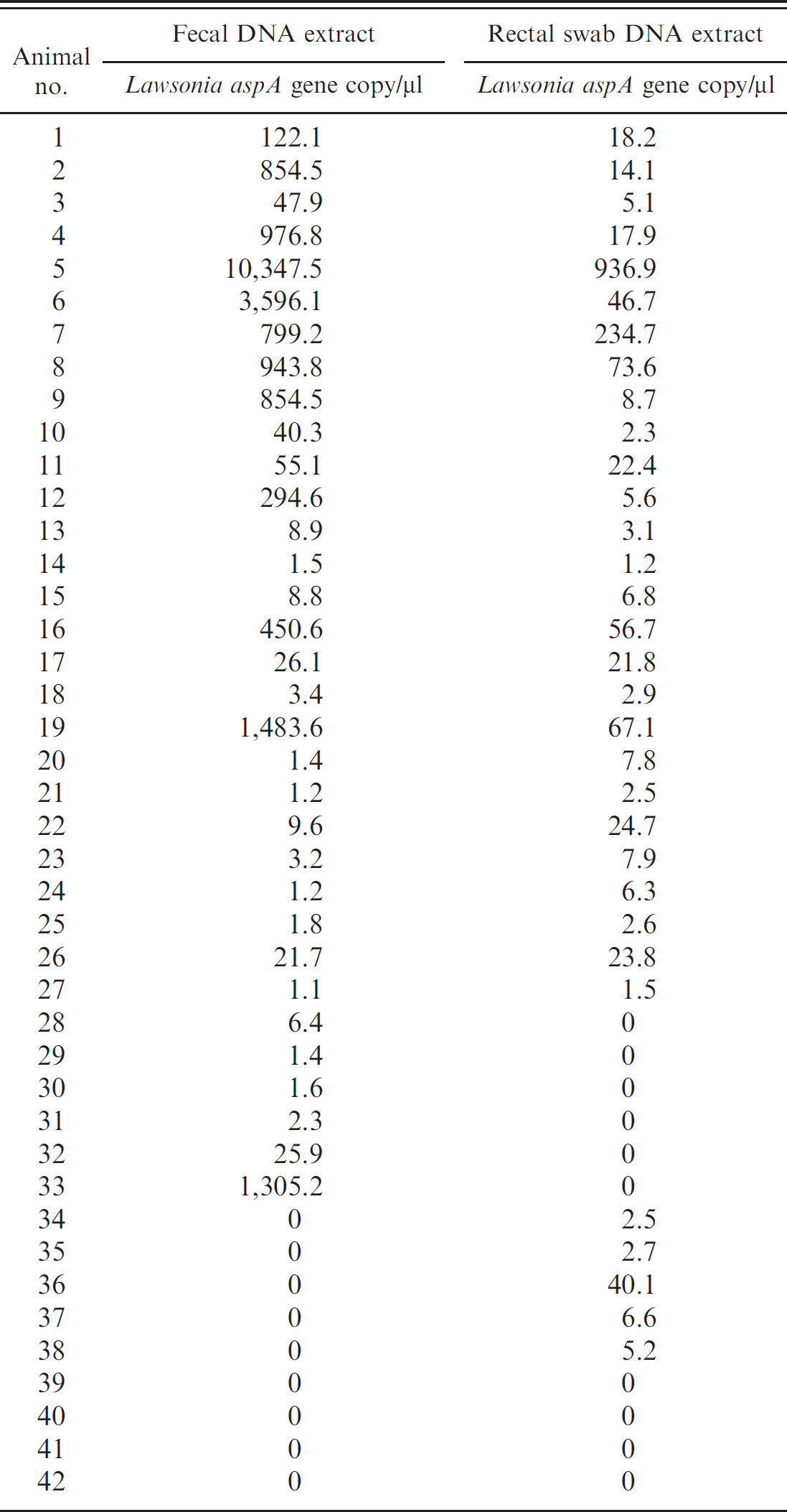
DNA was successfully extracted from all feces and rectal swabs based on positive PCR signals for the universal bacterial 16S rRNA gene. The Ct values for the universal bacterial 16S rRNA gene were similar between fecal samples (mean [SD]: 33.48 ± 0.89) and rectal swabs (31.34 ± 1.24; P < 0.05). The overall PCR accuracy for the detection of L. intracellularis was 90%, with 38 foals testing PCR positive in feces (33 samples), rectal swabs (32), or both (27). Feces yielded a significantly higher aspA gene copy number of L. intracellularis (mean [SD]: 675 ± 1,879 L. intracellularis aspA gene copies/μl DNA) than rectal swabs (52 ± 167 L. intracellularis aspA gene copies/μl DNA; P < 0.0001; Table 1). The aspA gene copy number of L. intracellularis in feces was positively correlated with the aspA gene copy number of L. intracellularis in rectal swabs (r = 0.65, P = 0.001). Six foals tested PCR positive only in feces (mean [SD]: 223 ± 530 L. intracellularis aspA gene copies/μl DNA), whereas 5 tested positive only in rectal swabs (11 ± 16 L. intracellularis aspA gene copies/μl DNA). The aspA gene copy numbers of L. intracellularis for these single positive samples were significantly lower (P < 0.01) when compared with the mean load of each corresponding sample type. Feces and rectal swabs tested PCR negative from 4 suspected EPE foals.
Lawsonia intracellularis aspartate ammonia lyase (aspA) gene copy number in fecal samples and rectal swabs collected from 42 foals with suspected equine proliferative enteropathy.
One of the limitations of the present study was the case definition, which used a positive IPMA titer as a prerequisite for case inclusion. A recent large-scale study showed that only 80% of foals with presumptive diagnosis of EPE also had a positive serum titer against L. intracellularis by IPMA. 2 One must keep in mind that serologic and molecular tests will never achieve 100% accuracy in diagnosing EPE antemortem. It is rather the integration of clinical, clinicopathologic, sonographic, serologic, and molecular results that will support or rule out an EPE diagnosis. PCR detection of L. intracellularis in feces and/or rectal swabs was achieved in 90% of the study foals. When the results were adjusted to the different sample types, 76% of rectal swabs and 79% of feces tested PCR positive for L. intracellularis. The later results are in agreement with previous work that showed that 38 of 51 foals (74%) with suspected EPE tested PCR positive for L. intracellularis in feces. 2
Fecal inhibition has been a major drawback associated with low sensitivity of PCR detection. Inhibitory substances in feces have been shown to interfere with nucleic acid purification and amplification. 11 Inhibition can be partially overcome by using solid-phase extraction methods and more-sensitive PCR platforms, such as real-time PCR. 10 Furthermore, only PCR assays using either an internal control or a calibrator should be used in an attempt to determine the quality of purified nucleic acid from fecal samples. 13,16 The latest approach was used in the present study by targeting the universal bacterial 16S rRNA gene. Study results displayed no statistical differences in Ct values for the universal bacterial 16S rRNA gene between feces and fecal swabs, which suggests that the amount of this housekeeping gene was similar between the 2 sample types. However, partial inhibition was not assessed in the present study because no internal calibrator was used in each sample.
Although the detection rate of L. intracellularis was similar between feces and rectal swabs, dual positive samples were only recorded in 27 foals (64%). In experimentally infected pigs, fecal shedding is generally first detected at 1 week and lasts, intermittently, for 12 weeks after exposure, with maximal shedding of up to 7 × 10 8 bacteria per gram of feces. 1,17 The onset, duration, and amount of L. intracellularis shed in the feces of naturally or experimentally infected foals have yet to be determined. The L. intracellularis gene load determined in the feces of the 42 study foals ranged from 1 to 1 × 10 4 aspA gene copies per PCR reaction, which equals to a range of 1 × 10 2 to 1 × 10 6 aspA gene copies per gram of feces. The L. intracellularis aspA gene load in feces was significantly higher and positively correlated with the L. intracellularis aspA gene load in rectal swabs. The higher load in feces was likely related to a higher antigen load or a larger amount of fecal material processed. A recent study performed in pigs determined that rectal swabs contained less fecal material than the average amount of fresh feces used for nucleic acid extraction and that inhibition was a lesser problem in fecal swabs (Jacobson M, Wennerbo S, Aspan A, et al.: 2006, The importance of faecal sampling techniques in the PCR diagnosis of Lawsonia intracellularis. Proceedings of the 19th International Pig Veterinary Society Congress, Copenhagen, Denmark). Unfortunately, the amount of feces contained on each rectal swab was not measured in the present study. One must keep in mind that swab type and size as well as collection method may be additional factors that influence the amount and quality of purified nucleic acid. In pigs, the number of L. intracellularis in feces is closely correlated to the severity of PE, with distinct histopathologic lesions being more frequent in PCR-positive animals. 4 Future work is needed in the equine field to determine if the same correlation applies to foals affected by EPE.
Differences in the L. intracellularis aspA gene load associated with sample type may potentially explain why 6 foals tested PCR positive for L. intracellularis in feces but PCR negative in rectal swabs. This is supported by the fact that the mentioned foals had significantly lower L. intracellularis aspA gene loads compared with the fecal load of foals testing positive in both sample types. Low antigen load, partial fecal inhibition, or unequal distribution of L. intracellularis in feces may have been associated with the PCR results of 5 foals that only tested PCR positive in rectal swabs. It is unknown why 4 foals with suspected EPE tested PCR negative in feces and rectal swabs. Potential factors that influence PCR results for L. intracellularis include the stage of disease (i.e., foals in late disease stages may shed L. intracellularis in numbers below the PCR detection limit), antimicrobial treatment before fecal analysis, and quality of fecal sample. 2,14,15
In conclusion, the results showed that feces yielded similar PCR-positive results with a higher L. intracellularis aspA gene load than rectal swabs. By analyzing dual samples, the PCR detection rate for L. intracellularis increased from 76% and 79% for rectal swabs and feces, respectively, to 90%. Rectal swabs should be considered an alternative sample type for EPE-suspected patients with decreased or no fecal output.
Footnotes
a.
Sterile Rayon Tipped Applicators, Puritan Products Company LLC, Guilford, ME.
b.
CAS-1820 X-tractor Gene, Corbett Life Science, Sydney, Australia.