
Abstract
Transmissible, spongiform encephalopathies including bovine spongiform encephalopathy (BSE) and scrapie are fatal neurodegenerative disorders associated with the presence of an infectious abnormal isoform of normal mammalian proteins called prions. Identification of the prion protein associated with scrapie (PrPSc) in the central nervous system is typically based upon immunoassays including immunohistochemistry (IHC) using formalin-fixed tissues or Western blot (WB) assays using fresh and/or frozen, non–formalin-fixed tissues. Each assay can discriminate between BSE, classical scrapie, and a previously reported strain of scrapie recently identified in the United States named Nor98 scrapie. Different tissue samples are required from the same animal to run these 2 different immunoassays. This may result in inconsistent test results for the same animal. Sampling problems such as collecting insufficient volumes of fresh tissue or less than optimal anatomic location of brainstem for IHC can affect the ability of the test procedures to offer definitive and discriminatory results. Recently, a WB method using formalin-fixed, paraffin-embedded (FFPE) tissue to identify PrPSc was developed that successfully identified PrPSc in sheep affected by classical scrapie. In the current study, the use of this technique to produce discriminatory results identifying classical BSE in bovine tissue and both classical and Nor98 scrapie in ovine tissue using paraffin-embedded brain samples is described. Protein-banding patterns from WB using FFPE tissue were similar to protein-banding patterns produced by WB assays utilizing fresh tissues from the same animals, and results correlated well with the IHC PrPSc-positive staining present in the cerebellum and obex regions of brain samples from these animals.
Keywords
Introduction
Transmissible spongiform encephalopathies (TSEs) are fatal neurodegenerative disorders associated with the accumulation of abnormal forms of a natural host protein known as prion proteins (PrPSc), and include scrapie in sheep and goats and bovine spongiform encephalopathy (BSE), which primarily affects cattle. When manifested clinically, these diseases may be differentiated based on signalment, epidemiology, and clinical history. 14,17,25,26 It is difficult, however, to identify subclinical cases in the live animal. Serologic tests used to detect PrPSc are not available because specific immune responses are not recognized in TSE-affected animals. The infectious agent associated with TSE diseases has no known nucleic acid component, so nucleic acid–based tests are not useful. The common hallmark features of TSEs are detectible most consistently through postmortem findings including vacuolar degeneration of the central nervous system (CNS) as detected by histopathology and the associated presence of PrPSc; however, PrPSc can be detected in some cases prior to the occurrence of CNS lesions. In cases of subclinical classical scrapie, PrPSc may be detectable in lymphoid tissue alone. Diagnosis is typically defined by the detection of PrPSc in affected tissues through the use of immunoassays such as immunohistochemistry (IHC), Western blot (WB), and enzyme-linked immunosorbent assay (ELISA). 25,26 The ELISA serves most often as a screening test because results can be obtained in a short period of time and the sensitivity of the test is high; however, ELISA provides no discriminatory detail on the molecular nature or anatomic location of the prion agent. Immunohistochemistry and WB are typically used to confirm the presence of PrPSc as well as provide specific information on the disease agent. 25,26
Multiple strains of scrapie agent have been identified through variation in incubation periods and neuropathology in rodent models. 4 In sheep, these various scrapie strains have also been found to differ in the distribution of PrPSc in host tissues, lesion profiles, and in prion protein conformation, glycosylation, protease resistance, and protein aggregation. 3,6,22 A distinct strain of scrapie, identified in Norway and called Nor98 scrapie, 2 has since been reported in sheep from Belgium, Germany, France, Ireland, United Kingdom, Sweden, Portugal, Netherlands, Lithuania, Italy, Estonia, Denmark, Falkland Islands, Switzerland, Iceland, and recently in the United States. 5,8,9,11–13,16,20,21 Nor98 scrapie has also been detected in goats from Switzerland, France, Spain, and Italy. 7,12,23 Classical scrapie and the Nor98 strain of scrapie can be differentiated based on epidemiology, signalment, clinical history, and genotype as well as through diagnostic assays including IHC and WB. 17 Definitive diagnosis of both classical and Nor98 scrapie is typically made with immunoassays that identify PrPSc in formalin-fixed tissue using IHC or in fresh or frozen, non-fixed tissues using the WB assay. Immunohistochemical staining of CNS tissues reveals differences in the distribution of PrPSc when comparing classical scrapie and Nor98 scrapie. In cases of Nor98 scrapie, regions of the brain including the cerebellum and spinal nucleus of the trigeminal nerve contain PrPSc, while areas typically affected in classical scrapie, such as the motor nucleus of the vagus nerve and lymphoid tissues, are spared. Western blot assays produce distinct protein band patterns for Nor98 scrapie that include at least 3 protein bands with an unglycosylated band measuring less than 15 kD. This differs from the banding profile of classical scrapie, which includes 3 protein bands ranging from 18–30 kD. 1 Bovine spongiform encephalopathy presents with diagnostic findings that are distinct from both classical and Nor98 scrapie. The earliest PrPSc deposition is found in the solitary nucleus and tract, making this the target area needed for identification of BSE by IHC. Results of WB assays for classical BSE include 3 protein bands with the molecular weight of the unglycosylated band being smaller than classical scrapie and greater than Nor98 scrapie.
With IHC, FFPE tissues are used, and results of these procedures provide information on the distribution of lesions and PrPSc within the various nuclei of brain tissue. Nuclei in the obex region of the brainstem, known to contain the earliest accumulation of PrPSc, must be present in histosections for samples to provide diagnostic and discriminatory results. Brain histosections that do not include the correct nuclei of the obex may not allow morphologic distinction between prion diseases. 10,25,26 Successful protocols for IHC detection of PrPSc may vary between diagnostic facilities, and one variation calls for immersion of tissue blocks in formic acid, post–formalin fixation prior to dehydration, and embedding in paraffin, which can affect prion epitope availability and inactivate prions. With WB assays, homogenization of the sample preempts the ability to document localization of PrPSc within appropriate nuclei. It does, however, provide the diagnostic capability of determining the molecular profile of the detected PrPSc. Routine WB assays require non-fixed tissue samples, which are often not available in sufficient quantities for testing. Retrospective studies can be limited by the difficulties of storing fresh, non-fixed tissues. Recently, a WB method utilizing immersion-fixed CNS samples was reported, which provided for PrPSc detection in homogenized, formalin-fixed tissue, creating the opportunity for WB diagnosis of prion diseases where fresh or frozen tissues were unavailable. 19 The FFPE tissue WB methodology used in the present study has been previously shown to identify PrPSc in sheep affected with classical scrapie. 15 The current study describes the use of this published technique to discriminate classical and Nor98 scrapie in ovine tissue and classical BSE in bovine tissue using paraffin-embedded brain samples (tissues from sheep with BSE and goats with Nor98 were unavailable).
Materials and methods
Archived FFPE tissue samples of brainstem (obex area) and cerebellum previously diagnosed by IHC as being positive for classical scrapie or Nor98 scrapie (case 1: Nor98-1, case 2: Nor98-2) were used in the current study along with bovine obex previously diagnosed by IHC as being positive for BSE. Matched samples of fresh tissue including obex and cerebellum for classical scrapie and Nor98-1, cerebrum for Nor98-2, and obex for BSE from the same animals were included for comparison. Formalin-fixed, paraffin-embedded ovine obex tissue, as well as fresh ovine obex tissue, from an animal determined to be negative for PrPSc by IHC were also included in the study as negative controls. Two additional cases of classical scrapie previously reported as PrPSc positive by IHC were included for a comparison of formalin-fixed tissue treated with formic acid prior to embedding in paraffin and non–formic acid-treated, paraffin-embedded samples from the same animal.
The archived samples that remained immersed in 10% neutral buffered formalin were embedded in paraffin wax and sectioned at 5-μm thickness. Formalin-fixed samples from the 2 additional positive classical scrapie cases were also placed in cassettes, incubated in 95% formic acid for 1 hr, and rinsed in tap water overnight prior to being embedded in paraffin wax and sectioned at 5-μm thickness. A total of 4 sections of each block were collected in individual 1.5-ml microcentrifuge tubes and processed for WB. A sequential fifth section from each block was processed by an automated IHC method to confirm the presence of PrPSc. 16 The WB assay used in the present study utilizes a tissue extraction technique, originally developed for the purpose of DNA isolation from FFPE tissue, later applied to PrPSc detection in FFPE tissue. 15,18 This WB method with minor modifications was utilized for detection of PrPSc. 15 Briefly, 150 μl of 0.05 M Tris (pH 7.5), 1 mM ethylenediamine tetra-acetic acid, and 0.5% Tween 20 was added to each 1.5-ml microcentrifuge tube. The tube was placed in a 100°C water bath for 10 min followed by snapfreezing (ethanol on dry ice). The 10-min boil and snapfreeze cycle was repeated, followed by one more 10-min boil, and then the tube was immediately centrifuged 16,000 × g for 10 min to separate the paraffin while pelleting the tissue. An additional 10-min boil followed by a repeat of the 10-min centrifugation was performed if separation of the paraffin was not complete. The tissue pellet was drawn from beneath the paraffin layer by using a 16-gauge needle affixed to a syringe and transferred to a sterile 1.5-ml centrifuge tube with 150 μl of the Tris buffer added. Mechanical sheering with the needle tip was performed to reduce the tissue pellet to small particles. Further disruption of the tissue pellet was accomplished using a sonicator a at 60-sec intervals repeated 10 times in an ice bath with brief vortex mixing between sonications. Proteinase K b was added to each 1.5-ml tube to a final concentration of 100 mg/ml and incubated at 37°C for 1 hr. Pefabloc c was added to a final concentration of 0.1 mg/ml in order to stop digestion. An equal volume of sample buffer was added, and the samples were incubated at 99°C for 30 min. For WB, 15 μl of sample was loaded on a 12% commercially prepared sodium dodecyl sulfate–polyacryl-amide electrophoresis gel d and run for 50 min. The samples were then blotted to a polyvinylidene difluoride membrane d and blocked with 3% bovine serum albumin. Detection of PrPSc by WB was accomplished by using mouse anti-PrP monoclonal antibody P4 e at 1:10,000 dilution (0.1 mg/ml), L42 e 1:2,500 dilution (1.0 mg/ml), or F99/97.6.1 f at manufacturer's dilution as the primary antibody. Incubation in primary antibody occurred at 4°C overnight. A biotinylated sheep anti-mouse secondary antibody g at 0.05 mg/ml and a streptavidin–horseradish peroxidase conjugate g were used in incubations conducted at room temperature for 1 hr. A commercial detection system g allowed visualization using a digital imaging system. d
Results
Classical scrapie, Nor98 scrapie, and BSE were accurately identified in FFPE tissue sections by WB assays utilizing P4 and L42 antibodies. Negative control FFPE tissue sections produced no banding patterns in the same assays. Banding patterns for each of the positive FFPE tissue samples were compared with the banding produced using fresh tissue from the same animal. Molecular weights for each band were approximated based on results obtained from the digital imaging software. d The P4 antibody was used when examining classical and Nor98 scrapie cases (Fig. 1). For classical scrapie, fresh obex banding was 18, 23, and 27 kD (Fig. 1, lane 2). The corresponding FFPE tissue sample banding was 19, 24, 28, and 48 kD (Fig. 1, lane 3). Classical scrapie cerebellum produced no bands when fresh tissue was tested (Fig. 1, lane 4) and very weak bands at 24 and 27 kD for the corresponding FFPE tissue sample (Fig. 1, lane 5). For Nor98 scrapie cases, fresh cerebellum was available for Nor98-1. The protein-banding pattern for this fresh cerebellum sample was 6.5, 17, 20, 22, and 27 kD (Fig. 1, lane 9). The protein-banding pattern for the corresponding FFPE tissue sample from Nor98-1 was similar at 6.5, 18, 24, and 28 kD (Fig. 1, lane 10). Formalin-fixed, paraffin-embedded tissue WB results for Nor98-2 cerebellum were identical (Fig. 1, lane 13). Fresh and FFPE obex tissue from Nor98-1 had similar protein-banding patterns with multiple bands ranging from 6.5 to 28 kD (Fig. 1, lanes 7 and 8), but the bands were moderately weaker in intensity than those produced when evaluating cerebellum. Fresh cerebrum available for Nor98-2 produced 1 weak band at 6.5 kD (Fig. 1, lane 11). Formalin-fixed, paraffin-embedded obex tissue from this same animal produced no protein bands (Fig. 1, lane 12). Based on these results, FFPE tissue is a comparable diagnostic sample for cases of classical and Nor98 scrapie. Furthermore, cerebellum provides the most diagnostic results for cases of Nor98 scrapie when compared with obex from these animals, while for classical scrapie, the obex remains the diagnostic sample needed.
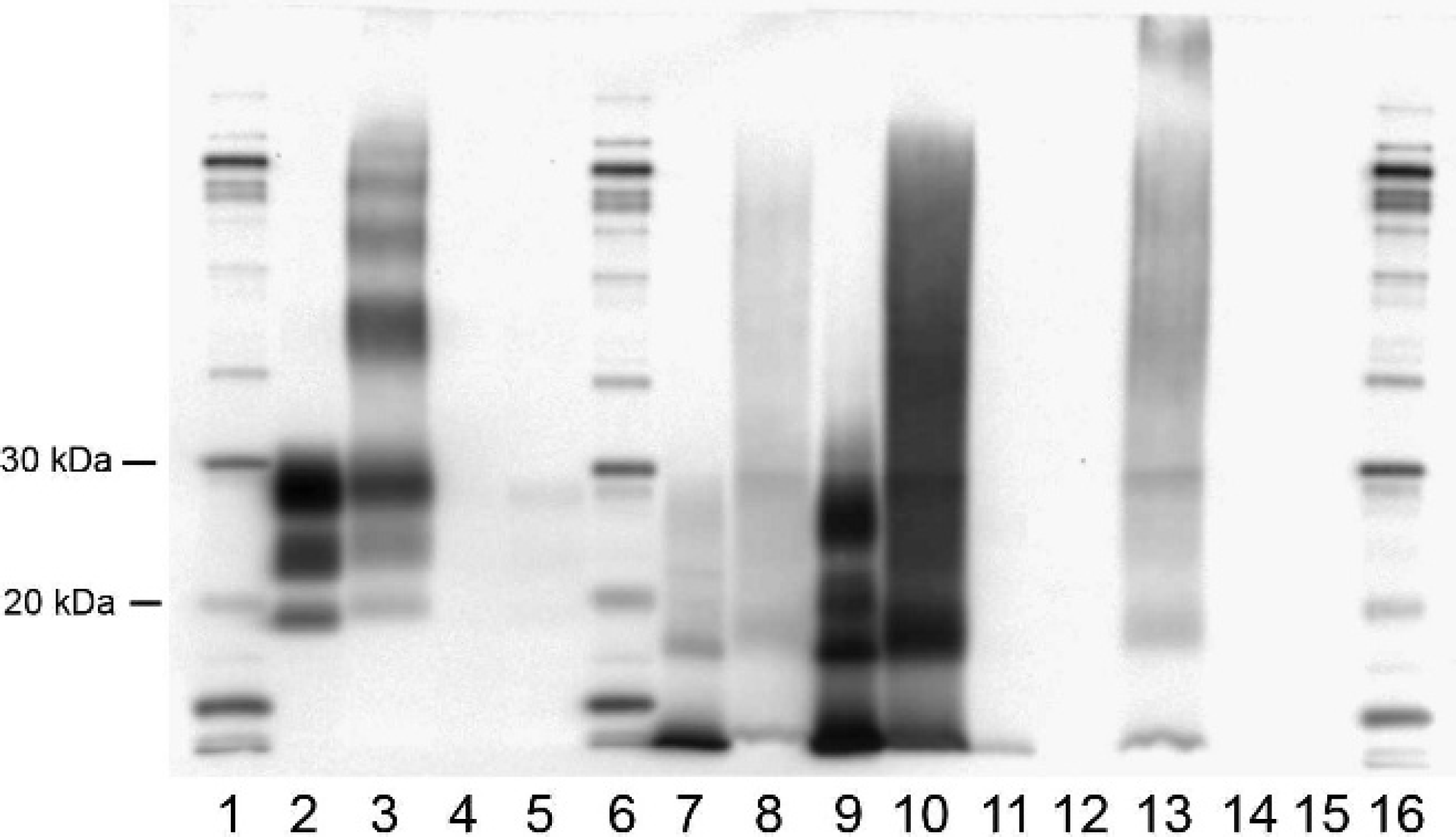
Western blot of paraffin-embedded tissues and fresh tissue from the same animals using P4 antibody, molecular weight marker (MWM), h diluted 1:100. Lane 1: MWM; lane 2: classical scrapie (CS) obex fresh; lane 3: CS obex formalin-fixed, paraffin-embedded (FFPE) tissue; lane 4: CS cerebellum fresh; lane 5: CS cerebellum FFPE tissue; lane 6: MWM; lane 7: Nor98-1 obex fresh; lane 8: Nor98-1 obex FFPE tissue; lane 9: Nor98-1 cerebellum fresh; lane 10: Nor98-1 cerebellum FFPE tissue; lane 11: Nor98-2 cerebrum fresh; lane 12: Nor98-2 obex FFPE tissue; lane 13: Nor98-2 cerebellum FFPE tissue; lane 14: negative control (NC) obex fresh; lane 15: NC obex FFPE tissue; lane 16: MWM.
Diagnostic Western blot identifying classical scrapie, Nor98 scrapie, and bovine spongiform encephalopathy (BSE) using fresh tissue and formalin-fixed, paraffin-embedded (FFPE) tissue.
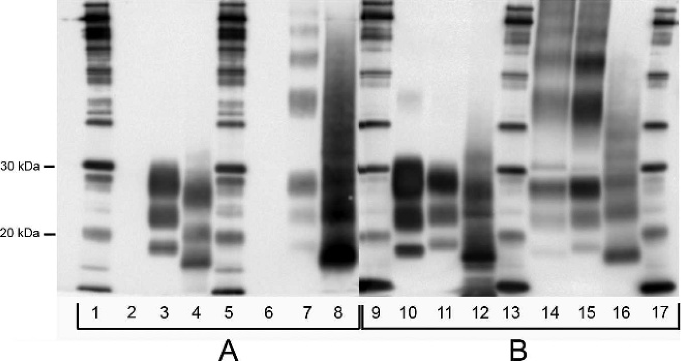
A second diagnostic comparison was made by FFPE tissue WB using classical scrapie–infected ovine obex, Nor98 scrapie–infected ovine cerebellum, and BSE-infected bovine obex (Fig. 2). Both P4 (Fig. 2, panel A) and L42 (Fig. 2, panel B) antibodies were used, and banding patterns were distinctive and diagnostic. The L42 antibody positively identified fresh and FFPE tissue samples of classical scrapie, Nor98 scrapie, and BSE. The P4 antibody, considered a discriminatory antibody, identified both classical and Nor98 scrapie with positive banding patterns, while, as expected, the fresh and FFPE tissue–positive BSE samples did not produce diagnostic banding (Fig. 2, lanes 2 and 6). 24 Classical scrapie and Nor98 scrapie cases produced bands similar in size to those described in Figure 1. Fresh obex from classical scrapie–infected animals produced 3 bands between 18 and 30 kD (Fig. 2, lanes 3 and 11). Fresh cerebellum from Nor98 scrapie–infected animals produced multiple bands with the smallest band measuring 6.5 kD (Fig. 2, lanes 4 and 12). Classical scrapie FFPE tissue banding included multiple bands, but the smallest 3 bands ranged from 18 to 30 kD (Fig. 2, lanes 7 and 15). Nor98 FFPE tissue also produced a banding pattern that approximated the fresh tissue samples. There were multiple bands, and the smallest measured less than 15 kD (Fig. 2, lanes 8 and 16). When using L42 antibody, fresh BSE-infected tissue produced 3 bands ranging from 17 to 30 kD (Fig. 2, lane 10) and BSE FFPE tissue produced multiple bands with the smallest 3 bands also ranging from 17 to 30 kD (Fig. 2, lane 14).
When brain samples from classical scrapie cases were treated with formic acid in the cassette prior to paraffin embedding, no bands were produced using the FFPE tissue for WB with P4, L42, and F99/97.6.1 as primary antibodies. Brain samples, from the same animals, not treated with formic acid did produce banding patterns typical for classical scrapie (data not shown).
Discussion
The WB assay using FFPE tissue differentiated classical scrapie, Nor98 scrapie, and BSE by producing distinctive molecular profiles for each of the prion proteins. The FFPE tissue WB technique also produced results that correlated well with the IHC PrPSc–positive staining present in cerebellum and obex regions of brain samples used in the study. Banding patterns were similar within each prion disease when comparing the fresh tissue WB results with the FFPE tissue WB results. Although more bands were typically present with the FFPE tissue WB, the molecular weights of the smallest bands matched well with the banding of the fresh tissue WB. Classical scrapie FFPE obex tissue produced 4–6 bands, while fresh tissue obex from the same animal produced 3 bands. In both instances, the smallest 3 bands ranged from 18 to 30 kD. Nor98 scrapie FFPE cerebellum tissue produced at least 4 bands for which the smallest bands had molecular weights that matched closely with the 3 bands produced using fresh cerebellum from the same animal (range: 6.5–28 kD). The diagnostic unglycosylated band measuring less than 15 kD was distinctly present in both Nor98 scrapie samples.
The diagnostic value of various regions of the brain differs depending on the strain of scrapie present. It is important to choose the most diagnostic samples to test when using FFPE tissue WB. High levels of PrPSc are not typically seen in the cerebellum of early classical scrapie cases. The FFPE tissue WB assay using cerebellum from classical scrapie cases produced weak to no banding, while this same location in Nor98 scrapie cases consistently produced diagnostic protein banding. So, while cerebellum is more diagnostic for Nor98 scrapie, obex is the preferred sample for classical scrapie when using the FFPE tissue WB technique.
The P4 antibody was chosen in the current study to evaluate classical and Nor98 scrapie cases. The banding results consistently differentiated these 2 strains of scrapie. Because P4 antibody is more selective for scrapie than for BSE, 24 a second antibody, L42, was chosen to confirm positive BSE samples (Fig. 2). Banding patterns using L42 for all positive PrPSc cases examined in the present study were similar to those seen using P4, with one exception. Nor98 scrapie FFPE cerebellum tissue produced 4 bands that ranged in molecular weight from 38 to 14.5 kD. These bands matched well with the banding pattern produced using P4 and L42 on fresh cerebellum; however, the 6.5-kD band seen with the fresh cerebellum was not present in the protein-banding pattern produced by the FFPE cerebellum tissue.
Treating brain tissue from classical scrapie–affected animals with formic acid prior to paraffin embedding reduces the ability of the FFPE tissue WB to detect protein banding using parameters of the method described in the present study. Therefore, formic acid treatment of brain tissue prior to paraffin embedding is not recommended for FFPE tissue WB.
The ability to evaluate FFPE tissue for the presence of PrPSc through WB assays offers a diagnostic platform when fresh tissues are limited or not available. For cases with ambiguous results from other testing methods including nonoptimal regions of the brain used for IHC or low volume homogenates for ELISA or fresh tissue WB, FFPE tissue WB assay could potentially provide a definitive answer. There is also potential for numerous retrospective studies to be performed based on the presence of often large libraries of FFPE tissue blocks. Comparisons can be made between samples from different geographic regions, different breeds, different genotypes within breeds representing resistance or susceptibility to prion disease, and even from different locations of the brain within the same animal. This technique may provide an additional testing method for use in the diagnosis and characterization of TSEs.
Acknowledgements
The authors would like to thank Dr. Eric Nicholson for helpful discussions and technical expertise, and Trudy Tatum, Jennifer Lamoreux, and Jim Fosse for technical support.
Disclaimer: Use of trade names or commercial products in this article is solely for the purpose of providing specific information and does not imply recommendation or endorsement by the U.S. Department of Agriculture.
Footnotes
a.
Branson Ultrasonic Corp., Danbury, CT.
b.
USB Corp., Cleveland, OH.
c.
Roche Diagnostics Corp., Indianapolis, IN.
d.
Bio-Rad Laboratories, Hercules, CA.
e.
R-Biopharm, Darmstadt, Germany.
f.
Anti-prion antibody (clone 99), Ventana Medical Systems Inc., Tucson, AZ.
g.
ECL™ Plus, GE Healthcare Technologies, Piscataway, NJ.
h.
Sigma-Aldrich, St. Louis, MO.