
Abstract
Scrapie is a naturally occurring fatal neurodegenerative disease of adult sheep and goats, one of a group of mammalian diseases known as transmissible spongiform encephalopathies (TSE) or prion diseases. Immunoassays that identify disease-associated prion protein (PrPSc) are integral to the diagnosis of scrapie and other prion diseases. Results obtained by either immunohistochemistry (IHC) or Western blot (WB) assay are generally adequate for the definitive diagnosis. Approved or accepted methods for WB diagnosis of TSEs requires the use of fresh or frozen nonfixed tissue samples, whereas formalin-fixed, paraffin-embedded tissue is required for the localization of PrPSc by IHC. Because disparate processing methods are used for these accepted diagnostic techniques, separate tissue samples are collected from the same animal. Occasions arise in which there is either insufficient quantity of tissue available to complete analysis by both techniques or initial tissue processing is incompatible with one of the assays. Also, results between the assays may differ because of the vagaries of sampling, especially in case material that contains moderate-to-low levels of PrPSc. The present article describes a method to conduct a WB assay from the same paraffin-embedded brainstem sample used for the IHC diagnosis of experimentally induced sheep scrapie.
Keywords
Scrapie is a progressive central nervous system (CNS) disease of adult sheep and goats. The disease was recognized as a clinical entity 2 or more centuries before its classification as the archetype of a family of disorders that affect mink, human, bovine, cervid, and feline, as well as ovine and caprine hosts, known as the transmissible spongiform encephalopathies (TSE). The TSEs are a class of fatal neurodegenerative diseases caused by abnormally folded prion proteins that induce templated refolding of normal cellular host prion protein (PrP c ) into an abnormal, infectious form (PrPSc). 7 Clinical signs of TSEs include less than specific neurologic abnormalities. Specific immune responses are not recognized in TSE-affected animals, thus serologic tests to obtain evidence for the presence of PrPSc are not available. Because the infectious agent is an abnormally folded protein with no known nucleic-acid component, nucleic-acid–based tests are not applicable. The common hallmark features of TSEs are progressive vacuolar degeneration of the CNS and associated presence of PrPSc. Definitive diagnosis is typically defined by postmortem pathology and the detection of PrPSc in affected tissues; however, a diagnosis is achieved solely by the detection of PrPSc in preclinical cases in which the microscopic lesions of spongiform encephalopathy are often inapparent. 6 Detection of PrPSc is generally made by means of immunoassay, with immunohistochemistry (IHC), Western blot (WB), and enzyme-linked immunosorbent assay (ELISA) based approaches available. 8,12 Whereas ELISA-based approaches serve as a rapid test, very little detail on the nature of the agent can be ascertained from them. Thus, IHC and/or WB are typically used as confirmatory tests that will also provide specific information on the disease agent.
Commonly used IHC procedures use formalin-fixed, paraffin-embedded tissues (PET), as do routine histopathologic examinations. Less commonly used, the tissue immunoblot is another method that uses PET to detect PrPSc in TSE-affected tissues. 9,10 The PET-based techniques provide information on the distribution of lesions and PrPSc at both the cellular and organ-system levels, whereas the diagnostic strength of the WB is in providing a molecular profile of the detected PrPSc. However, the WB lacks the power of localization of the agent, because sample preparation requires a homogenization step. Detection of PrPSc by WB is typically achieved by analysis of homogenized, nonfixed, fresh or frozen brainstem samples. A recent advance provides for WB analysis of homogenized, immersion-fixed tissues stored in formalin, thus expanding the opportunity of molecular strain typing in cases of TSE where fresh or frozen tissues are unavailable. 5
A method for the detection of PrPSc by WB with PET as source materials, by adaptation of the combination of techniques previously described for WB detection of PrPSc by using formalin-fixed tissues and for determination of sheep prion protein (PRNP) polymorphisms from PET, is presented in the current study. 3,5 This method affords 2 novel advantages: 1) only 1 brainstem sample processed as PET is required for the combined analyses of the molecular character of PrPSc by WB, the determination of cellular and tissue distribution of PrPSc by IHC, and PRNP sequencing; and 2) access to archival tissues only preserved as paraffin-embedded blocks that were previously unusable for WB analysis is established.
Comparison of immunohistochemistry (IHC) and Western blot (WB) intensity for positive samples.
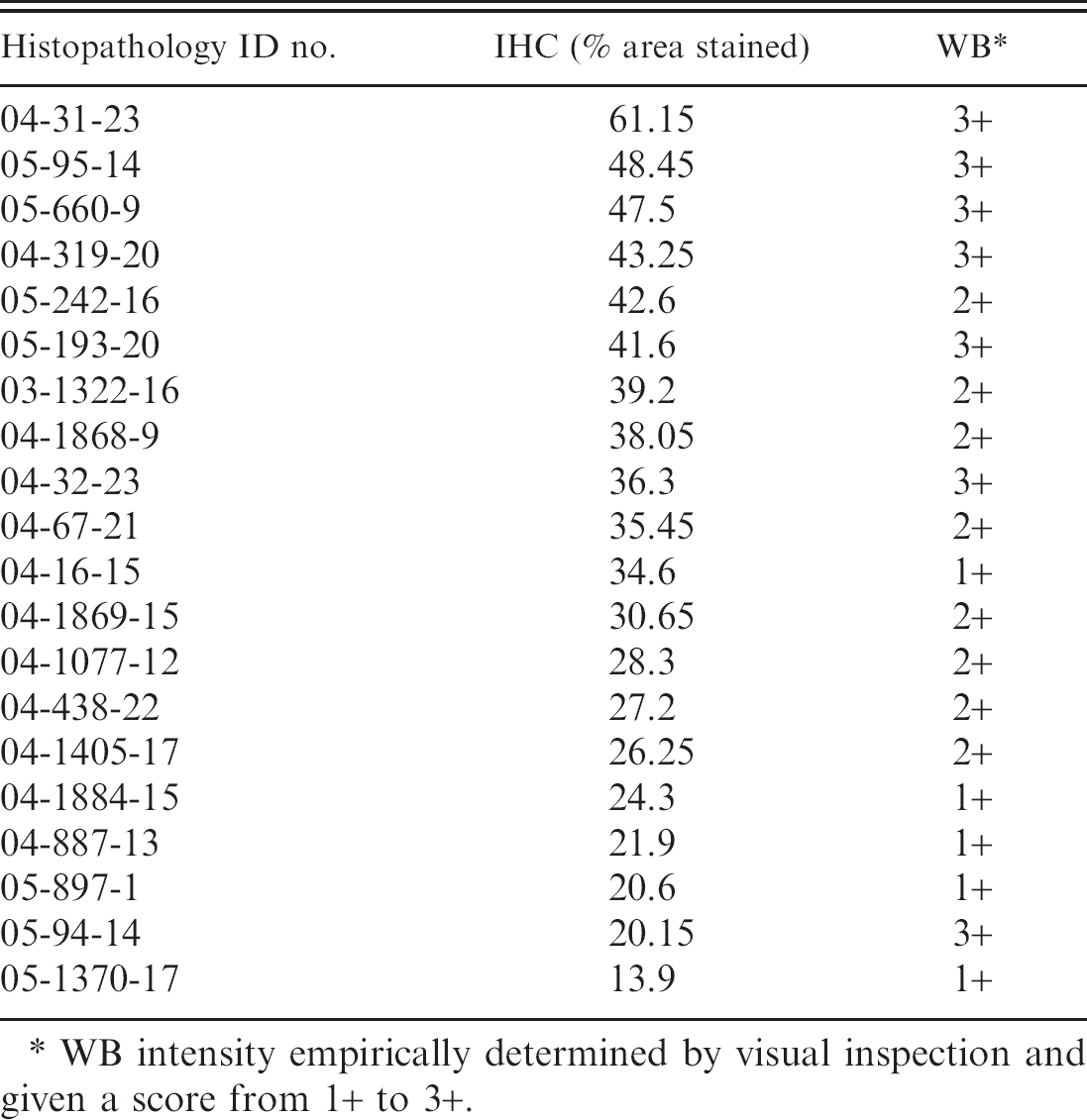
WB intensity empirically determined by visual inspection and given a score from 1+ to 3+.
The present study used archived PET samples of brainstem (obex area) collected from 27 sheep euthanized in the conductance of experimental scrapie transmission studies at the National Animal Disease Center (Ames, IA) during the time period of 2000–2005. Seventeen of the sheep were exposed to scrapie-affected brain inoculum (13–7) 2 and subsequently developed clinical signs consistent with the disease; 10 sheep served as negative controls in the transmission studies. The sheep were euthanized by an overdose of intravenously administered sodium pentobarbital, and complete necropsies were conducted. Brainstem samples collected in 10% buffered formalin were held in the fixative for a period of 3 days to 14 months and then embedded in paraffin by following routine methods. In composite, brainstem samples were immersed in formalin for a period of <30 days (n = 16), 1–6 months (n = 7), 7–12 months (n = 3), and >12 months (n = 1) before processing in paraffin blocks.
Each brainstem PET sample was sectioned at 5 μm to produce 8 serial sections. The first and eighth sections were affixed to glass slides for IHC processing; the fourth through seventh sections were collected into 1.5-ml microcentrifuge tubes for processing for WB analysis. As an aside, the second and third sections were used for DNA extraction, as previously described, 3,4 which confirmed the utility of a single tissue block as suitable for IHC, WB, and DNA extraction, which is a factor that is of significance not only for confirming the species of origin when needed but also for addressing archival TSE samples when considering the genetics of TSE susceptibility. 1
Immunohistochemistry was conducted by an automated alkaline phosphatase detection method as previously described. 2 Both the first and eighth sections obtained from each PET block were processed together to mitigate potential run-to-run variability of labeling. Computer-assisted morphometric analyses of the IHC-processed sections were performed for the purposes of determining the surface area of tissue present and the extent of PrPSc labeling on each slide microscopically examined. Magnified images of the brainstem sections were visualized on a monitor with an attached Model DC500 digital imaging camera. a The images were captured and analyzed by using Image Pro Plus version 5 software. b Brainstem surface area was automatically calculated subsequent to free-hand tracing of the tissue-section circumference. The percentage of the section immunolabeled for PrPSc was determined by color discrimination and automated calculation of labeled surface area. The results of the automated calculation are summarized in Table 1 as the mean for the 2 sections analyzed. The surface areas of the 27 brainstem sections ranged from 85 to 352 mm2, with an arithmetic mean of 189.4 mm2. The percentage of surface area of the scrapie-affected sheep (n = 17) brainstem sections labeled for PrPSc ranged from 5.3% to 61.7% with an arithmetic mean of 34.1%; the labeled area in sections from negative control sheep (n = 10) ranged from 0.05% to 6.02%, with an arithmetic mean of 0.82%. The surface area of brainstem tissue and percentage of PrPSc labeling present in the first and last serial sections obtained from each block of PET used in the study did not differ appreciably. The small amount of labeling present in the negative control sections was consistent with routinely encountered “background staining” observed with the IHC method as presented in the current report. The majority of background, or spurious, staining was localized to the walls and luminal contents of medium and small vascular elements in the brainstem tissue sections examined.
The fourth through seventh contiguous serial sections from each PET block were collected in a 1.5-ml microcentrifuge tube and processed for WB. The process parallels the approach used previously 3 and in the current study for extraction of DNA from PET for PCR amplification. Specifically, 150 μl of 0.05 M Tris (pH 7.5), 1 mM EDTA (ethylenediamine tetra-acetic acid), and 0.5% Tween 20 was added to each 1.5-ml microcentrifuge tube. The tube was placed in a 100°C water bath for 10 min followed by snap-freezing (ethanol on dry ice). The 10-min boil and snap-freeze were repeated, followed by one more 10-min boil, and then immediately centrifuged at 3,000 × g for 10 min to separate the paraffin while pelleting the tissue. If separation was incomplete, then the tube was boiled for an additional 10 min and centrifugation was repeated. The aqueous layer that contained the tissue pellet was drawn from beneath the paraffin layer by using a 16-gauge needle affixed to a syringe and was transferred to a clean 1.5-ml centrifuge tube. At this time, the tissue was further dispersed by mechanical sheering with the needle tip. The mechanically dispersed tissues were further disrupted with a Fisher Scientific Model 500 Ultrasonic Dismembrator c (40-sec intervals repeated 30 times in an ice bath with brief vortex mixing between sonication). To each sample, proteinase K d was added to a final concentration of 100 μg/ml and incubated at 37°C for 1 hr. The digestion was stopped by addition of Pefabloc e to a final concentration of 0.1 mg/ml. An equal volume of sodium dodecyl sulfate–polyacrylamide gel electrophoresis (SDS-PAGE) sample buffer was added, and the samples were placed at 99°C for 30–45 min. For WB, 10 μl of sample was loaded on a 4–20% commercially prepared SDS-PAGE gel f and run according to the manufacturer's instructions. The sample was then blotted to a polyvinylidene difluoride membrane g and blocked with 3% bovine serum albumin. Detection of PrPSc by WB was conducted by using mouse anti-PrP monoclonal antibody P4 h at a 1:10,000 dilution (0.1 μg/ml) as the primary antibody. A biotinylated sheep anti-mouse secondary antibody g at 0.05 μg/ml and a streptavidin-HRP (horseradish peroxidase) conjugate g were used in conjunction with the ECL Plus detection system g and were visualized on a Kodak In-Vivo Imaging System F. i Primary antibody incubations were conducted with the membrane at either room temperature for 1 hr or 4°C overnight (≥12 hr). Secondary antibody and streptavidin-HRP conjugate incubations were conducted at room temperature for 1 hr.
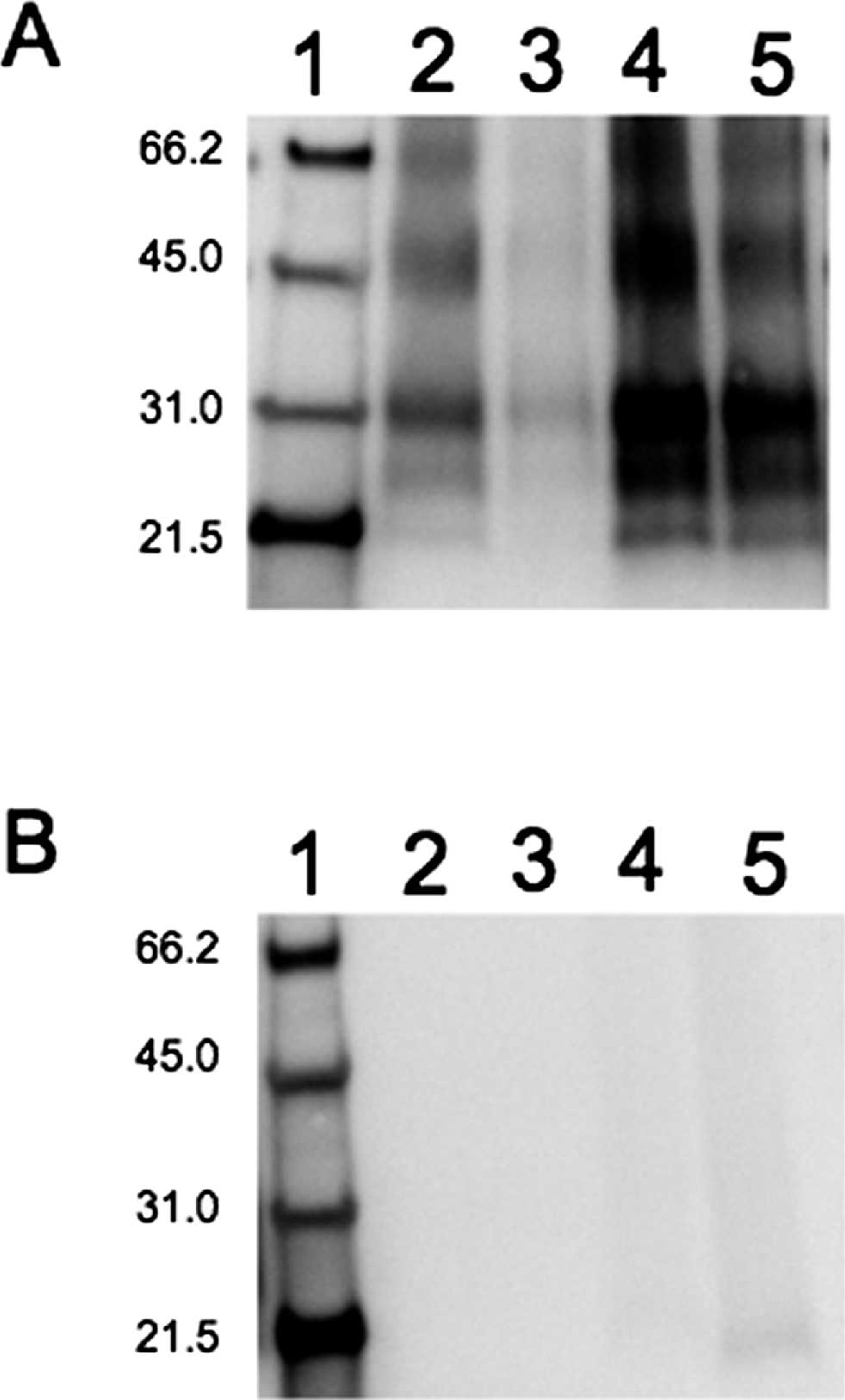
Representative Western blots of paraffin-embedded tissues.
The PET brainstem extracted for analysis by WB accurately defined the scrapie positive or negative status of all samples analyzed. Shown in Figure 1 are representative PET samples extracted as described. In total, 17 scrapie-affected and 10 negative control sheep brainstem samples were analyzed, and all 27 samples were readily categorized as to correct scrapie positive or negative status based upon the standard criteria associated with WB detection of a TSE-positive case. Specifically, the presence of the 3 bands that corresponded to di-, mono-, and unglycosylated PrPSc between 20 and 30 kD were observed in the scrapie positive samples, although not observed in negative samples (Fig. 1). These 3 bands, characteristic of proteinase K digested PrPSc and indicative of a TSE-positive sample, 11 were sufficient for an accurate WB determination of a scrapie-affected animal. As was observed for formalin-fixed tissues, proteinase K digestion was necessary only as part of the tissue disruption process. 5 The PrP c was not observed even in the absence of proteinase K digestion (data not shown). After formalin fixation, positive samples showed no consistent molecular weight shift upon proteinase K digestion migrating at a molecular weight near that expected for digested PrPSc, most likely indicating chemical or enzymatic cleavage during the fixation process. 5
A range of band intensities was observed. Qualitative assessment of the WB intensity was made and is summarized in Table 1. Overall, the band intensity score correlates well with IHC intensity by using the automated approach described here. Despite the good correlation, exceptions were observed. In 1 case, sample 05–94–14, was observed to have among the lowest IHC scores and a high WB score; although another case, sample 04–16–15, had a low WB score and an intermediate IHC score. The simplest explanation involves the combined effect of 2 variables. Namely, that labeling sensitivity varied between automated IHC runs and that, despite analysis of adjacent samples, the quantity of PrPSc may have varied even within the ∼40 μm of tissue sampled. The differences, however, were rare and of little concern, because in no case did a high IHC score pair with a low WB score, which would be indicative of reduced sensitivity of WB relative to IHC.
The methods described in the current study provided for relative quantitation of PrPSc by IHC, identification of PRNP polymorphisms, and WB confirmation of PrPSc in scrapie-affected sheep brainstem by using 8 serial 5-μm sections from PET. Thus, the aggregate of PET required for performance of the 3 assays was 40 μm from each archived paraffin-embedded block of brainstem obex. The outcome demonstrated that only 1 brainstem sample processed as PET was required for the combined analyses of the molecular character of PrPSc by WB, the determination of cellular and tissue distribution of PrPSc by IHC, and PRNP sequencing. The novel method presented may create new opportunities to study the molecular character of TSE-associated prion in archival tissues only preserved as paraffin-embedded blocks previously unusable for WB analysis.
The 27 PET samples, each from an individual sheep, processed by IHC and analyzed by image-analysis software, revealed variable amounts of PrPSc in the comparison of brainstem sections between the 17 scrapie-affected sheep; however, the PrPSc labeling did not differ dramatically in individual PET blocks as determined by IHC examination of the first and eighth contiguous sections collected. The PrPSc quantitation provided by the analysis of digital imaging corresponded to subjective assessments by light microscopy. The PrPSc labeling between individual sheep samples ranged from 5.3% to 61.7% of the tissue surface area. This demonstrated the variation of IHC labeling that can be anticipated when examining CNS tissues from TSE-affected animals. This variability may affect both sensitivity and specificity of PrPSc labeling. Variation in sensitivity between automated IHC runs is monitored by the inclusion of positive control tissue sections obtained from previously characterized PET blocks in each run. Some issues of specificity were minimized in the current study by the selection of brainstem at the level of the obex as the sample subject. In the authors' experience, relatively increased nonspecific staining is often encountered in other CNS sites, an example being the deep cerebral cortex in which beaded thread-like staining is routinely seen. The combination of cellular location and PrPSc labeling intensity assessments typically used for IHC diagnosis of scrapie, not the quantity of PrPSc in affected tissue, proved relevant in this study as predictors of WB results. Regardless of the percentage of brainstem section labeled for PrPSc, all 17 scrapie-affected, IHC-positive samples were confirmed by using the PET WB. Likewise, all 10 IHC-negative samples from healthy sheep were confirmed scrapie negative by using the PET WB.
Over the 14-month formalin fixation time frame addressed in the present study, the specific duration of time in which brainstem samples remained immersed in formalin before processing in paraffin did not affect the performance of the immunoassays, IHC and WB, in identifying scrapie-affected sheep. This is consistent with the 24-month formalin contact-time limit previously reported for WB analysis of immersion-fixed tissues stored in formalin. 5 The stability of PET for the purpose of microscopic examination is virtually unlimited, and institutionally maintained PET archives are common for this reason. The present article describes a method that provided for WB characterization of PrPSc in archived PET from scrapie-affected sheep. Contiguous sections from the same individual PET block also served as source material for sequencing PRNP amino-acid polymorphisms associated with scrapie and for quantitation of PrPSc by IHC. The benefit of using PET as the source tissue for WB and PRNP sequencing is especially relevant in instances in which scrapie is diagnosed by histopathology and/or IHC and fresh tissues are not available. Western blot and PRNP analyses of archived sheep PET could augment retrospective studies of the epidemiologic link between prion genetics and scrapie occurrence.
Acknowledgements. The authors thank Martha Church, Virginia Montgomery, and Joe Lesan for technical assistance. RAK and EMN contributed equally to this work. Mention of trade names or commercial products in this article is solely for the purpose of providing specific information and does not imply recommendation or endorsement by the U.S. Department of Agriculture.
Footnotes
a.
Leica Microsystems GmbH, Wetzlar, Germany.
b.
Media Cybernetics Inc., Bethesda, MD.
c.
Fisher Scientific Co., Waltham, MA.
d.
USB, Cleveland, OH.
e.
Roche Diagnostics Corp., Indianapolis, IN.
f.
Pierce Biotechnology Inc., Rockford, IL.
g.
GE Healthcare Technologies, Piscataway, NJ.
h.
R-Biopharm AG, Southmarshall, MI.
i.
Kodak, Rochester, NY.