
Abstract
Traditionally, diagnosis of Lawsonia intracellularis—associated proliferative enteropathy (PE) has depended on necropsy and histology. Since the establishment of the etiologic role of L. intracellularis, a number of specific polymerase chain reaction (PCR) assays have been developed for the detection of DNA in feces. The present article is a systematic review of peer-reviewed publications on the application of L. intracellularis specific fecal PCR as an antemortem diagnostic test for histologic lesions of PE in pigs. Based on this information, a range of diagnostic sensitivities (36-100%) and specificities (50-100%) of the published tests was calculated. Validity and confidence limits of the estimates varied considerably. The positive and negative predictive values of 6 different PCR assays were calculated for PE prevalence of 15%, 30%, 45%, 60%, 75%, and 90%, using a histologic case definition of PE and based on the reported test sensitivities and specificities. The simulated predictive values suggested that applying the fecal PCR assay as a diagnostic test is more likely to overestimate than underestimate the number of pigs having histologic lesions of PE under field conditions.
Introduction
Lawsonia intracellularis is an obligate intracellular bacterium and the causative agent of proliferative enteropathy (PE). Proliferative enteropathy was first described in pigs in 1931, 2 but it was not until 1973 that the presence of bacteria in the lesions was identified. 40 It took another 22 years before this bacterium was given the name Lawsonia intracellularis. 32 Proliferative enteropathy is characterized by proliferation of immature epithelial cells of intestinal crypts, resulting in crypt expansion and elongation, giving the mucosa a thickened appearance. Lawsonia intracellularis can be demonstrated in the proliferating epithelial cells. The pathology of PE has been reviewed previously. 26
Proliferative enteropathy is now recognized as one of the most economically important diseases in the swine industry worldwide. 26 Several different clinical manifestations of PE have been described in pigs. Proliferative hemorrhagic enteropathy (PHE) is classified as an acute form of PE, resulting in bloody diarrhea and sudden death. This type of PE is typically seen in adult pigs aged more than 16 weeks. In contrast, chronic PE is most common in pigs 6-20 weeks old. Chronic PE has traditionally been characterized by anorexia, reduced weight gain, wasting, diarrhea, and occasional death. 31 In 2005, a subclinical form of PE was recognized (subclinical ileitis) and defined as “when pigs become infected and have some intestinal lesions, but without clear diarrhea or weight loss.” 29
The gold standard for diagnosing PE is demonstration of proliferative intestinal lesions associated with intracellular curved bacteria in histologic sections. Several histologic techniques have been described, including staining with hematoxylin and eosin (HE), 39 Warthin—Starry (WS) silver stain, 46 modified Ziehl—Nielsen (ZN) stain, 46 in situ hybridization (ISH), 11 and immunohistochemistry (IHC). 30 Advantages and disadvantages of these and other diagnostic techniques for detection of L. intracellularis have been reviewed previously. 22
In the last 15 years, progress has been made in the development of antemortem diagnostic tests for L. intracellularis, including serologic and polymerase chain reaction (PCR) tests. Both techniques have been applied in veterinary practice and research. Lawsonia intracellularis cannot be cultured by routine methods, and PCR tests for detection of L. intracellularis in feces are now commonly used for routine diagnosis of PE in diagnostic laboratories around the world.
Diagnostic performance of a diagnostic test is characterized by its diagnostic sensitivity and specificity, positive predictive value, and negative predictive value. Diagnostic sensitivity and specificity must be differentiated from analytical sensitivity and specificity. Analytical sensitivity is the same as detection limit, which is the lowest detectable concentration of the substance or pathogen of interest. Analytical specificity is the ability of a test to give negative results in the presence of other substances or pathogens that may be considered potential cross-reactors. Analytical sensitivity and specificity do not have a mathematical definition. Analytical sensitivity and specificity depend on the conditions of the assay and the biologic properties of the test samples such as feces. They both contribute to a test's diagnostic sensitivity and specificity. The diagnostic sensitivity for a test is the proportion of test-positive animals in a population of diseased animals. The diagnostic specificity is the proportion of test-negative animals in a population of healthy animals.
Diagnostic sensitivity and specificity depend on the definition of diseased animals and occurrence of different disease manifestations and covariate factors in the population. 13 This is also the case for fecal PCR detection of L. intracellularis. Diagnostic sensitivity and specificity may be different for PCR detection of pigs subclinically infected with L. intracellularis, or detection of pigs having histologic or gross pathologic lesions of L. intracellularis—associated PE. The diagnostic sensitivity and specificity for detection of disease at the herd level also depends on disease prevalence, number of samples, and the number of test-positive samples before a herd is classified as positive.
The predictive values are very important test characteristics because they express how likely the test result reflects the true status of the animal. Positive predictive values represent the proportion of test-positive animals that are true positives. Negative predictive values represent the proportion of testnegative animals that are true negatives. Positive and negative predictive values depend on disease prevalence in the test population and the test's diagnostic sensitivity and specificity. The different aspects of diagnostic performance have to be taken into consideration for fecal PCR detection of L. intracellularis.
In clinical practice and epidemiologic field investigations, PCR detection of L. intracellularis in feces can be applied for several reasons. These include assessment of L. intracellularis as a possible cause of low productivity in a herd. Pigs can sometimes shed PCR-detectable levels of L. intracellularis without reduction in average daily gain. 6 This implies that interpretation of a positive fecal PCR result can be difficult in terms of importance to the pig or herd. The likely cause of the negative effect of L. intracellularis has been suggested as functional changes in the thickened intestinal mucosa. 31 Based on this suggestion, it can be hypothesized that histologic recognizable proliferative lesions of PE must be present for L. intracellularis to have a negative effect in a pig. In this context, it becomes important to know whether pigs with PCR-detectable levels of L. intracellularis in feces always have histologic lesions of PE or not.
The objective of the current study was to make a systematic review of diagnostic performance for PCR tests used for antemortem fecal detection of L. intracellularis—associated histologic lesions of PE in pigs. The diagnostic performance was reviewed in terms of diagnostic sensitivity, specificity, and positive and negative predictive values using a histologic case definition of PE and simulated PE prevalence. Aspects of analytical sensitivity and specificity were also reviewed.
Review methodology
Publications
A review of studies concerning PCR detection of L. intracellularis was conducted by searching available databases (up until December 13, 2009). The databases included Agricola (1970 to November 2009), Agris (1975 to June 2009), Biosis previews (1969-2009), CAB Abstracts (1973-2009 week 49), and Medline (1950 to November week 3, 2009). The search terms were “Lawsonia or proliferative enteropathy” and “PCR or polymerase chain reaction.”
The reference list of included publications was checked for other relevant publications. Duplicate records and non-peer-reviewed publications were removed.
Data extraction. Data concerning PCR evaluation were extracted from the publications. The data included the following:
use of PCR for detection of L. intracellularis in feces from pigs,
primers used,
PCR principle,
field or experimental samples,
number of samples tested,
use of spiked feces samples or purified DNA for analytical evaluation,
analytical sensitivity or detection limit,
bacteria species used to evaluate analytical specificity,
diagnostic criteria for PE or infection with L. intracellularis,
number of pigs in study,
number of PCR-positive test results among PE-positive pigs,
number of PCR-negative test results among PE-negative pigs,
age of pigs in the study,
statistical data analysis, and
authors and year of publication.
Inchusion criteria. To be included in the review, a publication had to contain data on analytical and/or diagnostic sensitivity and/or specificity for PCR tests applied for detection of L. intracellularis in fecal samples from pigs. For analytical sensitivity, only data for PCR tests applied on fecal samples are reported because feces may contain PCR inhibitors. 17 This may lead to a lower analytical sensitivity when the PCR test is applied on fecal samples, compared with samples containing purified DNA or samples from intestinal tissues. 18,28,44 For diagnostic sensitivity and specificity, only data for PCR tests applied on fecal samples and using one or more histologic technique as part of the diagnostic criteria for PE are reported.
Data analysis. To evaluate the performance of the published PCR tests, a histologic case definition of PE was applied to calculate diagnostic sensitivity and specificity with 95% confidence intervals. The necessary data were extracted from the publications. For the calculations, PE-positive pigs were defined as pigs with L. intracellularis-associated histologic lesions of PE. Various histologic techniques with different diagnostic abilities have been applied for diagnosis of PE. Currently, IHC detection of L. intracellularis and associated proliferative lesions are the reference method for histologic examinations. 16 This technique has not been applied for all PCR test evaluations. For the calculations in the current review, histology was defined as any histologic technique. The calculations for each PCR test were performed using the histologic technique applied in each PCR test evaluation study. The histologic technique used in each study is reported in the review.
The PCR test result of fecal samples obtained immediately before or at the time of necropsy was used in the calculations. Diagnostic sensitivity, specificity, and 95% confidence limits were calculated using the “Clinical Calculator 1” at VassarStats: Website for Statistical Computation (http://faculty.vassar.edu/lowry/VassarStats.html).
Positive and negative predictive values were simulated applying the calculated diagnostic sensitivities and specificities. Only those PCR tests where both diagnostic sensitivity and specificity could be calculated were included in the simulations. Assumed true prevalence for PE of 15%, 30%, 45%, 60%, 75%, and 90% was used. The positive and negative predictive values were simulated in Excel and calculated as described elsewhere 37 :
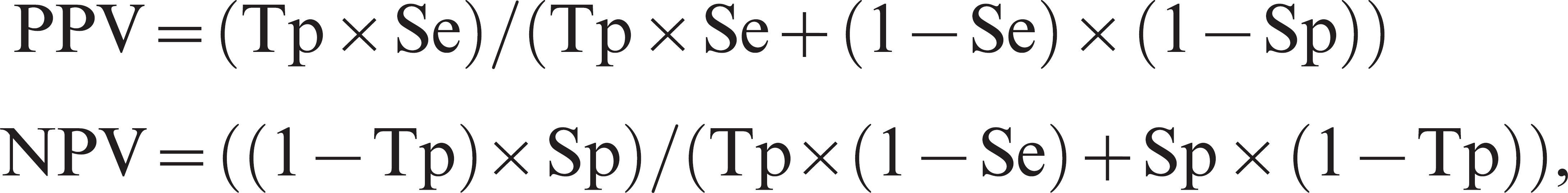
where PPV = positive predictive value, NPV = negative predictive value, Tp = true population prevalence of pigs with histologic lesions of proliferative enteropathy, Se = diagnostic sensitivity, and Sp = diagnostic specificity.
Finding
A total of 21 publications reported on the development of new PCR tests for detection of L. intracellularis in feces of pigs.
Primers. Seven different sets of primers have been applied for detection of L. intracellularis in feces of pigs. The first 2 sets of primers were based on sequences from a 375-base pair (bp) segment of a DNA fragment from the L. intracellularis DNA clone p78.18 These primers were later used in development of other new PCR tests for detection of L. intracellularis in feces of pigs. 1,3,8,10,17,21,25,28,33,34,36,38,47,48 The third set of primers was also directed towards a chromosomal DNA sequence. 44 Three sets of primers have been directed towards regions of the 16S ribosomal DNA gene. 7,24,27 The last reported primer set was directed towards a gene coding for a methylase involved in the ubiquinone/menaquinone biosynthesis. 35
Test principles. Five different PCR principles have been applied for detection of L. intracellularis in feces of pigs. The first principles were conventional and nested PCRs. 18 Conventional (simple) PCR refers to the original PCR principle, while nested PCR assay involves a second set of primers used to re-amplify the reaction products of the first PCR reaction. 18 These principles were followed by multiplex PCR assays having the ability to detect the DNA from a number of different intestinal pathogens, concurrently, in the one test sample. 10 Later, a PCR-enzyme-linked oligosorbent assay (PCR-ELOSA) was developed. 47
The ELOSA involved labeling PCR products with biotin during amplification followed by hybridization with an amine-modified internal oligonucleotide probe immobilized in microwell plates, addition of peroxidase–streptavidin complex, and measurement of spectrophotometric signal, reported as an optical density value. Finally a real-time PCR was described using a specific oligonucleotide probe labeled with 2 fluorescent dyes. This allows amplification and detection of an emission signal to occur simultaneously. 27 The real-time PCR technique has recently been applied to develop a method for quantification of L. intracellularis in porcine fecal samples. 35
A total of 8 publications reported analytical sensitivity and 16 publications reported analytical specificity for PCR tests applied on fecal samples (Table. 1). Microorganisms used to evaluate analytical specificity were Actinobacillus pleuropneumoniae, Brachyspira spp., Campylobacter spp., Clostridium perfringens types A–C, Desulfovibrio desulfuricans, Enterococcus faecalis, hemolytic and nonhemolytic Escherichia coli, Haemophilus parasuis, Mycoplasma hyopneumoniae, Myxococcus xanthus, Proteus mirabilis, Salmonella spp., Streptococcus suis.
After extraction of data, both diagnostic sensitivity and specificity from 7 publications representing 6 different PCR tests were calculated. These included 4 conventional PCRs, 1 nested PCR, and 1 real-time PCR. Three of these publications investigated cases and controls from natural sources, 25,27,44 3 publications investigated experimental cases and controls, 14,19,21 and 1 publication investigated a combination. 18 The age of the tested pigs ranged from 3 weeks to slaughter (approximately 25 weeks old). Results of the diagnostic sensitivity and specificity calculations, including 95% confidence limits, are displayed in Table 2. Diagnostic sensitivity and/or specificity could not be calculated from a total of 5 publications, though one or more histologic techniques were part of the diagnostic criteria for PE (Table. 2). Positive and negative predictive values were simulated using the reported combinations of diagnostic sensitivities and specificities. Results are displayed in Table 3.
Discussion
To the authors' knowledge, the current study is the first review of the analytical and diagnostic sensitivity and specificity of L. intracellularis–specific fecal PCR tests for PE in pigs. In the review, a clear distinction between analytical sensitivity and/or specificity and diagnostic sensitivity and/or specificity was made.
Specificity estimates were not clearly defined in a proportion of the published literature. Evaluation of a PCR test using negative fecal samples with or without additional DNA from other bacteria or samples of purified DNA from other bacteria species should be reported as analytical specificity. Diagnostic specificity should be restricted for evaluation of PCR tests in a population of healthy animals classified using a standard reference test.
Seven of the reviewed publications contained data from which it was possible to estimate both diagnostic sensitivity and specificity using histologic techniques for definition of case and control animals. Five publications reported studies where estimation would be possible based on the study design. However, diagnostic sensitivity and/or specificity were not reported in those publications, and the necessary data for calculation could not be extracted.
Other publications on PCR test evaluations used study designs without any histologic technique included in the diagnostic criteria for PE. Among these were agreement studies, where the evaluated PCR test was compared with another PCR test. In the reviewed publications, a large range in the estimates of both diagnostic sensitivity (36–100%) and specificity (50–100%) was observed. Different study designs may also explain some of the differences in diagnostic performance between the individual PCR tests.
One important explanation for observed differences may be the application of different histologic reference techniques in the different studies. The various histologic techniques have different diagnostic abilities. Hematoxylin and eosin examination may only pick up moderate to severe cases of PE in contrast to IHC. 16 Further, demonstration of L. intracellularis in tissues using IHC alone is different from demonstration of proliferative lesions alone and different from demonstration of lesions in combination with L. intracellularis. These differences between histologic techniques have to be taken into consideration in the interpretation of positive and negative test results for each PCR test. Application of different histologic techniques may explain those differences reported for the PCR tests that were evaluated in more than 1 study. However, large differences in diagnostic sensitivities and specificities for different PCR tests were observed between studies applying the same histologic techniques.
Four studies evaluated the PCR tests under experimental conditions. The experimental settings may not have reflected the situations where the tests will be applied in epidemiologic field studies and in veterinary practice. Therefore, the estimates of diagnostic sensitivity and specificity from those studies should be interpreted with caution. Studies using field samples are necessary for correct estimates for the field application of the PCR tests. These studies are most likely to include animals representing different disease manifestations of PE and include covariates with influence on diagnostic performance. That is, these studies may be closer to the situations where the tests will be applied in practice and research.
Analytical sensitivity (Se) and specificity (Sp) of fecal polymerase chain reaction (PCR) detection of Lawsonia intracellularis. *
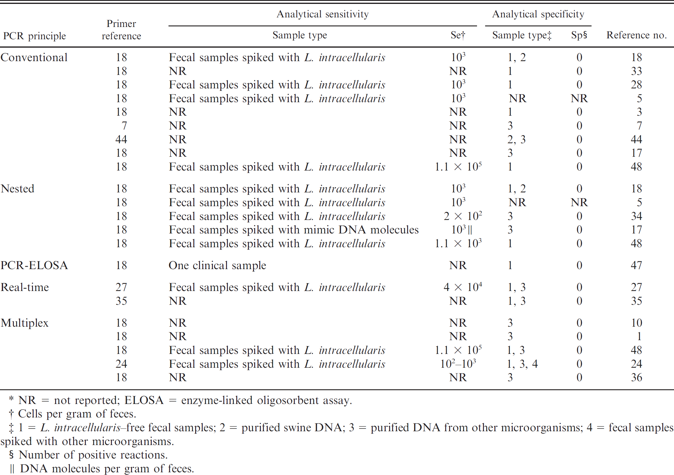
NR = not reported; ELOSA = enzyme-linked oligosorbent assay.
Cells per gram of feces.
1 = L. intracellularis–free fecal samples; 2 = purified swine DNA; 3 = purified DNA from other microorganisms; 4 = fecal samples spiked with other microorganisms.
Number of positive reactions.
DNA molecules per gram of feces.
Some of the reviewed studies were characterized by a small sample size. This is reflected in the wide 95% confidence limits for the estimates of diagnostic sensitivity and specificity. Interpretation of these studies should be done with caution. The small sample sizes could lead to underestimation or overestimation of the diagnostic sensitivity and/or specificity because of random error. Such random error concerning test evaluations should be taken into consideration when applying the tests for prevalence estimation or risk factor analysis. 12
Predictive values were calculated for prevalence ranging from 15% to 90% for the PCR assays where both diagnostic sensitivity and specificity were available. The 3 PCR tests with diagnostic specificity less than 1 had low positive predictive values at lower prevalence. Applying these tests in a herd with prevalence below 15% means that test-positive animals are roughly more than 40% likely not to have histologic lesions of PE.
The 4 PCR assays with diagnostic sensitivity less than 1 had low negative predictive values at higher prevalence. Three of these PCR tests had diagnostic sensitivity of 50% or less. Applying one of these tests in a herd with prevalence above 60% means that testnegative animals in fact are roughly more than 50% likely to have histologic lesions.
Previous studies in Denmark have reported within-herd prevalence for L. intracellularis of 5–100% with mean/mode around 15–30%. 42 From the simulated predictive values, it can be observed that the risk of false positives will usually exceed the risk of false negatives. This applies for the reported PCR tests used for antemortem detection of L. intracellularis–associated histologic lesions of PE under Danish conditions.
In practice, where the prevalence is unknown, a PCR test with high sensitivity could be combined with a PCR test with high specificity, increasing the validity of the diagnosis. At the herd level, another possibility is to raise the cutoff value for herd diagnosis (i.e., allow more positive individuals before classifying herds as positive).
Diagnostic sensitivity (Se) and specificity (Sp) of fecal polymerase chain reaction (PCR) detection of Lawsonia intracellularis-associated histologic lesions of proliferative enteropathy (PE).*
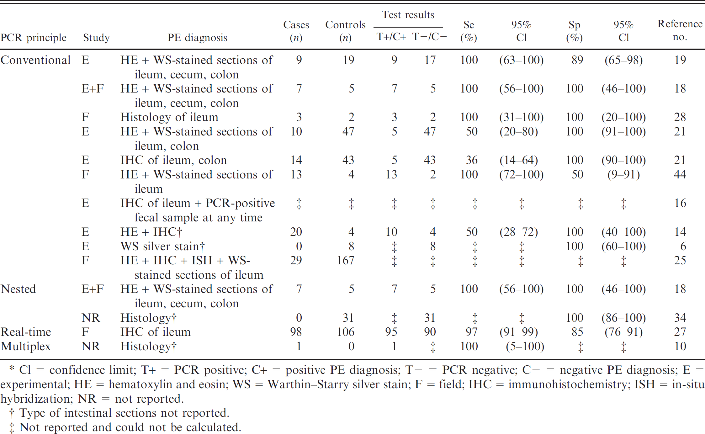
Cl = confidence limit; T+ = PCR positive; C+ = positive PE diagnosis; T- = PCR negative; C− = negative PE diagnosis; E = experimental; HE = hematoxylin and eosin; WS = Warthin-Starry silver stain; F = field; IHC = immunohistochemistry; ISH = in-situ hybridization; NR = not reported.
Type of intestinal sections not reported.
Not reported and could not be calculated.
The reported estimates of diagnostic sensitivity/ specificity and predictive values only imply for a case definition of PE based on histologic techniques as the standard reference tests. Other values may be obtained using other case definitions, such as occurrence of pigs having gross PE lesions, detection of pigs shedding L. intracellularis, or detection of pigs infected with L. intracellularis.
For detection of infected pigs, the diagnostic sensitivity will depend on the level of L. intracellularis cells per gram of feces in the positive pigs and the analytical sensitivity for the PCR test. The diagnostic specificity will depend on the analytical specificity. Analytical sensitivity has been reported in the range of 102–105 cells per gram of feces. Nested PCR techniques are in general considered to be the most sensitive. However, on average, the reported nested PCRs were equally sensitive compared with the conventional PCRs. This finding may reflect differences in the applied DNA extraction methods or PCR reaction conditions.
The level of L. intracellularis cells per gram of feces in positive pigs are to some extent unknown. Pigs with lesions of PE have been reported to excrete in the range of 5 × 10⁴ to 7 × 10⁸ L. intracellularis per gram of feces. 7,34,41 Recently, a minimum excretion of 2.2 × 10³ L. intracellularis per gram of feces in a pig without clinical signs under natural conditions has been detected by quantitative real-time PCR (Marie Stalh, unpublished data, 2009). These excretion levels are below the analytical sensitivity for some PCR tests. Others have reported that some pigs are PCR negative in feces but PCR positive in samples from intestinal mucosa. 21 More research is needed in this area to estimate diagnostic sensitivity for fecal PCR detection of pigs infected with L. intracellularis.
It seems that the analytical specificity is close to perfect for the PCR tests. In the reviewed publications, different strains of bacteria species and negative fecal samples were tested. No false-positive test results were reported. This could imply that the diagnostic specificity for detection of pigs infected with L. intracellularis is close to 100% (in contrast to detection of pigs having histologic lesions of PE). In 6 publications, analytical specificity was only evaluated using purified DNA from a number of different bacteria. This may not be sufficient when taking the large number of different microorganisms in a fecal sample into consideration. Testing a large number of negative fecal samples from different pigs could be the best evaluation of analytical specificity for future PCR tests.
Positive (PPV) and negative (NPV) predictive values of fecal polymerase chain reaction (PCR) detection of Lawsonia intracellularis–associated histologic lesions of proliferative enteropathy.*
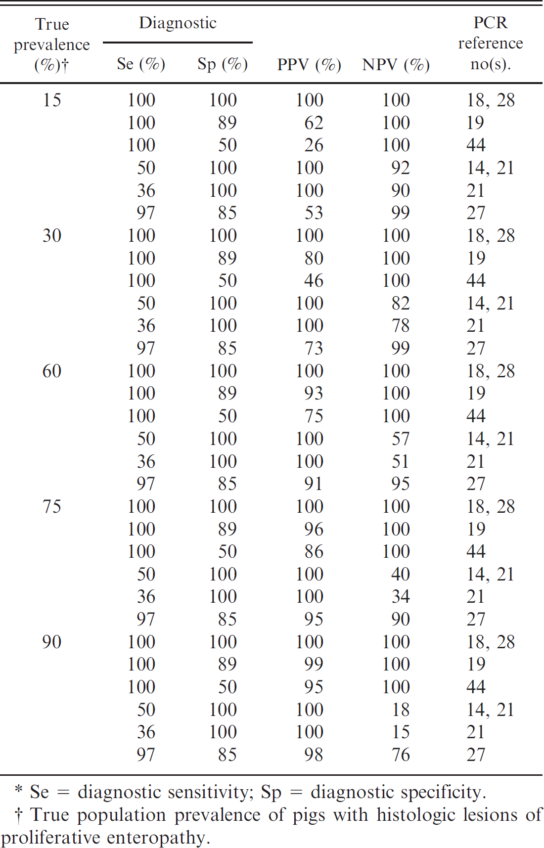
Se = diagnostic sensitivity; Sp = diagnostic specificity.
True population prevalence of pigs with histologic lesions of proliferative enteropathy.
A case definition where the PCR test and the histologic examination were applied to the same animal within a few days was applied in the current review. In this case definition, the PCR tests were evaluated for the ability to reflect the animal's disease status at the time of sampling. L. intracellularis can be detected in feces using PCR from day 7 to 8 after infection. Shedding of L. intracellularis continues on average for approximately 15 days. 4,14,15,19,21,23,43,45
Further, some have reported that pigs may shed L. intracellularis intermittently over a prolonged period of time (up to 8–12 weeks). 6,15,41 This implies that other estimates of diagnostic sensitivity/specificity and predictive values may be obtained if the PCR tests are evaluated for the ability to reflect the animal's disease history or future disease development. Such anamnestic and prognostic application for the PCR tests is important for interpretation of a positive test result's importance for the pig or herd. These aspects need further evaluation.
Conclusion
Estimates of the diagnostic sensitivity and specificity of L. intracellularis–specific fecal PCR as an antemortem diagnostic test for pigs with histologic lesions of PE were obtained from 7 publications. These represented 6 different PCR tests. A range in both diagnostic sensitivity (36–100%) and specificity (50–100%) were reported. Validity and confidence limits of the estimates varied considerably. From the simulated predictive values, it was concluded that the risk of false positives will usually exceed the risk of false negatives when applying the PCR tests under conditions where up to 30% of the pigs in a herd have histologic lesions of PE. The histologic technique applied for evaluation of each PCR test has to be taken into consideration in the interpretation of positive and negative test results for each PCR test.