
Abstract
Quantitative polymerase chain reaction (qPCR) tests for detection and quantification of Lawsonia intracellularis in feces from pigs have been developed. The objective of the current study was to evaluate the diagnostic performance of a fecal qPCR test for detection of nursery pigs with L. intracellularis–associated proliferative enteropathy (PE) under field conditions. Furthermore, the diagnostic performance for different subpopulations of pigs was investigated, including pigs infected or noninfected with Porcine circovirus-2, Brachyspira pilosicoli, and Escherichia coli. The diagnostic performance was evaluated in terms of diagnostic sensitivity and specificity. Data from pigs originating from 20 herds with antibiotic treatment requiring diarrhea outbreaks from a prior study were reused. Before treatment, pigs were randomly selected for histological and immunohistochemical examination of intestinal segments and fecal quantification of L. intracellularis by qPCR. A total of 313 pigs (157 without diarrhea, 156 with diarrhea) were included in the statistical analysis, and 37 pigs (11.8%) were classified as PE positives (defined as proliferative histological lesions in combination with L. intracellularis demonstration by immunohistochemistry). Lawsonia intracellularis was detected by qPCR in feces from 91 pigs (29.1%). A nonparametric receiver operating characteristic (ROC) analysis provided an area under the ROC curve (0.93) and an optimal cutoff value of 4.8 log10 L. intracellularis bacteria/g feces. This cutoff provided a diagnostic sensitivity of 0.84 and diagnostic specificity of 0.93. The diagnostic sensitivity and specificity were significantly different between herds (P < 0.0001). Numerically, diagnostic sensitivity and specificity were different between subpopulations of pigs, but no significant differences were demonstrated.
Introduction
Proliferative enteropathy (PE) is caused by Lawsonia intracellularis, which is a major cause of diarrhea and decreased productivity in weaned pigs and considered among the most economically important infections in the swine industry worldwide. 8 Confirmation of PE-associated clinical disease problems has traditionally been based on demonstration of proliferative intestinal lesions associated with intracellular curved bacteria in histological sections. Currently, immunohistochemistry (IHC) is considered the best reference test for diagnostic confirmation of PE by demonstration of L. intracellularis in proliferative intestinal lesions. 5 Fecal polymerase chain reaction (PCR) testing is now used routinely for diagnosis of L. intracellularis–associated diarrhea in diagnostic laboratories around the world. 13 Therefore, it is important to know how fecal PCR results relate to the traditional diagnosis of PE in the individual pig. Such comparison has previously been reviewed for traditional fecal PCR tests, 13 indicating diagnostic sensitivities of 36–100% and diagnostic specificities of 50–100%.
Quantification of bacteria by quantitative PCR (qPCR) tests has been used to establish levels of bacterial load, which are indicative of clinical disease rather than subclinical infection, for a number of pathogens.1,2,16 Quantitative PCR tests have been evaluated for quantification of L. intracellularis in fecal samples. 12 Quantification of the number of L. intracellularis bacteria in feces by such qPCR tests could potentially increase the diagnostic performance of fecal PCR testing.
The objective of the current study was to evaluate the diagnostic performance of fecal qPCR testing for detection of nursery pigs with L. intracellularis–associated PE under field conditions. Furthermore, the association between different covariables and the diagnostic performance of the qPCR testing was investigated. The diagnostic performance was evaluated in terms of diagnostic sensitivity and specificity.
Materials and methods
Design
A data set collected in 2008–2009 for investigation of outbreaks of diarrhea in pigs 10–70 days postweaning that required treatment was reused in the current investigation. The data originated from 313 pigs (157 without diarrhea, 156 with diarrhea) that were examined at the beginning of diarrhea outbreaks in 20 herds. Recruitment of herds, selection of diarrheic pigs, sample processing, and laboratory investigations have previously been reported. 14 Additional materials and methods not previously reported are presented below.
Herd examinations
Eight pigs without diarrhea were randomly selected using the same selection process as previously reported for the diarrheic pigs. 14 Information on herd, room, pen, and pigs was collected through questioning of the farmers and observations by the corresponding author. The average size of pigs in a pen compared to other pens in the same batch and the average size of a pig in a pen compared to pen mates were estimated as a subjective assessment by the observer. Pen hygiene was assessed as high (dry conditions in the entire pen), medium (wet and/or moisture conditions in part or the entire pen), or low (fecal contamination in the entire pen).
Microbiological examination of fecal samples
Fecal samples were examined for Salmonella spp., according to the Swedish Standards Institute ISO 6579:2002/Amd 1:2007, and for Escherichia coli and Brachyspira spp. as previously described. 11 Fecal samples were subjected to qPCR testing for L. intracellularis, B. pilosicoli, and Porcine circovirus-2. All qPCR assays were run as previously described.6,14,15
Statistical analysis
The diagnostic performance of the L. intracellularis qPCR test was evaluated relative to a traditional PE diagnosis in the individual pig. Pigs having histological proliferative intestinal lesions with demonstration of L. intracellularis by IHC within the proliferative lesions were classified as PE positive. Based on this classification, diagnostic sensitivity and specificity were calculated, and the relation to excretion load in feces was investigated by a nonparametric receiver operating characteristic (ROC) analysis. The excretion load providing the maximum diagnostic performance (maximum value of diagnostic sensitivity and specificity) was determined. The effect of covariables on diagnostic sensitivity and specificity was evaluated. The effect of covariables on diagnostic sensitivity was investigated in the population of diseased animals (positive by traditional PE diagnosis). The effect of covariables on diagnostic specificity was investigated in the population of nondiseased animals (negative by traditional PE diagnosis). Both analyses were performed in a similar way. First, an unconditional screening of covariables was performed using Fisher exact test (selection criteria: P < 0.25). Selected variables were included as fixed effects in 2 separate generalized mixed linear models (1 for sensitivity and 1 for specificity) with the herd included as a random effect. Nonsignificant variables (P < 0.05) were removed by manual backward stepwise regression. All statistical analyses were performed using commercial software. a
Results
From the data set, a total of 313 pigs (157 without diarrhea, 156 with diarrhea), from a single diarrhea outbreak in 20 different herds, were included in the statistical analysis. The occurrence of proliferative histological lesions, demonstration of L. intracellularis by IHC, and qPCR test results have previously been reported for the examined pigs with diarrhea. 14 The occurrence of proliferative histological lesions, demonstration of L. intracellularis by IHC, and qPCR test results have not previously been reported for the examined pigs without diarrhea.
In pigs without diarrhea, L. intracellularis was detected by qPCR in 51 of the pigs (32.5%), providing a total of 91 qPCR-positive pigs (29.1%) in feces for the nonparametric ROC analysis. Among the qPCR-positive pigs without diarrhea, 22 pigs (43.1%) were below the dynamic range (4.3 log10 bacteria/g feces) of the qPCR, 29 pigs (56.9%) were in the dynamic range, and no pigs were above the dynamic range (8.3 log10 bacteria/g feces). The median excretion for all positive pigs was 4.56 log10 bacteria/g feces. The occurrence of PE (proliferative histological lesions in combination with L. intracellularis demonstration by IHC) was not significantly different between pigs with diarrhea (20 pigs; 12.8%) and pigs without diarrhea (17 pigs; 10.8%; P = 0.59), providing a total of 37 PE-positive pigs (11.8%; proliferative histological lesions in combination with L. intracellularis demonstration by IHC) for the nonparametric ROC analysis. Among the PE-positive pigs, 14 (37.8%) were positive in ileum only, 1 (2.7%) was positive in jejunum only, and 1 (2.7%) was positive in colon only. Furthermore, 11 pigs (27.0%) were positive in both jejunum and ileum, 1 (2.7%) was positive in both ileum and colon, and 9 pigs (24.9%) were positive in jejunum, ileum, and colon.
The result of the nonparametric ROC analysis is displayed in Figure 1. The area under the ROC curve was 0.93. A cutoff value of 4.8 log10 L. intracellularis bacteria/g feces was the fecal load with the maximum diagnostic performance (maximum value of diagnostic sensitivity and specificity) providing a diagnostic sensitivity of 0.84 and specificity of 0.93. A total of 49 pigs (15.7%) were above the cutoff value. Using this cutoff, the diagnostic specificity ranged from 0.60 to 1.0 between herds (P < 0.0001) and the diagnostic sensitivity ranged from 0 to 1.0 (P < 0.004). Numerically, diagnostic sensitivity and specificity were different between different subpopulations of pigs (covariables). For diarrhea and bacterial coinfections, these differences were not statistically significant. For other covariables, diagnostic sensitivity and/or specificity were statistically significant different using Fisher exact test (Table 1). However, none of the covariables were significantly associated to either diagnostic sensitivity or specificity in the generalized linear mixed models (P > 0.05).
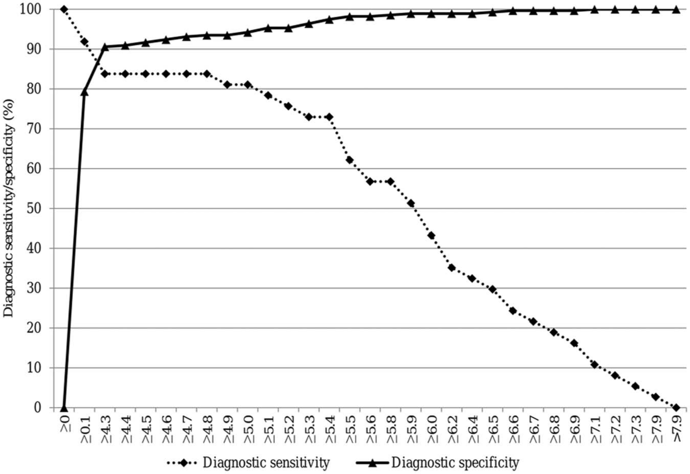
Two-way receiver operating characteristic curve displaying association between fecal load of Lawsonia intracellularis (x-axis) and diagnostic sensitivity/specificity (y-axis, %) relative to histological demonstration of proliferative intestinal lesions containing Lawsonia intracellularis within the same lesions (n = 313).
Investigation of differences in diagnostic sensitivity (DSn) and specificity (DSp) in subpopulations of pigs when using 4.8 log10 Lawsonia intracellularis bacteria/g feces as cutoff value for classifying a pig as test positive: diagnostic sensitivity and specificity determined relative to histological demonstration of proliferative intestinal lesions in combination with demonstration of L. intracellularis in the lesions.
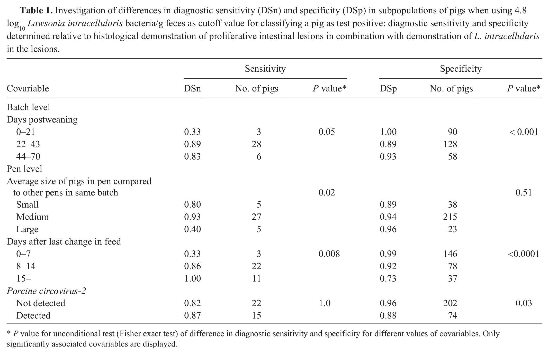
P value for unconditional test (Fisher exact test) of difference in diagnostic sensitivity and specificity for different values of covariables. Only significantly associated covariables are displayed.
Discussion
The diagnostic performance of the qPCR test was evaluated relative to the demonstration of proliferative histological lesions in combination with the demonstration of L. intracellularis by IHC in the same lesions. Using IHC as the reference test, diagnostic sensitivities of 0.36, 0.50, and 0.97 have previously been reported for fecal PCR test in pigs.4,7,9 Using the ROC determined cutoff value of 4.8 log10 L. intracellularis bacteria/g feces, the diagnostic sensitivity (0.84) of the qPCR falls within these limits, but was lower compared to the only other previously reported real-time PCR test (diagnostic sensitivity = 0.97). 9 Diagnostic specificities of 1.0, 1.0, and 0.85 have previously been reported.4,7,9 The diagnostic specificity (0.93) of the qPCR falls within these limits and was higher compared to the only previously reported real-time PCR (diagnostic specificity = 0.85). 9 Some of the differences in diagnostic performance between these PCR tests may be caused by differences in study designs. In the current study, randomly selected animals from natural outbreaks were examined, while previous studies were experimental or performed on pigs from laboratory submissions. Another possible explanation is differences in the PCR assays used, including different DNA extraction procedures. The most comparable study 9 used a similar (nonquantitative) real-time PCR test on field samples from laboratory submissions. Compared to this previously described test, 9 applying qPCR testing with a cutoff value of 4.8 log10 L. intracellularis bacteria/g feces (i.e., only pigs with >4.8 log10 L. intracellularis bacteria/g feces are classified as test positive) will increase the diagnostic specificity but decrease sensitivity. This will result in more false-negative test results but fewer false-positive test results. In other words, when using the reported cutoff, some test-negative pigs (excreting <4.8 log10 L. intracellularis bacteria/g feces) will actually have L. intracellularis–associated PE while some test-positive pigs (excreting >4.8 log10 L. intracellularis bacteria/g feces) will actually not have L. intracellularis–associated PE. However, these misclassifications will be reduced compared to the previously described test. 9 The prevalence of PE-positive pigs in the current study confirms the results of a previous report that found that false-positive test results are the largest concern under Danish conditions. 13 Therefore, using qPCR testing with a cutoff value of 4.8 log10 L. intracellularis bacteria/g feces under Danish conditions will result in fewer problems with overestimation of pigs with PE lesions.
The excretion load providing the maximum diagnostic performance (maximum value of diagnostic sensitivity and specificity) was selected as the cutoff in the current study. This was done based on the assumption that both false-positive and false-negative test results have equal practical importance. The ROC analysis demonstrated the significance of increasing or decreasing levels of L. intracellularis in feces compared to the traditional PE diagnosis. That is, the ROC analysis illustrated that increasing or decreasing the cutoff value will change diagnostic sensitivity and specificity in relation to the traditional PE diagnosis accordingly. If a higher diagnostic sensitivity is desired, the cutoff value can be lowered (pigs with less L. intracellularis bacteria in feces will be classified as test positive) or the opposite if a higher diagnostic specificity is optimal. This is similar to other traditional tests with continuous results such as optical density values obtained from serological enzyme-linked immunosorbent assays. In other words, if a practicing veterinarian is most concerned about identifying the occurrence of pigs without L. intracellularis–associated PE, a higher cutoff could be used (increasing diagnostic specificity with increasing bacteria cells per gram feces as the cutoff value) or vice versa. Furthermore, obtaining qPCR results with a higher number of bacteria per gram implies that the pigs are more likely to have L. intracellularis–associated PE compared to pigs with a lower number of bacteria. Such information and flexibility in diagnostic performance in relation to the diagnostic purpose should probably be considered one of the real benefits of quantifying L. intracellularis bacteria in feces.
Diagnostic sensitivity and specificity have previously been reported to vary between subpopulations of hosts for diagnostic tests. 3 Therefore, the effect of different covariables were investigated. Both diagnostic sensitivity and specificity were different between herds. This indicates that interpretation of positive and negative test results depends on the herd. Hypothetical explanations for such differences could be different strains of L. intracellularis, different levels of infection severity, or different timing in relation to progression of the infections between the individual herds. A number of potential herd-specific factors could explain such differences and currently it is not possible to predict the diagnostic performance in a specific herd, so the average values should be used. However, this aspect could be further evaluated or can at least be used to explain unexpected test results in practice. Apart from differences between herds, none of the other covariables were significantly associated with diagnostic sensitivity and specificity. Interestingly, this also included the diarrhea status and the simultaneous presence or absence of other infections. These results indicate that other infections do not interfere with the diagnostic performance of the qPCR in the field. However, for some covariables, numerical differences were demonstrated, indicating that interpretation of positive and negative test results may depend, for example, on simultaneous clinical signs. This could be an issue to further address in future studies.
The nonsignificant association between PE (proliferative histological lesions in combination with L. intracellularis demonstration by IHC) and diarrhea status was interesting from a pathogenesis point of view. It is possible that proliferative changes and/or L. intracellularis may have to occur above a certain level to induce clinical diarrhea. This aligns with the malabsorption mechanism believed to be linked with the pathogenesis of L. intracellularis–associated diarrhea. 10
In conclusion, a good diagnostic performance of fecal qPCR testing for detection of nursery pigs with L. intracellularis–associated PE under field conditions was demonstrated. Application of a ROC-determined, test-positive cutoff value of 4.8 log10 L. intracellularis bacteria/g feces will result in less overestimation of pigs with PE under Danish conditions. The diagnostic performance was significantly different between herds. No other covariables including simultaneous intestinal infections were associated with the diagnostic performance.
Footnotes
Acknowledgements
The authors thank the personnel at the National Veterinary Institute, Technical University of Denmark for their technical assistance.
a.
Stata/IC version 11, StataCorp LP, College Station, TX.
Declaration of Conflicting Interests
The author(s) declared no potential conflicts of interest with respect to the research, authorship, and/or publication of this article.
Funding
The author(s) disclosed receipt of the following financial support for the research, authorship, and/or publication of this article: This work was supported by the Danish Ministry of Food, Agriculture and Fisheries.